Author: ModernMedia

Cystic Fibrosis Damages the Immune System Early on

Photo by Robina Weermeijer on Unsplash

Despite new medication, cystic fibrosis often leads to permanent lung damage. Working with an international team, researchers from the Technical University of Munich (TUM) have discovered that the disease causes changes in the immune system early in life, presumably even in newborns. These changes lead to frequent inflammation and are not affected by drugs targeting the altered mucus production.

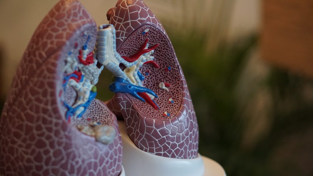
Cystic fibrosis is caused by hereditary genetic mutations that impair or halt the production of the CFTR protein. The respiratory tract is most severely affected. There, the mucus becomes so viscous that pathogens like bacteria cannot be removed by coughing. The result is often a deadly cycle of infection and inflammation.

In recent years, doctors have started using so-called CFTR modulator therapies to enhance the protein’s function. This reduces mucus formation and significantly improves the quality of life for those affected. However, clinical studies show that airway inflammation continues to occur frequently. In older patients, the decline in lung function seems unstoppable.

Current research aims to uncover additional processes in cystic fibrosis. “We specifically looked at how the immune system behaves in cystic fibrosis before the cycle of infection and inflammation begins,” said Prof. Nikolai Klymiuk from TUM. He is part of the international team that recently published a study on cystic fibrosis in Science Translational Medicine.

Immature immune cells in blood samples from children

The researchers found that in blood samples from children with cystic fibrosis and biological material from pigs with the same genetic defect, certain cells of the innate immune system are immature. This makes them less effective at fighting bacteria. Pigs with cystic fibrosis also showed an increased number and significantly altered composition of immune cells in the lungs at birth. The strong resemblance between the immune systems of pigs and humans suggests that this finding likely applies to human patients as well.

‘Emergency program’ responsible?

According to the authors, one possible explanation for the changes in the immune system could be a kind of “emergency program”. The program stimulates the body to produce a large number of immune cells particularly quickly or over a longer period of time. One consequence is the formation of immature immune cells, which could contribute to the fatal cycle of infections and inflammation in cystic fibrosis: Although immune cells are present in the lungs, they are ineffective and cause damage to the lung tissue without preventing infections in the long term.

Since immune cells generally produce only very small amounts of CFTR, the research team believes that the influence of cystic fibrosis on the immune system is indirect. This could explain why defective immune reactions cannot be treated well with novel CFTR modulator therapies.

Changes not a result of frequent infections

“We don’t yet know exactly why the immune cells in cystic fibrosis show such changes,” says Nikolai Klymiuk, Professor of Cardiovascular Translation in Large Animal Models. “However, we can show that these occur early in life. They then persist in the further course of life.” According to Klymiuk, although altered immune cells were known from blood samples of adults with cystic fibrosis, they were seen as a consequence of the numerous infections.

“To enable people with cystic fibrosis to live without symptoms, we probably need to tackle the disease on several levels,” said Klymiuk. “We hope our work will help us better understand the causes of the defective immune system and correct them in the future”

Source: Technical University of Munich (TUM)

Researchers Discover New Strength-boosting Mechanism in Androgens

Photo by Jonathan Borba on Unsplash

Researchers at Leipzig University’s Faculty of Medicine and Shandong University in China have discovered a new mechanism that is used by a key androgen essential for muscle and bone function. The findings could lead to the development of new drugs with fewer side effects, for use in applications such as strengthening the muscles of immobile patients. The researchers have published their findings in the prestigious journal Cell.

The most powerful of the androgens is called 5α-dihydrotestosterone (5α-DHT). Among other things, it is essential for bone and muscle function and for the development of secondary male sexual characteristics during puberty. As a driver of bone and muscle formation, 5α-DHT increases bone mineral density and promotes skeletal muscle growth to increase muscle strength. 

In this international study, the scientists were able to show that one of the adhesion G protein- coupled receptors – GPR133 – is activated by the androgenic steroid hormone 5α-DHT.

“This activation can, among other things, increase the contractile force of skeletal muscles, and our study also uses a newly developed, potent activator of this receptor to specifically trigger this effect,” says Professor Ines Liebscher, Professor of Signal Transduction at Leipzig University and co-leader of the study.

Increasing muscle strength with the chance of significantly fewer negative effects of androgens

Activation of GPR133 by the novel agonist AP503 increases muscle strength without triggering a specific negative effect that is otherwise observed when androgens are administered. For example, increased and prolonged exposure to testosterone can promote the development of prostate cancer, as evidenced by tissue changes in the prostate in mice after only two weeks of androgen administration. This side effect has not yet been observed with AP503.

In addition, the current study uses structural biology methods to elucidate the molecular basis of the interaction between the steroid hormone, the substance AP503 and GPR133. This will allow the activator to be specifically optimised and further developed into a new therapeutic agent. This could lead to the development of new muscle-strengthening drugs with a lower side-effect profile.

This publication is the result of a long-standing and successful collaboration between the Rudolf Schönheimer Institute of Biochemistry and the research group of Professor Jin-Peng Sun at Shandong University in China. The researchers are currently working on several follow-up studies to further investigate the use of AP503 in disease processes and the role of GPR133 in the organism. Here the data were analysed in animal models. Further studies are needed to investigate the applicability of the findings to humans.

Source: Universität Leipzig

Born to Heal: Why Babies Recover, but Adults scar, After Heart Damage

Photo by William Fortunato on Pexels

Newborns with heart complications can rely on their newly developed immune systems to regenerate cardiac tissues, but adults aren’t so lucky. After a heart attack, most adults struggle to regenerate healthy heart tissue, leading to scar-tissue buildup and, often, heart failure.

A new Northwestern Medicine study in experimental animals reveals a critical difference in how macrophages help repair the heart in newborns versus adults after a heart attack. The study highlights a fundamental difference in how the immune system drives healing based on age.

The study appears in the journal Immunity.

“Understanding why newborns can regenerate their hearts while adults cannot will open the door to developing treatments that could ‘reprogram’ adult macrophages,” said first and co-corresponding author Connor Lantz, lead scientist of the bioinformatics core at the Comprehensive Transplant Center at Northwestern University Feinberg School of Medicine.

In newborns, macrophages perform a process called efferocytosis, which recognizes and eats dying cells. This process triggers the production of a bioactive lipid called thromboxane, signaling nearby heart muscle cells to divide, and allowing the heart to regenerate damaged heart muscle, the study found. In adults, macrophages produce much less thromboxane, leading to a weaker repair signal.

“By mimicking the effects of thromboxane, we might one day improve tissue repair after a heart attack in adults,” Lantz said.

How the study worked

The study examined how the immune system responds to heart injury in mice of different ages, including newborn mice (one day old) and adult mice (eight weeks old). The researchers found the ability of macrophages to engulf dying cells was enhanced in newborn mice due to increased expression of MerTK, a receptor that recognizes dying cells. Therefore, when the scientists blocked this key receptor, newborn mice lost their ability to regenerate their hearts, resembling adult hearts after a heart attack.

Engulfment of dying cells by newborn macrophages triggered a chemical chain reaction that produced a molecule called thromboxane A2, which unexpectedly stimulated heart muscle cells to multiply and repair the damage, the study found. Additionally, nearby muscle heart cells in newborns are primed to respond to thromboxane A2, leading them to change their metabolism to support their growth and healing. But in adults, this process did not work the same way – after an injury, their macrophages did not produce enough thromboxane A2, limiting their ability to regenerate heart tissue.

Source: Northwestern University

Empathy for Other Peoples’ Pain Peaks in Young Adulthood

Photo by Alex Green on Pexels

Empathy responses to others in pain peak in young adulthood according to a new study led by Kent’s School of Psychology. Psychologists have discovered that young adults are especially sensitive to social pain, such as situations of embarrassment, grief and sadness, and empathise more strongly with others experiencing social pain than adolescents or older adults do.

Empathy is a critical component of social interaction that enables individuals to understand and share the emotions of others.

The research, published in the journal Social Cognitive and Affective Neuroscience, explored how empathy responses differ between adolescents (10-19 years old), young adults (20-40 years old) and older adults (60+ years old), by recording brain activity while participants viewed photographs of people in physically or socially painful situations. Findings showed that brain responses to painful situations increased from adolescence to young and older adulthood. This demonstrates that empathy responses develop throughout the lifespan as social experience and exposure to different social and pain-related situations increases.

While the research showed that people’s brain empathy responses get stronger as they age, the increased brain activity in older adults comes alongside reduced ratings of pain for others. Professor Heather Ferguson, lead researcher on the paper and Professor of Psychology at Kent, suggests that this is because older adults are less good at expressing empathy for others compared to young adults.

Professor Ferguson said: ‘This study provides valuable insights into the complex nature of empathic responses to others in pain. Empathy responses to others in pain peak in young adulthood, as seen in their behavioural ratings of pain intensity felt by others. However, the brain becomes increasingly reactive to seeing others in pain as we age, which suggests that older adults experienced empathy at the time of viewing the photographs of pain – but were less accurate later at rating the intensity of this pain.’

Source: University of Kent

Myth busted: Healthy Habits Take Longer than 21 Days to Set in

Photo by Ketut Subiyanto on Unsplash

Forming a healthy habit can take longer than you expect. In the first systematic review of its kind, University of South Australia researchers found that new habits can begin forming within about two months (median of 59-66 days) but can take up to 335 days to establish.

It’s an important finding that could inform health interventions to promote healthy behaviours and prevent chronic disease.

Many conditions, including type 2 diabetes, heart disease, lung diseases and stroke, can be prevented by changing unhealthy habits or lifestyle factors. University of South Australia researcher, Dr Ben Singh, says that contrary to popular belief, healthy habits take far longer than three weeks to lock down.

“Adopting healthy habits is essential for long-term well-being but forming these habits – and breaking unhealthy ones – can be challenging,” Dr Singh says.

“At the beginning of the year, many of us are setting goals and making plans for the months ahead –things like being more active, cutting back on sugar, or making healthier food choices – but while common wisdom suggests that it takes just 21 days to form such habits, these claims are not evidence-based.

“In our research, we’ve found that habit formation starts within around two months, but there is significant variability, with formation times ranging from four days to nearly a year.

“So, it’s important for people who are hoping to make healthier habits not to give up at that mythical three-week mark.”

The study of more than 2600 participants also found that certain factors can influence successful habit formation.

“When trying to establish a new healthy habit, success can be influenced by a range of things including how frequently we undertake the new activity, the timing of the practice, and whether we enjoy it or not,” Dr Singh says.

“If you add a new practice to your morning routine, the data shows that you’re more likely to achieve it. You’re also more likely to stick to a new habit if you enjoy it.

“Planning and intending to complete a new behaviour can also help solidify a new habit, so make sure you continue to make time to include your new healthy habits into your everyday activities. This could be as easy as laying out your gym clothes the night before a morning walk or having a healthy lunch ready to go in the fridge.

“Tailoring habit-building strategies into our day and making plans on how we can achieve them, will put you in a position for success.”

While more research is needed, researchers say that these findings can guide public health initiatives and personalised programs that support sustained and healthy behaviour change.

Source: University of South Australia

SA Health Research Facing Catastrophic Financing Cuts

Professor Ntobeko Ntusi is the president and CEO of the South African Medical Research Council. (Photo: SAMRC)

By Catherine Tomlinson

Cuts to United States funding of health research could have “catastrophic” consequences, says Professor Ntobeko Ntusi, who is at the helm of the country’s primary health research funder. He says the South African Medical Research Council is “heavily exposed” to the cuts, with around 28% of its budget coming from US federal agencies.

After an unprecedented two weeks of aid cuts by the United States government that left HIV programmes and research efforts across the world reeling, the Trump administration took the drastic step of freezing aid to South Africa in an executive order on 7 February.

The order – which is a directive to the executive branch of the US government and holds the weight of law – was issued to respond to what the White House called “egregious actions” by South Africa. It specifically points to the Expropriation Act and the country’s accusation of genocide against Israel at the International Court of Justice as the primary reasons for the funding freeze.

While there are some limited wavers and exceptions to the cuts, Spotlight understands that these have so far been poorly communicated and many HIV services remain in limbo.

The funding cuts, following an earlier executive order issued on 20 January,  are interrupting critical health research underway across South Africa and will ultimately undermine global efforts to stop HIV and TB.

The US is a major source of financing for health research in South Africa. Many of the country’s research institutes, groups, and universities receive funding from the US through the National Institutes of Health (NIH), the Centers for Disease Control and Prevention (CDC), USAID, and the President’s Emergency Plan for Aids Relief (PEPFAR).

Over the past few weeks, these funding sources have come under siege by the Trump administration resulting in a gaping, and most likely insurmountable financing gap, for many health research endeavors in the country.

US spending accounts for just over half (55%) of all spending on global health research around the world. In 2022, the super power spent $5.4 billion on global health research, according to Impact Global Health –  an NPO that tracks health research spending.

While the US gives money to global health research through several different government departments and programmes, the largest source of funding for global health research is the NIH. The NIH contributed 65% of global financing for HIV research between 2007 and 2022, according to Impact Global Health and 34% of tuberculosis research financing in 2023, according to New York-based policy think tank, the Treatment Action Group.

South Africa has the biggest HIV epidemic in the world in absolute terms and is among the top 10 countries in terms of TB cases per capita.

Catastrophic consequences

“South Africa is the biggest recipient of NIH funding outside of the US”, Professor Ntobeko Ntusi, president and CEO of the South African Medical Research Council (SAMRC), told Spotlight. “[T]he consequences will be catastrophic if [funding] is stopped… for science that is important for the whole world,” he said.

South Africa plays a critical role in advancing HIV science, said Ntusi, adding that “many of the major trials that have advanced our understanding of both the effective strategies for HIV management, as well as understanding the mechanisms of disease emanated from South Africa”.

People in the US, for example, are now able to access long-acting HIV prevention shots, largely because of research that was conducted in South Africa and Uganda. Research conducted in South Africa has also been critical to validating new tuberculosis treatments that are currently the standard of care across the world.

Heavily exposed

Stop work orders were sent to research groups receiving USAID funding at the end of January. These stop work orders coupled with the halting of funding have already interrupted critical HIV research efforts, including efforts to develop new vaccines against HIV.

Ntusi said that the SAMRC is currently “heavily exposed” to the halting of grants from USAID and the CDC, with research programmes supported by USAID and the CDC already being stopped.

The SAMRC’s research on infectious diseases, gender-based violence, health systems strengthening, as well as disease burden monitoring are also affected by the funding cuts.

“In addition to support for HIV research, we have significant CDC grant funding in our burden of disease research unit, the research unit that publishes weekly statistics on morbidity and mortality in South Africa,” said Ntusi. “Our health systems research unit has a number of CDC grants which have been stopped [and] in our gender and health research unit we had a portfolio of CDC funding which also has been stopped.”

Along with programmes being impacted by the halting of USAID and CDC funding, Ntusi said there will also be major staffing ramifications at the SAMRC as well as at universities.

He said that if funding from the NIH is stopped “there would be huge fallout, we just wouldn’t be able to cover the hundreds of staff that are employed through the NIH granting process”.

The SAMRC’s combined annual income from US grants (NIH, CDC and USAID) is 28% of its total earnings (including both the disbursement from the SA government as well as all external contracts) for the 2025/2026 financial year, according to Ntusi. “So, this is substantial – effectively a third of our income is from US federal agencies,” he said.

Pivot away from infectious disease?

In addition to the executive order freezing funding to South Africa, it is unknown whether the NIH will remain a dominant funder of global health. Robert F. Kennedy Jr., the US health secretary nominee, has called for cutting to the NIH’s infectious disease research spending to focus more on chronic diseases.

Looking beyond health, Ntusi said the executive order halting aid to South Africa will be felt across a range of different development initiatives such as water and sanitation, and climate change.

Republished from Spotlight under a Creative Commons licence.

Read the original article.

Skeletal Muscle Health Amid Growing use of Weight Loss Medications

Photo by Andres Ayrton on Pexels

A recent commentary published in The Lancet highlights the critical importance of skeletal muscle mass in the context of medically induced weight loss, particularly with the widespread use of GLP-1 receptor agonists. These medications, celebrated for their effectiveness in treating obesity, have raised concerns regarding the potential for substantial muscle loss as part of the weight loss process. 

Dr Steven Heymsfield, professor of metabolism and body composition, and Dr M. Cristina Gonzalez, adjunct professor in metabolism-body composition, both of Pennington Biomedical Research Center joined colleagues Dr Carla Prado of the University of Alberta, and Dr Stuart Phillips of McMaster University on authoring the commentary, titled “Muscle Matters: The Effects of Medically Induced Weight Loss on Skeletal Muscle.”  

The authors emphasise that muscle loss, as measured by decreases in fat-free mass, can account for 25 to 39% of total weight lost over a period of 36 to 72 weeks. This rate of muscle decline is significantly higher than what is typically observed with non-pharmacological caloric restriction or normal aging and could lead to unintended negative health consequences. 

Despite the promising metabolic benefits associated with GLP-1 receptor agonists, including improvements in fat-to-fat-free tissue ratios, the potential adverse effects of muscle loss are gaining attention. Skeletal muscle plays critical roles not only in physical strength and function but also in metabolic health and immune system regulation.  

A decline in muscle mass has been linked to decreased immunity, increased risk of infections, poor glucose regulation, and other health risks. The authors suggest that muscle loss due to weight reduction may exacerbate conditions like sarcopenic obesity, which is prevalent among individuals with obesity and contributes to poorer health outcomes, including cardiovascular disease and higher mortality rates. 

While the short-term effects of muscle loss on physical strength and function remain unclear, the commentary calls for future research to explore how reductions in muscle mass might improve muscle composition and quality. The authors stress the need for a multimodal approach to weight loss treatment, combining GLP-1 receptor agonists with exercise and nutritional interventions to preserve muscle mass. 

“We have to be mindful of the side effects that we are seeing with the new weight loss medications, such as a person eating less while on the medications and not getting the appropriate amount of dietary vitamins and minerals,” Dr Heymsfield said. “Also, when a person loses weight, they are not only losing fat, they also lose muscle. We are looking at how that muscle loss can be better managed with consumption of an adequate amount of protein along with an optimum amount of exercise.” 

This evolving conversation underscores the importance of ensuring that weight loss interventions promote overall health, including muscle preservation, as part of a comprehensive strategy for treating obesity. 

For more information, please refer to the full commentary in The Lancet at https://www.thelancet.com/journals/landia/article/PIIS2213-8587(24)00272-9/fulltext.  

Source: Pennington Biomedical Research Center

The Cycle of Pain and Substance Use in Cancer Survivors

Photo by Elsa Olofsson on Unsplash

Experiencing pain may increase the odds that cancer survivors will use cigarettes and cannabis, according to a recent study published by Wiley online in CANCER, a peer-reviewed journal of the American Cancer Society. The study also found that cigarette smoking and pain are linked to more treatment-related side effects and worse health among cancer survivors.

Pain and use of cigarettes, e-cigarettes, alcohol, and cannabis commonly occur together in the general population. To characterise pain in relation to such non-opioid substance use specifically among cancer survivors (who often experience pain), investigators analysed data from two national samples of individuals with a past diagnosis of cancer in the United States: 1252 adults from Wave 6 (2021) of the Population Assessment of Tobacco and Health (PATH) Study and 4130 adults from the 2020 National Health Interview Survey.

PATH data indicated that higher past-week pain intensity was associated with a greater likelihood that cancer survivors would use cigarettes, e-cigarettes, and cannabis, and a lower likelihood that they would drink alcohol. National Health Interview Survey data indicated that chronic pain was associated with a greater likelihood of cigarette smoking and a lower likelihood of alcohol use. In both studies, cigarette smoking and pain were linked to fatigue, sleep difficulties, poorer mental/physical health, and lower quality of life.

“These findings show that because pain and substance use are interconnected among cancer survivors, it’s important to focus on treating both together in cancer care. Pain can drive substance use, and substance use can worsen pain, creating a cycle that’s hard to break,” said lead author Jessica M. Powers, PhD, of Northwestern University’s Feinberg School of Medicine. “While cancer survivors might smoke cigarettes or use substances to get immediate relief from their pain and cope with other symptoms, this can be incredibly harmful for their health by reducing the effectiveness of cancer treatments and increasing risk for cancer recurrence.”

Source: Wiley

Spinal Cord Stimulation Slows Loss of Function in Spinal Muscle Atrophy

Photography by: Nate Langer/UPMC

Electrical stimulation of the sensory spinal nerves targets the root cause of progressive loss of neural function in spinal muscle atrophy (SMA), an inherited neuromuscular disease. The intervention can gradually reawaken functionally silent motor neurons in the spinal cord and improve leg muscle strength and walking in adults with SMA. The findings were reported by University of Pittsburgh School of Medicine researchers in Nature Medicine.  

Early results from a pilot clinical trial in three human volunteers with SMA show that one month of regular neurostimulation sessions improved motoneuron function, reduced fatigue and improved strength and walking in all participants, regardless of the severity of their symptoms.

“To counteract neurodegeneration, we need two things – stop neuron death and restore function of surviving neurons,” said co-corresponding author Marco Capogrosso, assistant professor of neurological surgery at Pitt School of Medicine. “In this study we proposed an approach to treat the root cause of neural dysfunction, complementing existing neuroprotective treatments with a new approach that reverses nerve cell dysfunction.” 

Doug McCullough, one of three participants in the study, says his SMA had progressed to the point that even walking on smooth surfaces was difficult when he started the trial in 2023. The research team kept him blind to most of the quantitative data but showed him video to reveal how effective the treatment was proving to be. The team captured footage of McCullough at various points during the trial to monitor his progress. 

“Because my hip flexors are so weak, I basically have this waddling gait where my hips sway back and forth and I swing my legs out to the side because I can’t pick them straight up,” he says. “You could clearly see from the video that my walk was improved and that I was walking faster. I had a little more natural gait. It still wasn’t completely normal, but it was better than what it was before the study.” 

SMA is a genetic neurodegenerative disease that manifests in progressive death and functional decline of motor neurons – nerve cells that control movement by transmitting signals from the brain and the spinal cord to the muscles. Over time, the loss of motor neurons causes gradual muscle weakness and leads to a variety of motor deficits, including for the participants in this trial, difficulty in walking, climbing stairs and standing up from chairs.  

While there is no cure for SMA, several promising neuroprotective treatments have become available in the last decade. These include gene replacement therapies and medications, both of which stimulate the production of motoneuron-supporting proteins that prevent neuronal death and that slow down, though not reverse, disease progression. 

Studies show that movement deficits in SMA emerge before widespread motoneuron death, suggesting that underlying dysfunction in spinal nerve circuitry may contribute to disease onset and symptom development. Earlier research on animal models of SMA by study coauthor George Mentis of Columbia University, showed that surviving motor neurons receive fewer stimulation inputs from sensory nerves. Compensating for this deficit in neural feedback could, therefore, improve communication between the nervous system and the muscles, aid muscle movement and combat muscle wasting. 

Pitt researchers hypothesised that a targeted epidural electrical stimulation therapy could be used to rescue lost nerve cell function by amplifying sensory inputs to the motor neurons and engaging the degenerated neural circuits. These cellular changes could, in turn, translate into functional improvements in movement capacity.  

The Pitt study was conducted as part of a pilot clinical trial that enrolled three adults with milder forms of SMA (Type 3 or 4 SMA). During a study period of 29 days, participants were implanted with two spinal cord stimulation (SCS) electrodes that were placed in the lower-back region on each side of the spinal cord, directing the stimulation exclusively to sensory nerve roots. Testing sessions lasted four hours each and were conducted five times a week for a total of 19 sessions, until the stimulation device was explanted. 

After confirming that the stimulation worked as intended and engaged spinal motor neurons, researchers performed a battery of tests to measure muscle strength and fatigue, changes in gait, range of motion and walking distance, as well as motoneuron function. 

“Because SMA is a progressive disease, patients do not expect to get better as time goes on. But that is not what we saw in our study. Over the four weeks of treatment, our study participants improved in several clinical outcomes with improvements in activities of daily living. For instance, toward the end of the study, one patient reported being able to walk from their home to the lab without becoming tired,” said co-corresponding author Elvira Pirondini, assistant professor of physical medicine and rehabilitation at Pitt School of Medicine. 

 All participants increased their 6-Minute Walk Test score (a measure of muscle endurance and fatigue) by at least 20m, compared to a mean improvement of 1.4m over three months of comparable exercise regimen unaided by SCS and a median increase of 20m after 15 months of SMA-specific neuroprotective pharmacologic therapy. 

These functional gains were mirrored by improved neural function, including a boost in motoneurons’ capacity to generate electrical impulses and transmit them to the muscles. 

Source: University of Pittsburgh

A New Way of Destroying Superbugs – a Combination of Turmeric and Light

Photo by TUMERI on Unsplash

Joining the effort to fight these deadly pathogens, researchers at Texas A&M have now shown that curcumin, the compound that gives turmeric its characteristic bright yellow colour, can potentially be used to reduce antibiotic resistance. 

The researchers showed that when curcumin is intentionally given to bacteria as food and then activated by light, it can trigger deleterious reactions within these microbes, eventually killing them. This process, they demonstrated, reduces the number of antibiotic-resistant strains and renders conventional antibiotics effective again.

The results of the study are published in the journal Scientific Reports.

Antibiotics have increased the human lifespan by 23 years on average. But as the development of new antibiotics has tapered off, antibiotic resistance has grown. Infectious diseases are now projected to be the main causes of human mortality once again, claiming up to 10 million lives annually. 

“When bacteria start becoming resistant to conventional antibiotics, we have what we call an antibiotic catastrophe,” said Dr Vanderlei Bagnato, professor in the Department of Biomedical Engineering and senior author on the study. “To overcome this challenge, we need alternative ways to either kill the superbugs or find a novel way to modify natural processes within the bacteria so that antibiotics start to act again.” 

Bacteria display natural variation within a given population. This heterogeneity introduces variations in cell behaviours, including response to antibiotics, which can directly contribute to treatment resistance if some strains survive antimicrobial medication and continue replicating. Thus, the researchers wanted to curb bacterial heterogeneity to control bacterial resistance.

Photodynamic inactivation, a technique that has shown promise in combating bacterial resistance, uses light and light-sensitive molecules, called photosensitisers, to produce reactive oxygen species that can kill microorganisms by disrupting their metabolic processes. In their experiments, the team used curcumin, which is also a natural food for bacteria. They tested this technique on strains of Staphylococcus aureus that are resistant to amoxicillin, erythromycin, and gentamicin. 

The researchers exposed the bacteria to many cycles of light exposure and then compared the minimum concentration of antibiotics needed to kill the bacteria after light exposure versus those that did not get light exposure. 

“When we have a mixed population of bacteria where some are resistant, we can use photodynamic inactivation to narrow the bacterial distribution, leaving behind strains that are more or less similar in their response to antibiotics,” said Bagnato. “It’s much easier now to predict the precise antibiotic dose needed to remove the infection.”

The team noted that photodynamic inactivation using curcumin has tremendous potential as an adjuvant or additional therapy with antibiotics for diseases, like pneumonia, caused by antibiotic-resistant bacteria.

“Photodynamic inactivation offers a cost-effective treatment option, which is crucial for reducing medical expenses not only in developing countries but also in the United States,” said Dr Vladislav Yakovlev, professor in the Department of Biomedical Engineering and author on the study. “It also has potential applications in military medicine, where this technology could be used to treat battlefield wounds and prevent the development and spread of antimicrobial resistance, a significant concern in combat situations.”

Source: Texas A&M University