Year: 2025

Immunotherapy Lets Children with High-threshold Allergy Safely Eat Peanut Butter

Photo by Corleto on Unsplash

Children with high-threshold peanut allergy who ate gradually larger doses of store-bought peanut butter achieved significantly higher and long-lasting rates of desensitisation compared to those who avoided peanuts, according to a new study led by researchers at the Icahn School of Medicine at Mount Sinai.  

Results of the trial appear in NEJM Evidence

“Our study results suggest a safe, inexpensive and effective pathway for allergists to treat children with peanut allergy who can already tolerate the equivalent of at least half a peanut, considered a high-threshold peanut allergy,” said Scott Sicherer, MD, director of a food allergy institute at Mount Sinai and lead author of the paper. “Our findings open the gateway to personalised threshold-based treatments of food allergy and will encourage additional studies that delve deeper into peanut and other foods for this approach that might be a game-changer for the majority of people with food allergies.”   

The most common approach to a food allergy is to avoid the food, but in recent years peanut oral immunotherapy – medically supervised, very gradual daily feeding of increasing amounts of pharmaceutical-grade peanut protein – has become an option for individuals with peanut allergies.. However, studies that led to Food and Drug Administration approval of an injected biologic and oral peanut immunotherapy have specifically focused on people who react to very small amounts of food allergens, such as half a peanut or less (considered a low-threshold peanut allergy).

“Years ago, when people with milk and egg allergies were advised to undertake strict avoidance, our team initiated studies that found most people with milk and egg allergies could tolerate these foods in baked goods, which changed the global approach to these allergies,” said Julie Wang, MD, Professor of Pediatrics at the Icahn School of Medicine, clinical researcher at the Jaffe Food Allergy Institute, and co-senior author of the paper. “The research team recognised that more than half of people with peanut allergy can tolerate half a peanut or more, and thought that this group of people might be treatable if we took a different approach to peanut oral immunotherapy. We were thrilled to find that this treatment strategy was even more successful than we had anticipated.”  

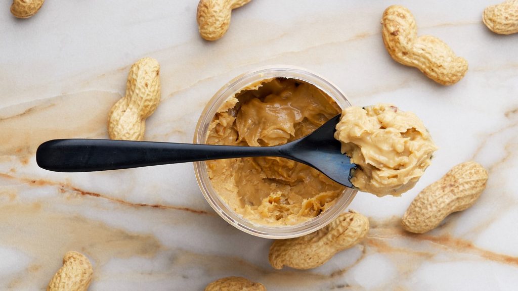
To test this hypothesis, the study team recruited 73 children ages 4 to 14 years old. Study participants were assigned, at random, to either test the new treatment strategy or continue avoiding peanuts. The children in the peanut-ingestion group began with a minimum daily dose of 1/8 teaspoon of peanut butter and gradually increased their dose every eight weeks over the course of 18 months, ending at one tablespoon of peanut butter or an equivalent amount of a different peanut product. All dose increases took place under medical supervision. None of the study participants in the peanut-ingestion group had severe reactions or needed epinephrine during home dosing  and only one received epinephrine during a supervised dosing visit.  

Following the treatment regimen, children from the peanut-consuming cohort participated in a feeding test, carefully supervised by the study team, to evaluate how much peanut they could eat without an allergic reaction. All 32 children from the peanut-consuming group who participated in the feeding test could tolerate the maximum amount of 9 grams of peanut protein, or three tablespoons of peanut butter. By contrast, only three of the 30 children from the avoidance group who underwent the feeding test after avoiding peanuts for the duration of the study could tolerate this amount.  

Because the trial took place during the COVID-19 pandemic and some families preferred avoiding close encounters indoors, some did not return to the study site for the feeding test. Using a common statistical technique to account for the children who missed the feeding test, the team reported that 100 percent of the ingestion group and 21 percent of the avoidance group tolerated an oral food challenge that was at least two doses more than they could tolerate at the beginning of the study.  

To test if the response to treatment was durable, children in the peanut-ingestion group who could tolerate nine grams of protein during the feeding test went on to consume at least two tablespoons of peanut butter weekly for 16 weeks and then avoided peanuts entirely for eight weeks. Twenty-six of the 30 treated children who participated in a final feeding test after the eight-week abstinence period continued to tolerate nine grams of peanut protein, indicating that they had achieved sustained unresponsiveness to peanuts. The three participants from the avoidance group who could eat nine grams of peanut protein without reaction at the earlier food test were considered to have developed natural tolerance to peanuts. A comprehensive analysis of data collected from all 73 study participants revealed that 68.4 percent of the peanut-ingestion group achieved sustained unresponsiveness, while only 8.6 percent of the avoidance group developed a natural tolerance.   

“These study results are very exciting and a huge step forward in personalizing food allergy treatment,” concluded Dr. Sicherer, the Elliot and Roslyn Jaffe Professor in Pediatric Allergy and Immunology at Mount Sinai. “My hope is that this study will eventually change practice to help these children and encourage additional research that includes this approach for more foods.”  

In addition to expanding the work to more foods and validation studies of their approach, the Mount Sinai study team aims to determine a better way of identifying individuals with higher thresholds, because the best way to do so currently requires a feeding test that is bound to cause an allergic reaction.  

Source: The Mount Sinai Hospital / Mount Sinai School of Medicine

Drug may Counteract the Muscle Loss and Osteoporosis after Rapid Weight Loss

Photo by I Yunmai on Unsplash

Weight loss medication has taken the world by storm and helped many overweight people. But for some, significant weight loss also comes with a loss of muscle mass and can lead to an increased risk of osteoporosis.

New research now suggests that the monoclonal antibody drug bimagrumab may be able to alleviate some of this risk, says PhD student Frederik Duch Bromer and postdoc Andreas Lodberg from the Department of Biomedicine at Aarhus University, who are behind the study published in the Journal of Cachexia, Sarcopenia and Muscle.

“We are the first to study how certain drugs affect bones, and the results show that bimagrumab can increase the amount of bone tissue while building muscle mass, and this could be very important for the many people currently taking weight loss medication.”

Bimagrumab was originally developed to treat muscle loss and dysfunction, but since then, it has beome apparent that it also has a fat “burning” component to it. So, if approved, it could be part of a second-generation weight loss drug on the market.

Therefore, it’s relevant to research how this particular patient group reacts to the drug,” says Andreas Lodberg.

“An estimated two billion people will be categorised as overweight by 2035, so it’s also important that we research the drugs that come on the market for this particular patient group in order to better understand their long-term impact on the body.”

Osteoporosis can prove costly for patients and society

Patients on weight loss medication often have a history of weight fluctuation, which can contribute to the development of osteoporosis. Brittle bones increase the risk of serious fractures, and this is costly for both patients and society.

Therefore, the research results could be good news for patients on weight loss medication. And according to Frederik Duch Bromer, the study shows that bimagrumab not only counteracts the breakdown of bone and muscle tissue, it actually promotes the build-up of both.

“Bimagrumab slightly increases the calcium content in bones and promotes the formation of new bone in what we call the shell (cortex) of the long bones. We also saw a significant build-up of bone tissue in the area around the femoral head, which is typically where many older people incur fractures.”

According to Frederik Duch Bromer, the results also showed that bimagrumab has no effect on the blood. Similar drugs have previously been shown to increase red blood cell production, increasing the risk of blood clots.

The study is based on mice with both osteoporosis and reduced muscle mass, and the drug is now being tested in several phase 2 clinical trials. Andreas Lodberg emphasises that more research is needed.

“Our study shows that bimagrumab has a positive effect in many areas, but we also have indications that the drug may have other side effects, and we’ll now investigate this further to get a clearer picture of the implications of using the drug for patients.”

Andreas Lodberg and Frederik Duch Bromer hope to be able to continue with further research to investigate both the positive results and possible side effects.

Source: Aarhus University

New Treatment Offers a Quick Cure for Common Cause of Hypertension

Credit: Pixabay CC0

Doctors at Queen Mary University of London, Barts Health NHS Trust and University College London have developed a groundbreaking, minimally invasive treatment, Triple T, offering hope for millions of people with high blood pressure caused by a commonly overlooked condition. 

The treatment, which could transform blood pressure management, has been published in The Lancet.

Triple T, also known as endoscopic ultrasound-guided radiofrequency ablation, is poised to change the way we address primary aldosteronism (PA) – a hormonal disorder that causes high blood pressure in one in 20 patients with blood pressure yet is often undiagnosed and untreated. This treatment has shown promising results in clinical trials and could become an accessible alternative to surgery, offering relief to those who suffer from this condition. 

The hidden cause of high blood pressure  

High blood pressure, affecting one in three adults, has several underlying causes, with PA being one of the most common yet underdiagnosed. In this condition, benign nodules in the adrenal glands produce excess aldosterone, a hormone that raises blood pressure by increasing salt levels in the body. Patients with PA often do not respond to standard medications and face increased risks of heart attacks, strokes and kidney failure. 

Until now, the only effective treatment for PA has been the surgical removal of the affected adrenal gland. However, this procedure requires general anaesthesia, a hospital stay, and weeks of recovery, causing many patients to go untreated. Triple T provides a faster, safer, and less invasive alternative by targeting and destroying the malfunctioning adrenal nodule without removing the gland.  

How Triple T works 

The procedure uses a combination of radiofrequency or microwaves and ultrasound to deliver targeted heat to the adrenal nodule. A fine needle is inserted through the stomach to the adrenal gland, guided by real-time ultrasound imaging, where short bursts of heat are used to destroy the problematic tissue. This targeted approach ensures minimal damage to surrounding healthy tissues. The entire procedure lasts only 20 minutes and requires no incision.   

Triple T’s success stems from recent advances in diagnostic scans, which use molecular dyes to accurately locate even the smallest adrenal nodules. These breakthroughs, combines with the ability to directly target nodules adjacent to the stomach, have enabled this minimally invasive approach. 

Successful trial and promising results  

The Feasibility study of radiofrequency endoscopic ABlation, with ULtrasound guidance (FABULAS) trial which tested Triple T on 28 patients with PA, showed excellent results. The procedure was found to be safe and effective, with most patients experiencing normalised hormones levels within six months. Many participants were able to stop all blood pressure medications, and the condition did not recur. 

Professor Morris Brown, co-senior author of the study and Professor of Endocrine Hypertension at Queen Mary University of London, reflected on the significance of this milestone: “It is 70 years since the discovery in London of the hormone aldosterone, and, a year later, of the first patient in USA with severe hypertension due to an aldosterone-producing tumour. This patient’s doctor, Jerome Conn, predicted, with perhaps only minor exaggeration, that 10-20% of all hypertensions might one day be traced to curable nodules in one or both glands. We are now able to realise this prospect, offering 21st-century breakthroughs in diagnosis and treatment.”  

One trial participant, Michelina Alfieri, shared her experience: “Before the study, I suffered from debilitating headaches for years despite multiple GP visits. As a full-time worker and single parent, my daily life was severely affected. This non-invasive treatment provided an immediate recovery—I was back to my normal routine straight away. I’m incredibly grateful to the team for giving me this choice.”  

What’s next?  

The success of the FABULAs trial has led to a larger study, WAVE, which will compare Triple T with traditional surgery in 120 patients. Results are expected in 2027. 

Professor Stephen Pereira, Chief Investigator of FABULAS, emphasised the potential global impact of Triple T. he said: “This less invasive technique could be widely offered in endoscopy units across the UK and internationally.”  

Clinical Endocrinology Lead at Addenbrooke’s Hospital and Professor of Clinical Endocrinology at the University of Cambridge, Professor Mark Gurnell, said: “Thanks to this work, we may finally be able to diagnose and treat more people with primary aldosteronism, lowering their risk of developing cardiovascular diseases and other complications, and reducing the number of people dependent on long-term blood pressure medication.” 

The research was primarily supported by Barts Charity, National Institute for Health and Care Research (NIHR) through the Barts and Cambridge Biomedical Research Centres (BRCs), and the British Heart Foundation.  

It is being followed by a larger randomised trial, called ‘WAVE’, which will compare TTT to traditional surgery in 120 patients. The results are expected in 2027.  

Source: Queen Mary University of London

Over 15 000 South African Health Workers’ Jobs are at Risk as US Cuts Aid

Photo by Scott Graham on Unsplash

By Jesse Copelyn

If the US President’s Emergency Plan for AIDS Relief (PEPFAR) is halted, the South African public health system “will face a severe crisis” that could endanger millions of lives. This is according to a coalition of 17 health service organisations in South Africa, including large ones such as Anova Health, Health Systems Trust, TB HIV Care, The Aurum Institute and Wits RHI.

In a statement, they appealed to private sector donors and “high net-worth individuals” to help fund the shortfall caused by US aid cuts.

Read the statement

PEPFAR is a multi-billion dollar US initiative that supports HIV and TB-related health services around the world. In South Africa alone, over 15 000 staff (mostly health workers) are funded by PEPFAR, according to the national health department.

But a series of executive orders issued by US President Donald Trump has suspended some of this funding and the rest remains precarious. The orders include a 90-day pause on all US foreign development assistance and another that explicitly bars South Africa from aid (with some leeway allowed).

Some health service providers in South Africa continue to receive money from PEPFAR under a limited waiver that allows for the continuation of certain “life-saving HIV services”. But the waiver hasn’t protected all PEPFAR beneficiaries. As a result, some organisations have had to close their doors, while many others have had to curtail what they can provide.

The waiver doesn’t cover all health services, and many health programs that target high-risk groups (such as people who use drugs) have not been protected. This is even if they provide life-saving HIV services.

Services suspended for the most vulnerable

Under the waiver, PEPFAR can continue to fund programs that offer treatment and testing for HIV, including antiretroviral (ARV) services. Projects can also continue to provide condoms and HIV prevention medication, known as PrEP, but only to pregnant and breastfeeding women.

The waiver does not allow for continued funding of PrEP medication or condoms to anyone else. It also doesn’t cover crucial research, like population surveys which tell us how many people have HIV and where they’re located. Additionally, it doesn’t allow for continued funding of methadone maintenance programs for people who use heroin. This is despite the fact that this is the most effective way to help people to stop using heroin and to curb the sharing of drug needles (something which contributes to the spread of HIV).

Dr Gloria Maimela, who represents the coalition of organisations behind the statement, told GroundUp and Spotlight: “The staff who are providing [HIV] testing and treatment [are] back at facilities to provide those services, but staff that are providing other services not included in the waiver have been stopped, and are waiting for further guidance.”

In addition, organisations that help key populations have not been protected by the waiver, according to Maimela. Key populations are groups that are more at risk of becoming infected with HIV, such as people who inject drugs, sex workers, transgender people, and men who have sex with men. South African policy documents and the World Health Organisation recommend that health programs focus on these groups since they’re more likely to acquire and transmit HIV.

Despite this, US-funded organisations that target key populations have been forced to shut their doors in South Africa. Maimela says that this is even in cases where they were offering the kind of life-saving ARV treatment covered in the waiver.

“For us, this is of grave concern,” Maimela says, “because we know that right now that is where most of the [HIV] infections lie”.

So far, organisations which provide HIV treatment and prevention services to LGBTI people have been forced to shut down, including the Ivan Toms Centre and Engage Men’s Health.

Additionally, GroundUp and Spotlight have identified two PEPFAR-supported harm reduction centres that have had to close. These centres provided methadone and clean needles to people who inject drugs (when drug users have access to clean needles, they’re less likely to resort to sharing them, which brings down HIV transmission).

Ricardo Walters, who provides consulting services to health service organisations across Africa, told Spotlight and GroundUp that a similar trend could be seen across the continent.

“Many organisations that were specifically offering services to key populations were not suspended; their project funding was terminated,” he said. “They will not be coming back.”

These organisations were assisting patients “who often could not access services in a general [health] setting”.

Walters says the reasons given for the termination of these programs vary across organisations and countries.

“Where there are reasons, it’s often [stated] that it’s because the program contains components of DEIA [Diversity, Equity, Inclusion and Accessibility] and gender ideology, which is directly from a previous executive order [in which the Trump administration terminated all federal funding for DEIA]. The terms are never defined … no one says don’t treat gay men.”

Appeal to private sector

Beyond the shuttering of existing organisations, providers that are covered under the waiver remain unsure about whether funding will restart after the 90-day period. Also large sections of the US aid establishment have been gutted.

The recent statement by health organisations argues that if this aid is terminated “patients, including children, will lose access to life-saving antiretroviral treatment, while thousands of healthcare workers will be unable to provide essential HIV care. The consequences will be immediate. Fewer people will receive timely testing and treatment, leading to more undiagnosed cases, rising infections, and the spread of drug resistance. Mortality will increase, opportunistic infections will surge, and TB rates will escalate – putting the entire population at risk.”

As such, the statement calls on private corporations, donors and philanthropists to assist in supporting these health services.

“We encourage people to get in touch with us,” says Maimela, “so that even as we hold dialogues with the government, [those people] could be part of [the conversation] and step in and say how they want to help.”

To find out how to support organisations that provide HIV and TB related health services in South Africa contact Gloria Maimela at gloriam@foundation.co.za.

Published jointly by GroundUp and Spotlight.

Republished from GroundUp under a Creative Commons Attribution-NoDerivatives 4.0 International License.

The Surprising Link between Muscle Signalling and Brain Memory

New research shows that how a network of subcellular structures is responsible for transmitting signals in neurons. This movie shows 3D renderings of these structures in high-resolution 3D electron microscopy images of fruit fly neurons. The endoplasmic reticulum (green), plasma membrane (blue), mitochondria (pink), microtubules (tan), and ER-plasma membrane contacts (magenta) are segmented from FIB-SEM datasets of a Drosophila melanogaster MBON1 neuron. Credit: Benedetti et al.

New research led by the Lippincott-Schwartz Lab shows that a network of subcellular structures similar to those responsible for propagating molecular signals that make muscles contract are also responsible for transmitting signals in the brain that may facilitate learning and memory.

“Einstein said that when he uses his brain, it is like he is using a muscle, and in that respect, there is some parallel here,” says Janelia Senior Group Leader Jennifer Lippincott-Schwartz. “The same machinery is operating in both cases but with different readouts.” The research appears in the journal Cell.

The first clue about the possible connection between brain and muscle cells came when Janelia scientists noticed something strange about the endoplasmic reticulum, or ER – the membranous sheets and folds inside cells that are crucial for many cellular functions.

Research scientist Lorena Benedetti was tracking molecules at high resolution along the surface of the ER in mammalian neurons when she saw that the molecules were tracing a repeating, ladder-like pattern along the entire length of the dendrites.

Around the same time, Senior Group Leader Stephan Saalfeld alerted Lippincott-Schwartz to high-resolution 3D electron microscopy images of neurons in the fly brain where the ER was also forming regularly spaced, transversal structures.

This movie shows time-lapse high-resolution imaging in neurons, revealing the dynamic behavior of ER tubules contrasted with the persistence of ER-PM junctional sites over time. Time-lapse acquired using 2D lattice-SIM in burst mode of HaloTag-Sec61β (labeled with JF585 HaloTag-ligand) expressing neurons. Scale bars: 0.5 μm. Credit: Benedetti et al.

The ER normally appears like a huge, dynamic net, so as soon as Lippincott-Schwartz saw the structures, she knew her lab needed to figure out what they were for.

“In science, structure is function,” says Lippincott-Schwartz, who also heads Janelia’s 4D Cellular Physiology research area. “This is an unusual, beautiful structure that we are seeing throughout the whole dendrite, so we just had this feeling that it must have some important function.”

The researchers, led by Benedetti, started by looking at the only other area of the body known to have similar, ladder-like ER structures: muscle tissue. In muscle cells, the ER and the plasma membrane – the outer membrane of the cell – meet at periodic contact sites, an arrangement controlled by a molecule called junctophilin.

Using high-resolution imaging, the researchers discovered that dendrites also contain a form of junctophilin that controls contact sites between their ER and plasma membrane. Further, the team found that the same molecular machinery controlling calcium release at muscle cells’ contact sites – where calcium drives muscle contraction – was also present at dendrite contact sites – where calcium regulates neuronal signalling.

Because of these clues, the researchers had a hunch that the molecular machinery at the dendritic contact sites must also be important for transmitting calcium signals, which cells use to communicate. They suspected that the contact sites along the dendrites might act like a repeater on a telegraph machine: receiving, amplifying, and propagating signals over long distances. In neurons, this could explain how signals received at specific sites on dendrites are relayed to the cell body hundreds of micrometres away.  

“How that information travels over long distances and how the calcium signal gets specifically amplified was not known,” says Benedetti. “We thought that ER could play that role, and that these regularly distributed contact sites are spatially and temporally localised amplifiers: they can receive this calcium signal, locally amplify this calcium signal, and relay this calcium signal over a distance.”

The researchers found that this process is triggered when a neuronal signal causes calcium to enter the dendrite through voltage-gated ion channel proteins, which are positioned at the contact sites. Although this initial calcium signal dissipates quickly, it triggers the release of additional calcium from the ER at the contact site.

Source: Howard Hughes Medical Institute

Nearly 5000 Children are Living with Blood Cancer: The Toll on SA’s Caregivers

Credit: National Cancer Institute

Blood cancer, a term covering several malignant diseases of the bone marrow or blood-forming system, accounts for 33% of all childhood cancers in South Africa. Currently, nearly 5000 children are living with the condition. For parents and caregivers, the emotional and financial strain can be overwhelming, often leaving them struggling to cope.

Ahead of International Childhood Cancer Day, on the 15th of February, Palesa Mokomele, Head of Community Engagement and Communications at DKMS Africa, explains that with the childhood cancer survival rate as low as 20%, a diagnosis is often a devastating blow for families.

“One thousand four hundred South African children are diagnosed with blood cancer annually,” she continues. “While the diagnosis is traumatic for the child, caregivers experience immense psychological distress which can severely impact their quality of life.”

A Mother’s Story

Elizabeth, whose son Ntsako was diagnosed with blood cancer in August 2024, describes the experience as “a bolt of lightning” that turned her world upside down. “I try not to cry in front of my son, even when I feel like I am falling apart. The treatment phase has been brutal. I want to stay strong for him but knowing there’s only so much I can do is heartbreaking.”

Mokomele notes that Elizabeth’s experience is shared by many. “Prolonged treatment, high stress, sleep deprivation, and financial strain take a heavy toll. Many caregivers struggle with anxiety, depression, and burnout, affecting their well-being, family dynamics, and social lives.”

Coping With the Emotional Impact

While every parent handles these challenges differently, there are ways to manage the emotional burden:

·        Fear and anxiety: The unknown can be debilitating. Engaging with doctors and learning about the treatment and outcomes, which, while still stressful, can remove much of the uncertainty. Your child’s care team is not only there for your child but also to help you; enlist their support and lean on them.

·        Denial and anger: In the short term, denial may help you adjust to the reality of your child’s diagnosis, but staying in denial for too long can cause isolation and delay treatment. Once this wears off, it can give way to anger, and without a proper outlet, it may build up inside. This can lead to you misdirecting it toward other loved ones, co-workers, and even doctors. Look for support from other parents who are going through the same process. Communicate your feelings with those close to you and explore ways to help you cope, like exercise, journaling, mindful meditation, or even just giving yourself private time to vent your feelings.

·        Guilt and blame: It is natural to look for someone or something to blame. You may look inward to find something you think you did wrong; maybe you feel you didn’t act soon enough, or you’re angry that you didn’t get to the doctor earlier. Acknowledging these feelings and allowing yourself to process them is important. If these feelings become too overwhelming, seek support from a professional or even from your child’s care team.

·        Sadness and loss: Give yourself the space to acknowledge grief and adapt to your new reality. If these feelings start to impact your ability to function, get support to work through them because they will affect your ability to help your child and other family members cope.

A Life-Saving Solution

More than 500 South African children die from blood cancer annually – a number that can be reduced with early detection and timely intervention.

“Blood cancer patients can often overcome the disease with the help of a stem cell transplant from a suitable donor,” highlights Mokomele. “DKMS provides a second chance at life for more than 22 patients every day, but doctors still struggle to find matches. Registering as a donor takes just five minutes but could save a child’s life and offer some much-needed relief for those caregivers who are doing their best to hold their families together.”

Register at https://www.dkms-africa.org/register-now.

For further information, get in touch with DKMS Africa at 0800 12 10 82.

Research Reveals New Insights into how LDL Cholesterol Works in the Body

Image by Scientific Animations, CC4.0

National Institute of Health (NIH) scientists have made a significant breakthrough in understanding how “bad” cholesterol, known as low-density lipoprotein-cholesterol or LDL-C, builds up in the body. The researchers were able to show for the first time how the main structural protein of LDL binds to its receptor – a process that starts the clearing of LDL from the blood – and what happens when that process gets impaired.

The findings, published in Nature, further the understanding of how LDL contributes to heart disease, the world’s leading cause of death, and could open the door to personalising LDL-lowering treatments like statins to make them even more effective.

“LDL is one of the main drivers of cardiovascular disease which kills one person every 33 seconds, so if you want to understand your enemy, you want to know what it looks like,” said Alan Remaley, MD, PhD, co-senior author on the study who runs the Lipoprotein Metabolism Laboratory at NIH’s National Heart, Lung, and Blood Institute.

Until now scientists have been unable to visualise the structure of LDL, specifically what happens when it links up with its receptor, a protein known as LDLR. Typically, when LDL binds to LDLR, the process of clearing LDL from the blood begins. But genetic mutations can prevent that work, causing LDL to build up in the blood and get deposited into the arteries as plaque, which can lead to atherosclerosis, a precursor for heart disease.

In the new study, the researchers were able to use high-end technology to get a view of what’s happening at a critical stage of that process and see LDL in a new light.

“LDL is enormous and varies in size, making it very complex,” explained Joseph Marcotrigiano, PhD, chief of the Structural Virology Section in the Laboratory of Infectious Diseases at NIH’s National Institute of Allergy and Infectious Diseases and co-senior author on the study. “No one’s ever gotten to the resolution we have. We could see so much detail and start to tease apart how it works in the body.”

Using cryo-electron microscopy, the researchers were able to see the entirety of the structural protein of LDL when it bound to LDLR. Then, with AI-driven protein prediction software, they were able to model the structure and locate the known genetic mutations that result in increased LDL.

The researchers found that many of the mutations that mapped to the location where LDL and LDLR connected, were associated with familial hypercholesterolaemia (FH). FH is marked by defects in how the body uptakes LDL into its cells, and people with it have extremely high levels of LDL and can have heart attacks at a very young age. They found that FH-associated variants tended to cluster in particular regions on LDL.

The study findings could open new avenues to develop targeted therapies aimed at correcting these kinds of dysfunctional interactions caused by mutations. But, as importantly, the researchers said, they could also help people who do not have genetic mutations, but who have high cholesterol and are on statins, which lower LDL by increasing LDLR in cells. By knowing precisely where and how LDLR binds to LDL, the researchers say they may now be able to target those connection points to design new drugs for lowering LDL from the blood.

Source: NIH/National Heart, Lung and Blood Institute

Air Pollution Linked to More Lower Respiratory Infection Hospital Admissions

Photo by Kouji Tsuru on Pexels

Air pollution is a well-known risk factor for respiratory diseases such as asthma and chronic obstructive pulmonary disease (COPD) – but its contribution to lower respiratory infections is less well known, especially in adults. To address this, a team from the Barcelona Institute for Global Health (ISGlobal), a centre supported by the ”la Caixa” Foundation, assessed the effect of air pollution on hospital admissions for lower respiratory infections in adults, and which subgroups that could be particularly vulnerable to these infections. The results have been published in the journal Environment International.

The research shows that long-term exposure to particulate matter (PM2.5 and PM10), nitrogen dioxide (NO2) and ozone (O3) air pollution is associated with more hospital admissions for lower respiratory tract infections in adults. The associations were stronger in men, people over 65 years of age and those diagnosed with hypertension.

The study involved 3 800 000 adults from the COVAIR-CAT cohort, a large cohort of 7.7 million people based on the health system of Catalonia. The research team used exposure models to estimate annual average concentrations of PM2.5, PM10, NO2 and ozone during the warm season (May-September) between 2018 and 2020 at the participants’ residences. Information on hospital admissions, mortality and comorbidities was obtained from various administrative databases. The study included hospital admissions for all lower respiratory infections and, separately, the subgroup of hospital admissions for influenza and pneumonia. A statistical model was then used to assess the association between air pollution and hospital admissions.

“The association between air pollution and hospital admissions for lower respiratory tract infections was observed even at pollution levels below current EU air quality standards,” says Anna Alari, ISGlobal researcher and first author of the study. “It is crucial to adopt stricter air quality standards, as more ambitious measures to reduce air pollution would decrease hospital admissions and protect vulnerable populations,” she adds.

Stronger association in men and people over 65

The association between air pollution and hospitalisations for lower respiratory tract infections was more pronounced in people over 65 years of age or with comorbidities, compared with younger people or those without comorbidities. Specifically, elevated levels of air pollution were associated with approximately three times higher rates of hospital admissions for lower respiratory infections among people aged 65 years and older compared with younger people.

In addition, exposure to elevated levels of NO2, PM2.5 or PM10 (but not O3) was associated with about a 50% increase in hospital admissions in men, while the association was about 3% higher in women.

The team observed the same pattern for hospital admissions for influenza or pneumonia, but with smaller associations compared to lower respiratory infections. “This may be due to the influence of available vaccines against the pathogens responsible for influenza and most cases of pneumonia,” says Cathryn Tonne, senior author of the study.

Source: Barcelona Institute for Global Health (ISGlobal)

Thrombolytic Drug Still Effective up to 24 Hours after Ischaemic Stroke Onset

Credit: American Heart Association

The thrombolytic medication, alteplase, improved stroke patients’ recovery by more than 50% when given up to 24 hours after the beginning of an ischaemic stroke, according to preliminary late-breaking science presented at the American Stroke Association’s International Stroke Conference 2025.

These results give hope to stroke patients worldwide who may not be able to access thrombolytic medications within the approved time window, which in China is within 4.5 hours, said the trial’s principal investigator Min Lou, MD, PhD, a professor at the Second Affiliated Hospital of Zhejiang University’s School of Medicine in China.

In the US, alteplase is approved to treat stroke within three hours of symptom onset and is recommended for use up to 4.5 hours for select patients. Other research has indicated it may also work well in some patients 4.5 to 9 hours after stroke onset.

The American Heart Association/American Stroke Association 2019 Guidelines for the Early Management of Patients with Acute Ischemic Stroke note that IV alteplase within 4.5 hours of stroke onset is the standard of care for most ischaemic stroke patients in the United States.

Researchers enrolled 372 stroke patients whose symptoms began 4.5 hours to 24 hours earlier. They used widely available CT perfusion imaging (advanced brain scanning) to confirm that these patients still had brain tissue that could recover with treatment. Participants were randomised to receive alteplase, while the other received standard stroke care of antiplatelet therapy at the discretion of the investigator, based on the Chinese Guidelines for Diagnosis and Treatment of Acute Ischemic Stroke 2018. Functional recovery was assessed at 90 days.

“We believe these findings mean more people may return to normal or near-normal lives after a stroke, even if they receive treatment later than originally thought beneficial,” Lou said. “This method of treatment could become the new standard, especially in hospitals that use CT perfusion imaging. This technology helps health care professionals see how blood flows in different parts of the brain after an ischemic stroke. This could extend treatment eligibility to millions more patients across the globe.”

The study found:

  • 40% of participants treated with alteplase had little to no disability after 90 days, compared to 26% of those who received standard care – a 54% higher chance of functional recovery.
  • Less than 3% of participants in either group received rescue mechanical clot removal as an additional treatment.
  • Rates of death were the same (10.8%) for both groups.
  • The risk of brain bleeding was higher among those who received alteplase than among participants who did not (3.8% vs. 0.5%), but researchers believe this is a manageable risk.

“We also need to look more closely at how safe and effective other clot-dissolving medications, like tenecteplase, are when given after a stroke, especially beyond the usual time frames. It’s also important to learn if our findings apply to other groups of people, especially in areas with different stroke risks and health care resources,” Lou explained.

Study limitations include the that both participants and researcher knew which treatment was being given, which could have introduced bias, and results may not be generalizable to patients outside of China.

Study design, background and details:

  • The study enrolled 372 stroke patients in a multicenter, prospective, randomized trial at 26 stroke centers in China.
  • The patient’s average age was 72 years, and 43% were women.
  • The trial used widely available CT perfusion imaging software to gauge salvageable brain tissue, making the findings more applicable to real-world clinical settings.
  • Enrolled patients were assigned to the alteplase group or a standard medical treatment group.
  • The primary outcome was a score of 0 or 1 on the modified Rankin scale, which scores disability from 0 (no symptoms) to 6 (death) at 90 days.

Study co-authors, funding and disclosures are available in the abstract.

Source: American Heart Association

Coping with the Fear of Breast Cancer Recurrence

Photo by Michelle Leman on Pexels

Breast cancer is the world’s most prevalent cancer. Although earlier detection and targeted treatment have resulted in high survival rates, many breast cancer survivors experience fear of cancer recurrence. For some survivors this fear is occasional, for others it is persistent and often debilitating.

A new study of breast cancer survivors has found this psychosocial challenge impacts almost every important domain of their lives – the emotional, behavioural, cognitive, relational and professional. A larger number of domains was affected, and they were affected more frequently in those with greater fear of recurrence.

“Study participants were reportedly disease free and trying to rebuild their lives during their post-treatment survivorship,” said senior author Shelley Johns, PsyD, a researcher-clinician with the Regenstrief Institute, the Indiana University School of Medicine and the IU Melvin and Bren Simon Comprehensive Cancer Center. “Our findings provide clarity about how breast cancer survivors are impacted by fear of recurrence and insight into how they cope with this understandable fear.”

The study was published in Supportive Care in Cancer.

The impact of fear of recurrence ranged from mildly to severely disruptive. Women experiencing mild fear reported sporadic occurrences. Those with significant fear described it as persistent and/or easily triggered across multiple life domains.

Disturbed sleep prior to mammograms was reported by survivors with mild fear, while frequent need to absent themselves from social activities, get into bed and pull the blanket over their eyes to avoid thinking about cancer was an example of severe, also known as clinical, fear of recurrence. Approximately 74 percent of study participants were experiencing clinical fear of recurrence.

347 women completed the study’s open-ended survey:

  • Many reported feelings of stress, irritability and sadness.
  • Some said fear of recurrence frequently interrupted their train of thought, for example interfering with their job when their disease popped into their mind.
  • Survivors who thought that they were more worried than they should be compared to other breast cancer survivors reported feelings of embarrassment.
  • Some indicated it was too hard to be around their family because they were constantly wondering how many more Christmases and birthdays they were going to have with their children.

The paper’s title includes the phrase, “out of a dark place,” a direct quote from a breast cancer survivor who said that she joined the study to support “getting out of a dark place.”

Other survivors noted the specific impact of fear of cancer recurrence on daily life:

  • “It motivates me to maintain healthy habits. Such as eating five servings of fruits and vegetables, working out and drinking less alcohol. It also motivates me to maintain mental health and physical health.”
  • “Whenever I feel any kind of pain or discomfort in the area where I had cancer it concerns me and I feel anxious and irritable.”
  • “Cancer is all around us. Everything is a trigger. Anniversaries, other family/friends’ diagnosis, commercials about drugs, social media, etc. …it’s a daily thought or a daily emotion.”
  • “Sit for hours doing nothing, do not turn on TV, sleepless, find hours pass by and I am in the same place just thinking, do not participate in activities, get lost driving because I’m deep in thought, compulsive online shopping, collecting things.”

Survivors offered specifics on their coping mechanisms:

  • “Just trying to be positive, eat healthy, take my meds, get enough sleep, exercise three times a week, and hope for the best.”
  • “I try to avoid things that make me think about recurrence. For example, unfollowing social media accounts, fast forwarding or leaving the room when commercials about cancer medications are on.”
  • “I try not to focus on it. I also speak with family members who have lived with cancer longer than myself.”
  • “Prayer, meditation, staying in the moment, and focusing on making the best of each day.”

While many survivors cited avoidance of thoughts and feelings as their primary coping behaviour, Dr Johns, a health services researcher and clinical health psychologist, observes that research is needed to probe the function of various coping behaviours’ to determine if they are helpful.

In a question seldom posed to participants in a clinical trial, when asked what they hoped to gain by participating in the study, the majority indicated that they sought senses of purpose, belonging, control and connection with others.

The paper concludes, “Fear of cancer recurrence is one of the most common psychological challenges for cancer survivors. Understanding affected life domains, coping strategies employed prior to intervention, and reasons for seeking guidance can inform the development and implementation of evidence-based interventions to effectively address fear of cancer recurrence among persons living with breast cancer.”

Source: Regenstrief Institute