Month: March 2025

My Five-hour Wait for Treatment at Mamelodi Hospital

Gauteng Health MEC has said Mamelodi Regional Hospital meets National Health Insurance standards, but my experience was not good

The writer waited five hours for treatment for a broken wrist and head injuries at Mamelodi Regional Hospital in Tshwane. Photo: Warren Mabona.

By Warren Mabona

I waited five hours to get medical treatment at Mamelodi Regional Hospital in Tshwane, with a broken wrist and an injured head.

On 19 February 2025 at about 4pm I was walking in Mamelodi West. I was on a journalism assignment, heading to informal settlements that are prone to flooding.

The street was quiet, but I felt safe because I had walked there before. Suddenly, a car stopped in front of me, and two men got out of it and tried to rob me. I ran away and jumped into the stormwater passage, but slipped and fell, hitting my face against the concrete.

When I managed to stand up, I was dizzy and my vision was blurred. I was drenched in dirty water and my belongings — my cell phone, my wallet and my camera bag — were wet.

The men who attacked me were no longer on the street. My right wrist was swollen and painful, an injury above my eye was bleeding profusely, and my head was aching. But I was relieved that I was still alive and I still had all my belongings.

I decided not to call an ambulance, but to walk about 800 metres to Mamelodi Regional Hospital.

I went to the casualty unit, expecting that I would receive treatment quickly. At the front desk, a clerk took more than 20 minutes to fill in my file. He said the hospital’s computer system was offline and he had to fill in the file with a pen. I then went to sit at the reception area. My head was aching and I repeatedly requested headache tablets from the nurses, who gave me two tablets after 30 minutes. But my pain lingered.

The wound on my face was still bleeding and my wrist was swollen and bent. About 40 minutes after my arrival, a nurse cleaned my wound and wrapped it with a bandage, stopping the bleeding.

At about 8pm, a man sitting next to me said he had arrived at the hospital at 2pm after falling from scaffolding at a construction site. He was still waiting for his X-ray results.

I went for X-rays and long afterwards, at about 10pm, I had a cast put on my wrist. I was given injections which helped with the pain. I was discharged at 11pm and went home.

In September last year, the Gauteng MEC for Health Nomantu Nkomo-Ralehoko said that Mamelodi Regional Hospital was the first hospital in Gauteng ready to meet National Health Insurance (NHI) standards.

In response to GroundUp’s questions, Gauteng Department of Health spokesperson Motalatale Modiba said a triage priority system is followed at the hospital, meaning that four patients with critical wounds that required life-saving emergencies were attended to first. He said this affected my waiting time for wound care and the application of a cast.

“You were classified as Orange P2, that is a person who is in a stable condition and is not in any immediate danger, but requires observation,” said Modiba.

“At the time of your arrival, the casualty unit had 31 other patients to be seen. These include four critical cases in the resuscitation unit, ten trauma cases, 16 medical cases and four pediatric cases,” he said.

Modiba confirmed that the hospital’s computer system was offline when I arrived.

I asked Modiba whether the Gauteng Department of Health can still confidently regard this hospital as NHI-ready despite the slow delivery of medical services I experienced. Modiba said: “Mamelodi Regional Hospital remains committed to provide best healthcare services.”

Republished from GroundUp under a Creative Commons Attribution-NoDerivatives 4.0 International License.

Read the original article.

Oxytocin Can ‘Pause’ Pregnancy After Conception

Photo by Pavel Danilyuk on Pexels

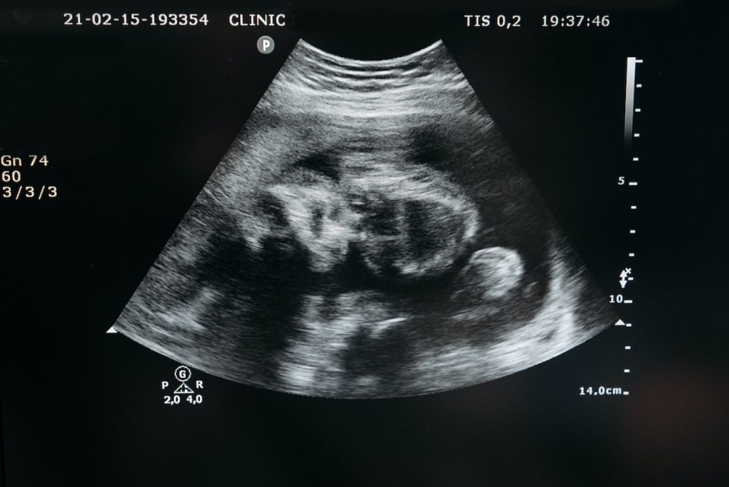
Oxytocin, a hormone already known for its role in childbirth, milk release, and mother–infant bonding, may have a newfound purpose in mammalian reproduction. In times of maternal stress, the hormone can delay an embryo’s development for days to weeks after conception, a new study in rodents shows. According to the authors, the findings about so-called “diapause” may offer new insights into pregnancy and fertility issues faced by humans.

Led by researchers at NYU Langone Health, the study explored diapause, in which an embryo temporarily stops growing early in its development before it attaches to the lining of its mother’s uterus, a key step leading to the formation of the placenta. Known to occur in species as varied as armadillos, giant pandas, and seals, diapause is thought to have evolved to help expectant mothers preserve scarce resources (e.g., breast milk) by delaying birth until they have enough to successfully take care of their offspring.

Although recent studies have uncovered evidence that a form of diapause may occur in humans, the underlying mechanisms behind it have until now remained unclear.

The findings in mice showed that one type of stress that may cause diapause is milk production and release (lactation), as it requires a mother to expend bodily resources nursing already-born pups, and supplying nutrients to pups growing in the womb, at the same time. The study revealed that the time between conception and birth, typically 20 days for these animals, was delayed by about a week in pregnant rodents that were already nursing a litter.

Further, the research team showed that this delay was brought about by a rise in the production of oxytocin, levels of which are known to go up as a mother lactates. To confirm this role for the hormone, the researchers exposed mouse embryos in the lab to a single dose (either 1 microgram or 10 micrograms) of oxytocin, and found that even these small amounts delayed their implantation in the uterus by as much as three days. The team found that the chemical did more than just pause pregnancy: oxytocin surges that were large enough to mimic the amounts and timing measured during nursing caused loss of pregnancy in the mice in nearly all cases.

“Our findings shed light on the role of oxytocin in diapause,” said study co-author Moses V. Chao, PhD, a professor at NYU Grossman School of Medicine. “Because of this newfound connection, it is possible that abnormalities in the production of this hormone could play roles in infertility, premature or delayed birth, and miscarriage.”

A report on the findings appears in Science Advances in a special issue focused on women’s health.

In another part of the study, the team searched for a mechanism that would allow embryos to react to an oxytocin surge. They found that the hormone can bind to special proteins called receptors on the surface of a layer of cells known as the trophectoderm, which surrounds the early embryo and eventually forms the placenta.

Notably, mouse embryos that were genetically altered to disable oxytocin receptors lived long enough to implant into their mother’s placenta at much lower rates than normal embryos. This suggests that the ability to respond to oxytocin spikes and therefore go into diapause is somehow important for the developing pups’ survival, says Dr. Chao, who plans to examine this protective function in more detail.

“Despite being extremely common, infertility and developmental issues that can arise during pregnancy remain poorly understood and can have a lasting, devastating impact on parents and their children,” said study senior author Robert Froemke, PhD, professor of genetics. “Having a deeper understanding of the factors that contribute to these problems may allow experts to better address them in the future.”

The researchers next plan to examine how cell growth gets turned back on after diapause, Dr Froemke says. In addition, the team plans to explore how diapause may affect offsprings’ health and development after birth, and determine whether and how their discoveries can inform reproductive medicine.

Dr Froemke cautions that while the study results are promising, mice and humans have significant differences in their reproductive processes, despite both being mammals. He adds that the current investigation did not assess the role that other pregnancy-related hormones, such as oestrogen and progesterone, may play in diapause.

Source: NYU Langone Health / NYU Grossman School of Medicine

Leakiness of First Blood–brain Barrier Layer Results in Cognitive Deficits

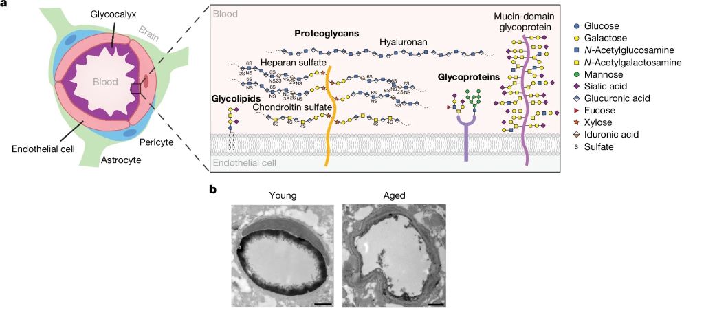
a, Diagram of the BBB and brain endothelial glycocalyx layer. b, TEM of cortical capillaries with lanthanum nitrate staining from young (3-month-old) and aged (21-month-old) mice. Scale bars, 1 µm. Source: Shi et al., Nature, 2025.

A study published in Nature reveals how a key component of the blood–brain barrier (BBB), the endothelial glycocalyx layer, becomes dysregulated in ageing, causing the BBB to become compromised. The researchers also investigated the possibility of to restore this layer’s integrity, reducing neuroinflammation and restoring cognitive function.

The BBB is a highly specialised safeguard keeping the brain separate from harmful factors, such as toxins and also albumin, IgG and fibrinogen (and, unfortunately, many medications which could otherwise treat brain disease). The leakage of such blood-derived molecules into the brain has been shown to trigger neuroinflammatory changes and create a neurotoxic brain environment. The part of the BBB directly in contact with the blood is the endothelial glycocalyx layer, a carbohydrate-rich meshwork mostly composed of proteoglycans, glycoproteins and glycolipids that coats the BBB lumen. Yet the endothelial glycocalyx’s composition and role is poorly understood despite it being the first layer of interface between the blood and brain.

The researchers found that the brain endothelial glycocalyx is highly dysregulated during ageing and neurodegenerative disease. Two mucin-type O-glycan biosynthetic enzymes, C1GALT1 and B3GNT3 were also found to be downregulated mouse models of ageing and in the brains of Alzheimer’s and Huntington’s disease patients. To test these, the researchers used adeno-associated viruses (AAV) in young mice to turn down the expression of C1GALT1 and B3GNT3. These mice showed signs of BBB leakage and in severe cases, brain haemorrhaging occurred in mice.

In samples from the brains of Alzheimer’s patients, the researchers also observed reduced C1GALT1 in microvessels.

To test if it was possible to restore the BBB’s ability to protect the brain against harmful blood-borne molecules, they administered AAVs in aged mice to restore levels of B3GNT3 and C1GALT1.

Assessing cognitive function, they found that aged mice treated with B3GNT3 via an AAV displayed improvements in spatial working memory in a maze test and hippocampal-dependent learning and memory in a fear conditioning test. Aged mice treated with C1GALT1 did not improve in the maze test, and no significant difference was observed in cued freezing in the fear conditioning among the three aged groups.

Although the study shows that increasing C1GALT1 and B3GNT3 reduces BBB permeability and improves brain health, the precise mechanisms that underlie these beneficial effects remain unclear. The researchers believe that by limiting the nonspecific uptake of blood-derived molecules, the brain can be protected. But C1GALT1 and B3GNT3 are also likely to influence a wide range of proteins and glycan structures and in order to further understand brain ageing and rejuvenation it is therefore crucial to understand the molecular pathways affected by them.

The authors concluded: “Cumulatively, our findings provide a detailed compositional and structural mapping of the ageing brain endothelial glycocalyx layer and reveal important consequences of ageing- and disease-associated glycocalyx dysregulation on BBB integrity and brain health.”

In Younger Women, Stress is Associated with an Increased Stroke Risk

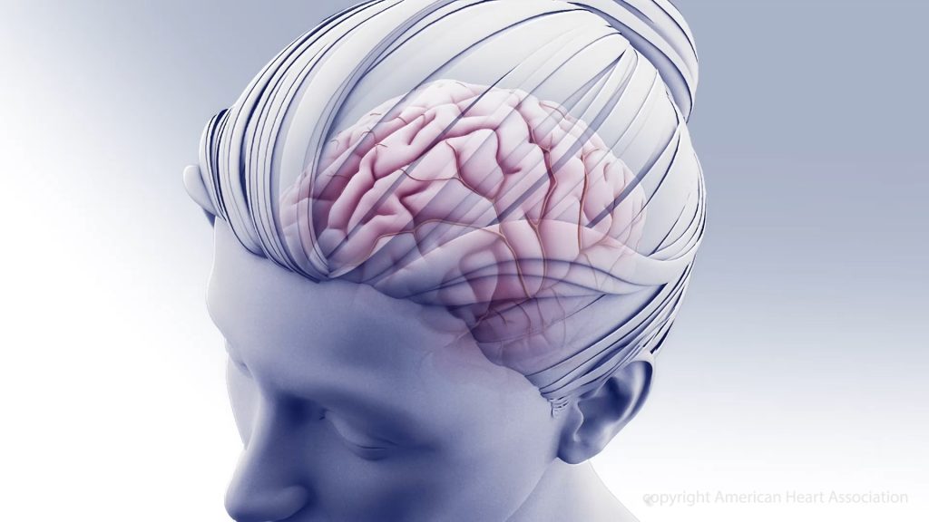
Credit: American Heart Association

Some people living with chronic stress have a higher risk of stroke, according to a study published on online in Neurology®, the medical journal of the American Academy of Neurology. The study looked at younger adults and found a correlation between stress and stroke, with no known cause, in female participants, but not male participants.

“Younger people often experience stress due to the demands and pressures associated with work, including long hours and job insecurity, as well as financial burdens,” said Nicolas Martinez-Majander, MD, PhD, of the Helsinki University Hospital in Finland.

“Previous research has shown that chronic stress can negatively affect physical and mental health. Our study found it may increase the risk of stroke in younger women.”

For the study, researchers looked at 426 people aged 18 to 49 who had an ischaemic stroke with no known cause. They were matched for age and sex with 426 people who did not have stroke. Participants completed a questionnaire about stress levels over a one-month period. Those with stroke were asked after their stroke to record stress levels in the month prior to their stroke.

Participants were asked 10 questions, such as “In the last month, how often have you felt that you were unable to control the important things in your life?” Scores for each question ranged from zero to four, with four meaning “very often.” A total score of 0 to 13 represented low stress; 14 to 26, moderate stress; and 27 to 40, high stress.

Those with stroke had an average score of 13 compared to those without stroke who had an average score of 10. People with stroke were more likely to have at least moderate stress levels. Of those with stroke, 46% had moderate or high stress levels compared to 33% of those who did not have stroke. After adjusting for factors that could affect risk of stroke such as education level, alcohol use and blood pressure, researchers found for female participants, moderate stress was associated with a 78% increased risk of stroke and high stress was associated with a 6% increased risk.

Researchers did not find a link between stress and stroke in male participants. “More research is needed to understand why women who feel stressed, but not men, may have a higher risk of stroke,” said Martinez-Majander.

“In addition, we need to further explore why the risk of stroke in women was higher for moderate stress than high stress. Knowing more about how stress plays a role could help us to create better ways to prevent these strokes.”

A limitation of the study was that people experiencing higher levels of stress may have been less likely to enrol in the study, which could have affected the results.

Source: American Academy of Neurology

Side-effect Free Cannabinoid Offers Hope for Pain Relief

Researchers at WashU Medicine and Stanford University developed a compound that relieves pain in mice but doesn’t affect the brain, thereby avoiding mind-altering side effects and abuse potential. The custom-designed molecule, derived from cannabis, may provide an alternative to opioids for treating chronic pain. The compound is illustrated here in cyan, nestled within a protein (green and purple) involved in sensing pain. Credit: Tasnia Tarana

In the quest to develop a safe, effective alternative to opioids, researchers have developed a compound that mimics a natural molecule found in the cannabis plant, harnessing its pain-relieving properties without causing addiction or mind-altering side effects in mice.

While more studies are needed, the compound shows promise as a nonaddictive pain reliever. The study, from Washington University School of Medicine in St. Louis and Stanford University, appears in Nature.

“There is an urgent need to develop nonaddictive treatments for chronic pain, and that’s been a major focus of my lab for the past 15 years,” said the study’s senior author Susruta Majumdar, PhD, a professor of anaesthesiology at WashU Medicine. “The custom-designed compound we created attaches to pain-reducing receptors in the body but by design, it can’t reach the brain. This means the compound avoids psychoactive side effects such as mood changes and isn’t addictive because it doesn’t act on the brain’s reward centre.”

Opioids dull the sensation of pain in the brain and hijack the brain’s reward system, triggering the release of dopamine and feelings of pleasure, which make the drugs so addictive. Despite widespread public health warnings and media attention focused on the dangers of opioid addiction, numerous overdose deaths still occur. In 2022, some 82 000 deaths in the U.S. were linked to opioids.

“For millennia, people have turned to marijuana as a treatment for pain,” explained co-corresponding author Robert W. Gereau, PhD, professor of anaesthesiology and director of the WashU Medicine Pain Center. “Clinical trials also have evaluated whether cannabis provides long-term pain relief. But inevitably the psychoactive side effects of cannabis have been problematic, preventing cannabis from being considered as a viable treatment option for pain. However, we were able to overcome that issue.”

The mind-altering properties of marijuana stem from natural molecules found in the cannabis plant referred to as cannabinoid molecules. They bind to a receptor, called cannabinoid receptor one (CB1), on the surface of brain cells and on pain-sensing nerve cells throughout the body.

Working with collaborators at Stanford University, co-first author Vipin Rangari, PhD, a WashU Medicine postdoctoral research associate in Majumdar’s laboratory, designed a cannabinoid molecule with a positive charge, preventing it from crossing the blood-brain barrier into the brain while allowing the molecule to engage CB1 receptors elsewhere in the body. By modifying the molecule such that it only binds to pain-sensing nerve cells outside of the brain, the researchers achieved pain relief without mind-altering side effects.

They tested the modified synthetic cannabinoid compound in mouse models of nerve-injury pain and migraine headaches, measuring hypersensitivity to touch as a proxy for pain. Applying a normally non-painful stimulus allows researchers to indirectly assess pain in mice. In both mouse models, injections of the modified compound eliminated touch hypersensitivity.

For many pain relievers, particularly opioids, tolerance to the medications over time can limit their long-term effectiveness and require higher doses of medication to achieve the same level of pain relief. In this study, the modified compound offered prolonged pain relief – the animals showed no signs of developing tolerance despite twice-daily treatments with the compound over the course of nine days. This is a promising sign that the molecule could be used as a nonaddictive drug for relief of chronic pain, which requires continued treatment over time.

Eliminating the compound’s tolerance resulted from the bespoke design of the compound. The Stanford collaborators performed sophisticated computational modeling that revealed a hidden pocket on the CB1 receptor that could serve as an additional binding site. The hidden pocket, confirmed by structural models, leads to reduced cellular activity related to developing tolerance compared to the conventional binding site, but it had been considered inaccessible to cannabinoids. The researchers found that the pocket opens for short periods of time, allowing the modified cannabinoid compound to bind, thus minimizing tolerance.

Designing molecules that relieve pain with minimal side effects is challenging to accomplish, said Majumdar. The researchers plan to further develop the compound into an oral drug that could be evaluated in clinical trials.

Source: WashU Medicine

Substituting NHS Doctors with Physician Associates is not Necessarily Safe

Source: Pixabay CC0

Researchers say they can find no convincing evidence that physician associates add value in UK primary care or that anaesthetic associates add value in anaesthetics, and some evidence suggested that they do not.

In a special paper published by The BMJ, Professors Trisha Greenhalgh and Martin McKee say the absence of safety incidents in a handful of small studies “should not be taken as evidence that deployment of physician associates and anaesthetic associates is safe.”

New research is urgently needed “to explore staff concerns, examine safety incidents, and inform a national scope of practice for these relatively new and contested staff roles,” they add.

Physician associates and anaesthetic associates are being introduced in the UK to work alongside doctors and nurses. They are graduates – usually with a health or life sciences degree – who complete two years of extra training, but there has been much debate about the effectiveness and safety of these new roles.

As a result, the UK government has commissioned an independent review into the scope and safety of these roles in the NHS and their place in providing care to patients.

To inform this review, the researchers trawled three electronic research databases (PubMed, CINAHL, Cochrane Library) for any studies of physician associates and anaesthetic associates in UK healthcare published between 2015 and January 2025.

In all, 52 papers were eligible (48 on physician associates, 4 on anaesthetic associates), of which 29 (all from England) met their inclusion criteria of trustworthiness, generalisability, and relevance to current UK policy.

They found that the total number of physician associates studied was very small, especially in primary care, and no studies reported direct assessment of anaesthetic associates.

Only one study, of four physician associates, involved any assessment by a doctor of their clinical competence by direct observation, and no studies examined safety incidents.

Some studies among the 29 suggested that physician associates could support the work of ward based teams and work in emergency departments when appropriately deployed and supervised in low risk clinical settings, but the number of individuals and settings studied was small, so these findings should be considered preliminary.

However, studies reported that physician associates seemed to struggle in primary care because the role was more autonomous, the case mix was more diverse, decisions were more uncertain, institutional support was more limited, and supervision arrangements were more challenging.

Patients’ views of physician associates were mostly positive or neutral, whereas staff expressed concern about physician associates’ and anaesthetic associates’ competence to manage undifferentiated, clinically complex, or high dependency patients; order scans; or prescribe. Physician associates reported a range of experiences and desired a clear role within the team.

Overall, the researchers found no evidence that physician associates add value in primary care or that anaesthetic associates add value in anaesthetics and some evidence suggested that they do not.

They acknowledge some limitations, such as not including evidence on similar roles in other countries, and stress that their findings should be interpreted in the context of the wider international evidence base. But say their focus on UK based research, detailed search and analysis of the most influential papers, and identification of gaps in existing research, provide robust conclusions to help inform this policy review.

“Very few UK studies have assessed the clinical competence and safety of physician associates or anaesthetic associates,” they write. “Findings of apparent non-inferiority in non-randomised studies may obscure important unmeasured differences in quality of care.”

In a linked editorial, Professor Kieran Walshe at the University of Manchester, asks how did the NHS end up in this mess, and what should we do about it?

He points to massive underinvestment in research on the healthcare workforce, ambiguous and largely uncosted future plans for workforce expansion, and statutory arrangements for regulating the health professions that are not fit for purpose.

“It seems likely that a messy compromise will be found to resolve the debacle over physician associates and anaesthetic associates,” he writes. But says “we need to do these kinds of workforce reforms much better in the future—both for the safety of patients and for the wellbeing of staff.”

Source: The BMJ

Drug More than Doubles Survival Time for Glioblastoma Patients

MRI scan showing brain cancer. Credit: Michelle Monje, MD, PhD, Stanford University

A drug developed at The University of Texas Health Science Center at San Antonio (UT Health San Antonio) has been shown to extend survival for patients with glioblastoma, the most common primary brain tumour in adults.

Results of a trial led by the university and reported in Nature Communications revealed that a unique investigational drug formulation called Rhenium Obisbemeda (186RNL) more than doubled median survival and progression-free time, compared with standard median survival and progression rates, and with no dose-limiting toxic effects.

“As a disease with a pattern of recurrence, resistance to chemotherapies and difficulty to treat, glioblastoma has needed durable treatments that can directly target the tumour while sparing healthy tissue,” said lead author Andrew J. Brenner, MD, PhD, professor and chair of neuro-oncology research with Mays Cancer Center at UT Health San Antonio. “This trial provides hope, with a second trial under way and planned for completion by the end of this year.”

Brenner said that the median overall survival time for patients with glioblastoma after standard treatment fails with surgery, radiation and chemotherapy is only about 8 months. More than 90% of patients have a recurrence of the disease at its original location.

Rhenium Obisbemeda enables very high levels of a specific activity of rhenium-186 (186Re), a beta-emitting radioisotope, to be delivered by tiny liposomes, referring to artificial vesicles or sacs having at least one lipid bilayer. The researchers used a custom molecule known as BMEDA to chelate or attach 186Re and transport it into the interior of a liposome where it is irreversibly trapped.

In this trial, known as the phase 1 ReSPECT-GBM trial, scientists set out to determine the maximum tolerated dose of the drug, as well as safety, overall response rate, disease progression-free survival and overall survival.

After failing one to three therapies, 21 patients who were enrolled in the study between March 5, 2015, and April 22, 2021, were treated with the drug administered directly to the tumours using neuronavigation and convection catheters.

The researchers observed a significant improvement in survival compared with historical controls, especially in patients with the highest absorbed doses, with a median survival and progression-free time of 17 months and 6 months, respectively, for doses greater than 100Gy.

Importantly, they did not observe any dose-limiting toxic effects, with most adverse effects deemed unrelated to the study treatment.

“The combination of a novel nanoliposome radiotherapeutic delivered by convection-enhanced delivery, facilitated by neuronavigational tools, catheter design and imaging solutions, can successfully and safely provide high absorbed radiation doses to tumours with minimal toxicity and potential survival benefit,” Brenner concluded.

Source: University of Texas Health Science Center at San Antonio

Scientists Upend the Current Understanding of How PARP Inhibitors Kill Cancer

Breast cancer cells. Image by National Cancer Institute

Research by UMass Chan Medical School scientists poses a new explanation for how PARP inhibitor drugs attack and destroy BRCA1 and BRCA2 tumour cells. Published in Nature Cancer, this study illustrates how a small DNA nick – a break in one strand of the DNA – can expand into a large single-stranded DNA gap, killing BRCA mutant cancer cells, including drug-resistant breast cancer cells. These findings identify a novel vulnerability that may be a potential target for new therapeutics. 

Mutations in BRCA1 and BRCA2, tumour suppressor genes that play a crucial role in DNA repair, substantially increase the likelihood of cancer. These cancers are, however, quite sensitive to anticancer drugs such as poly (ADP-ribose) polymerase inhibitors (PARPi). When successful, these cancer treatments cause enough DNA damage to trigger cancer cell death. However, the array of different damages potentially induced by these drugs makes it difficult to pinpoint the exact cause of cell death. Additionally, PARPi resistance does occur, complicating treatment and leading to recurrent cancer.

“The conventional thinking has been that single-stranded DNA breaks from PARPi ultimately generated DNA double-strand breaks, and that was what was killing the BRCA mutant cancer cells,” said Sharon Cantor, PhD, professor of molecular, cell and cancer biology. “Yet, there wasn’t much in the literature that experimentally confirmed this belief. We decided to go back to the beginning and use genome engineering tools to see how these cells dealt with single-strand nicks to their DNA.” 

Using CRISPR technology, Cantor and Jenna M. Whalen, PhD, a postdoctoral researcher in the Cantor lab, introduced small, single strand breaks into several breast cancer cell lines, such as those with the BRCA1 and BRCA2 mutation, as well BRCA-proficient cells. They found that cells with BRCA1 or BRCA2 deficiency were uniquely sensitive to nicks. They also found that breast cancer cells that lose components of the complex that protects DNA from unnecessary DNA end cuts become resistant to chemotherapy drugs such as PARP inhibitors. However, restoring double strand DNA repair functions in breast cancer cells did not save the cells from dying, thus demonstrating that these repair functions are not critical for breast cancer cell survival. Instead, the cells become even more sensitive to single strand nicks, which then accumulate and form large gaps.  

“Our findings reveal that it is the resection of a nick into a single-stranded DNA gap that drives this cellular lethality,” said Whalen. “This highlights a distinct mechanism of cytotoxicity, where excessive resection, rather than failed DNA repair by homologous recombination, underpins the vulnerability of BRCA-deficient cells to nick-induced damage.” 

The findings suggest that PARPi may also work by generating nicks in BRCA1 and BRCA2 cancer cells, exploiting their inability to effectively process these lesions. For cancers that have developed PARPi-resistance, nick-inducing therapies provide a promising mechanism to bypass resistance and selectively target resection-dependent vulnerabilities.  

“Importantly, our findings suggest a path forward for treating PARPi-resistant cells that regained homologous recombination repair: to kill these cells, nicks could be induced such as through ionizing radiation,” said Cantor. “By targeting nicks in this way, therapies could effectively exploit the persistent vulnerabilities of these resistant cancer cells.”

Source: UMass Chan Medical School

Low-carb Diet’s Colorectal Cancer Risk is Mediated by the Gut Microbiome

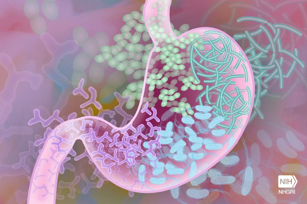
Gut Microbiome. Credit Darryl Leja National Human Genome Research Institute National Institutes Of Health

Researchers from the University of Toronto have shown how a low-carbohydrate diet can worsen the DNA-damaging effects of some gut microbes to cause colorectal cancer.

The study, published in the journal Nature Microbiology, compared the effects of three different diets: normal, low-carb, or Western-style with high fat and high sugar, each in combination with specific gut bacteria on colorectal cancer development in mice.

They found that a unique strain of E. coli bacteria, when paired with a diet low in carbs and soluble fibre, drives the growth of polyps in the colon, which can be a precursor to cancer.

“Colorectal cancer has always been thought of as being caused by a number of different factors including diet, gut microbiome, environment and genetics,” says senior author Alberto Martin, a professor of immunology at U of T’.

“Our question was, does diet influence the ability of specific bacteria to cause cancer?”

To answer this question, the researchers, led by postdoctoral fellow Bhupesh Thakur, examined mice that were colonized with one of three bacterial species that had been previously linked to colorectal cancer and fed either a normal, low-carb or Western-style diet.

Only one combination, a low-carb diet paired with a strain of E. coli that produces the DNA-damaging compound colibactin, led to the development of colorectal cancer.

The researchers found that a diet deficient in fibre increased inflammation in the gut and altered the community of microbes that typically reside there, creating an environment that allowed the colibactin-producing E. coli to thrive.

They also showed that the mice fed a low-carb diet had a thinner layer of mucus separating the gut microbes from the colon epithelial cells. The mucus layer acts as a protective shield between the bacteria in the gut and the cells underneath. With a weakened barrier, more colibactin could reach the colon cells to cause genetic damage and drive tumour growth. These effects were especially strong in mice with genetic mutations in the mismatch repair pathway that hindered their ability to fix damaged DNA.

While both Thakur and Martin emphasize the need to confirm these findings in humans, they are also excited about the numerous ways in which their research can be applied to prevent cancer.

Defects in DNA mismatch repair are frequently found in colorectal cancer, which is the fourth most commonly diagnosed cancer in Canada. An estimated 15 per cent of these tumours having mutations in mismatch repair genes. Mutations in these genes also underlie Lynch syndrome, a genetic condition that significantly increases a person’s risk of developing certain cancers, including colorectal cancer.

“Can we identify which Lynch syndrome patients harbour these colibactin-producing microbes?” asks Martin. He notes that for these individuals, their findings suggest that avoiding a low-carb diet or taking a specific antibiotic treatment to get rid of the colibactin-producing bacteria could help reduce their risk of colorectal cancer.

Martin points out that a strain of E. coli called Nissle, which is commonly found in probiotics, also produces colibactin. Ongoing work in his lab is exploring whether long-term use of this probiotic is safe for people with Lynch syndrome or those who are on a low-carb diet.

Thakur is keen to follow up on an interesting result from their study showing that the addition of soluble fibre to the low-carb diet led to lower levels of the cancer-causing E. coli, less DNA damage and fewer tumours.

“We supplemented fibre and saw that it reduced the effects of the low-carb diet,” he says. “Now we are trying to find out which fibre sources are more beneficial, and which are less beneficial.”

To do this, Thakur and Martin are teaming up with Heather Armstrong, a researcher at the University of Alberta, to test whether supplementation with a soluble fibre called inulin can reduce colibactin-producing E. coli and improve gut health in high-risk individuals, like people with inflammatory bowel disease.

 “Our study highlights the potential dangers associated with long-term use of a low-carb, low-fibre diet, which is a common weight-reducing diet,” says Martin.

“More work is needed but we hope that it at least raises awareness.”

Source: University of Toronto

Prosthetic Hand ‘Knows’ What it’s Touching, Grasps Like a Human

Sriramana Sankar/Johns Hopkins University.

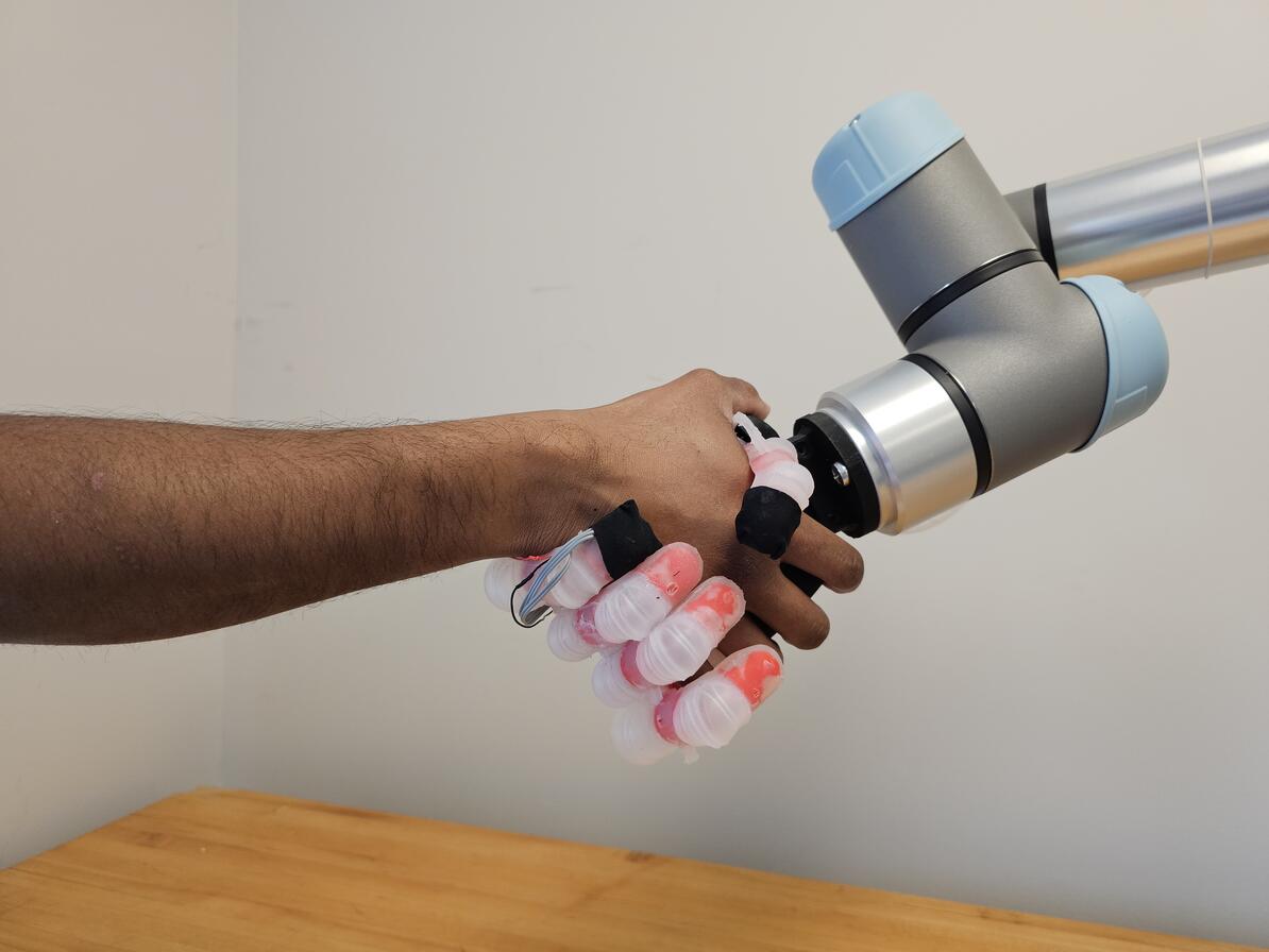
Johns Hopkins University engineers have developed a pioneering prosthetic hand that can grip plush toys, water bottles, and other everyday objects like a human, carefully conforming and adjusting its grasp to avoid damaging or mishandling whatever it holds.

The system’s hybrid design is a first for robotic hands, which have typically been too rigid or too soft to replicate a human’s touch when handling objects of varying textures and materials. The innovation offers a promising solution for people with hand loss and could improve how robotic arms interact with their environment.

Details about the device appear in Science Advances.

“The goal from the beginning has been to create a prosthetic hand that we model based on the human hand’s physical and sensing capabilities—a more natural prosthetic that functions and feels like a lost limb,” said Sriramana Sankar, a Johns Hopkins PhD student in biomedical engineering who led the work. “We want to give people with upper-limb loss the ability to safely and freely interact with their environment, to feel and hold their loved ones without concern of hurting them.”

The device, developed by the same Neuroengineering and Biomedical Instrumentations Lab that in 2018 created the world’s first electronic “skin” with a humanlike sense of pain, features a multifinger system with rubberlike polymers and a rigid 3D-printed internal skeleton. Its three layers of tactile sensors, inspired by the layers of human skin, allow it to grasp and distinguish objects of various shapes and surface textures, rather than just detect touch. Each of its soft air-filled finger joints can be controlled with the forearm’s muscles, and machine learning algorithms focus the signals from the artificial touch receptors to create a realistic sense of touch, Sankar said.

“The sensory information from its fingers is translated into the language of nerves to provide naturalistic sensory feedback through electrical nerve stimulation,” Sankar said.

In the lab, the hand identified and manipulated 15 everyday objects, including delicate stuffed toys, dish sponges, and cardboard boxes, as well as pineapples, metal water bottles, and other sturdier items. In the experiments, the device achieved the best performance compared with the alternatives, successfully handling objects with 99.69% accuracy and adjusting its grip as needed to prevent mishaps. The best example was when it nimbly picked up a thin, fragile plastic cup filled with water, using only three fingers without denting it.

“We’re combining the strengths of both rigid and soft robotics to mimic the human hand,” Sankar said. “The human hand isn’t completely rigid or purely soft—it’s a hybrid system, with bones, soft joints, and tissue working together. That’s what we want our prosthetic hand to achieve. This is new territory for robotics and prosthetics, which haven’t fully embraced this hybrid technology before. It’s being able to give a firm handshake or pick up a soft object without fear of crushing it.”

To help amputees regain the ability to feel objects while grasping, prostheses will need three key components: sensors to detect the environment, a system to translate that data into nerve-like signals, and a way to stimulate nerves so the person can feel the sensation, said Nitish Thakor, a Johns Hopkins biomedical engineering professor who directed the work.

“The goal from the beginning has been to create a prosthetic hand that we model based on the human hand’s physical and sensing capabilities—a more natural prosthetic that functions and feels like a lost limb.”

Sriramana Sankar

PhD student, Biomedial engineering

The bioinspired technology allows the hand to function this way, using muscle signals from the forearm, like most hand prostheses. These signals bridge the brain and nerves, allowing the hand to flex, release, or react based on its sense of touch. The result is a robotic hand that intuitively “knows” what it’s touching, much like the nervous system does, Thakor said.

“If you’re holding a cup of coffee, how do you know you’re about to drop it? Your palm and fingertips send signals to your brain that the cup is slipping,” Thakor said. “Our system is neurally inspired—it models the hand’s touch receptors to produce nervelike messages so the prosthetics’ ‘brain,’ or its computer, understands if something is hot or cold, soft or hard, or slipping from the grip.”

While the research is an early breakthrough for hybrid robotic technology that could transform both prosthetics and robotics, more work is needed to refine the system, Thakor said. Future improvements could include stronger grip forces, additional sensors, and industrial-grade materials.

“This hybrid dexterity isn’t just essential for next-generation prostheses,” Thakor said. “It’s what the robotic hands of the future need because they won’t just be handling large, heavy objects. They’ll need to work with delicate materials such as glass, fabric, or soft toys. That’s why a hybrid robot, designed like the human hand, is so valuable—it combines soft and rigid structures, just like our skin, tissue, and bones.”