Month: February 2025

Caner Signals may Promote Blood Clot Formation in the Lungs

Thrombophilia. Credit: Scientific Animations CC4.0.

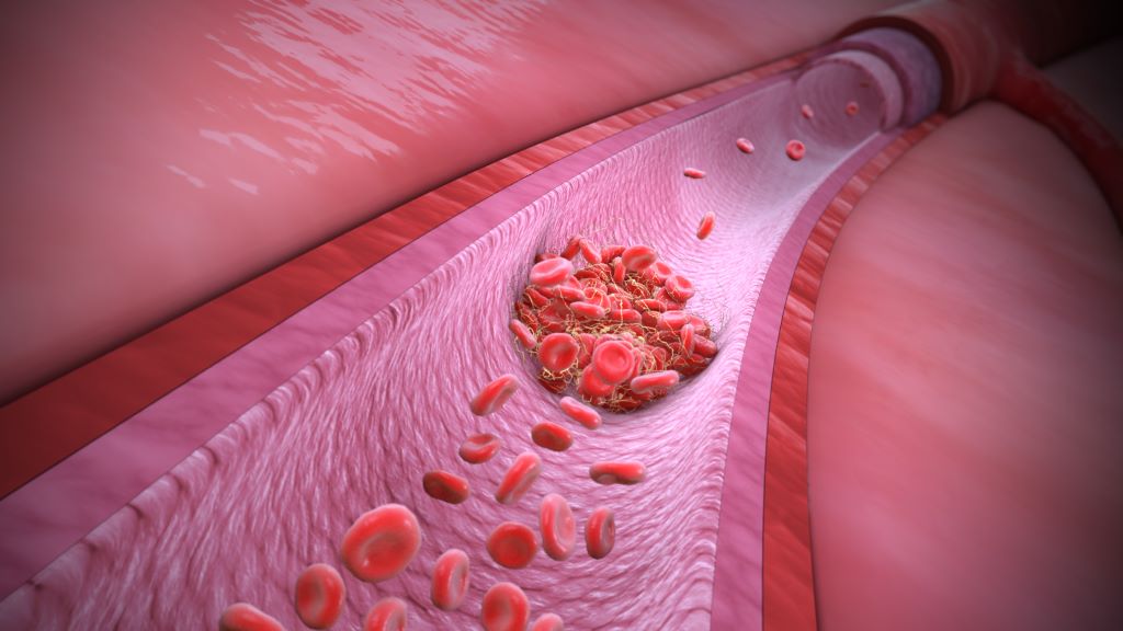
Blood clots form in response to signals from the lungs of cancer patients—not from other organ sites, as previously thought—according to a preclinical study. Clots are the second-leading cause of death among cancer patients with advanced disease or aggressive tumours.

While blood clots usually form to stop a wound from bleeding, cancer patients can form clots without injury, plugging up vessels and cutting off circulation to organs. The study, published in Cell, shows that tumours drive thrombosis by releasing chemokines, secreted proteins which then circulate to the lung. Once there, the chemokines prompt macrophages to release small vesicles that attach to platelets, forming life-threatening clots.

The findings may lead to diagnostic tests to determine blood clotting risk and safer therapies that target the root of the problem to prevent blood clots.

“This work redefines the concept of how thrombosis develops in cancer patients, compared to the traditional view that factors on blood vessel walls or tumour cells themselves are responsible,” said study lead Dr David Lyden, professor in paediatric cardiology, and cell and developmental biology at Weill Cornell Medicine. “It’s a revolutionary concept that thrombosis is initiated in the lung, which wasn’t appreciated before.”

Tumors in the Driver’s Seat

“We reviewed post-mortem studies and found that up to 60% of cancer patients died because of clots rather than cancer itself,” said first author Dr Serena Lucotti,  instructor of cell biology in paediatrics at Weill Cornell Medicine. “It is unfortunate because we have drugs that can prevent clots, but we can’t give them unconditionally to all patients because they can cause excessive bleeding in some. At the same time, we can’t predict who is at high risk for clots and would benefit from the drugs.”

In a series of experiments in mice and human tissues, the researchers showed that different tumours release varying amounts of the chemokine CXCL13. Breast cancers and melanomas release relatively small quantities of CXCL13. However, if these tumour cells spread to the lung, they can trigger clot formation by releasing CXCL13 and locally influencing interstitial macrophages. “In contrast, pancreatic cancer secretes high levels of CXCL13 into the bloodstream,” said Dr Lyden. “It’s so high that it circulates all the way to the macrophages in the lung, so these tumour cells don’t need to be close by.”

Blocking Clots—and Metastases

Other experiments revealed that after interacting with CXCL13, lung interstitial macrophages send out small vesicles loaded with an adhesion molecule, integrin β2, on their surface. The integrin β2 is in an open conformation that can attach to platelets and trigger clot formation.

Mice treated with an antibody that blocks vesicle-bound integrin β2 from binding to platelets had no side effects and didn’t have excessive bleeding. Strikingly, mice with early or advanced cancers that were treated with the antibody not only had fewer clots, but also had significantly fewer metastases than untreated controls. “This is important because there aren’t effective treatments for patients with metastases and will be further investigated,” Dr Lucotti said. She is developing a human antibody to block the integrin β2-platelet interaction in patients.

The researchers are also hopeful that integrin β2 can be a biomarker that indicates a patient’s risk for developing clots. As proof of concept, the team analyzed blood samples from some pancreatic cancer patients from Moores Cancer Center at the University of California San Diego Health. By analyzing blood samples collected before and after the patients experienced blood clots, the authors could easily and accurately distinguish between low-risk and high-risk patients based on integrin β2 levels on extracellular vesicles in the blood.

The study highlights that cancer is a disease that can affect many parts of the body. “Cancer is a systemic disease. We have to pay attention to not only future sites of metastasis, but other organs that may be affected independent of metastasis by systemic complications such as thrombosis, leading to morbidity and mortality,” Dr Lyden said.

Source: Weill Cornell Medicine

Continuation of Opioids for Chronic Pain: Experts Divided

Photo by Usman Yousaf on Unsplash

Chronic pain is complex and difficult to treat. Prescribing opioid pain medications has become controversial but may help some patients.

With the goal of informing clinician practice, a new study explores the harms and benefits of continuing and of discontinuing the long-term prescription of opioid medicines to adults with chronic pain. The authors analysed the opinions of 28 experts on the harms versus benefits of maintaining, tapering or terminating opioid pain medication prescriptions for chronic pain, a common condition worldwide that is typically quite difficult to treat.

The study authors found a lack of consensus among the experts on how to treat chronic (lasting three or more months) non-cancer pain. Slightly more than a third of the experts (36%) believed that long-term opioid therapy is beneficial, while an equal percentage indicated that it should be discontinued.

More than half of the experts believed that patients can experience harm from overly rapid tapering and discontinuation, while some recommended attempting a slow taper (even with a prior unsuccessful taper), possibly with addition of medications to manage withdrawal) in order not to maintain opioid therapy.

Some of the experts advocated for switching patients to buprenorphine, which diminishes the effects of physical dependency to opioids, such as withdrawal symptoms and cravings, and is used to treat pain. Some considered adding non-opioid pain therapies (including re-trying these therapies even if they were unhelpful in the past) as well as engaging in shared decision-making with the patient, although there was little consensus on how to accomplish these options.

Some, but not all of the experts, noted the benefit of addressing co-occurring conditions related to patient safety, such as alcohol use, mental health symptoms and opioid side effects.

Few of the experts brought up assessing or addressing opioid use disorder or overdose risk.

“The potential harms of opioid pain medication are well known, nevertheless patients can become habituated to them and want their physicians to continue prescribing them. Taking patients off opiates may result in return or worsening of chronic pain, mental health issues, drug seeking and potentially overdose and death. Additionally, these drugs could be used by someone else, possibly winding up on the street,” said study co-author Kurt Kroenke, MD of the Regenstrief Institute and the Indiana University School of Medicine. “On the benefit side, these drugs may be helping relieve the patient’s often debilitating pain which can impact the ability to interact with family, to hold a job, participate in social activities and many other aspects of life.”

A substantial number of people who are prescribed opioid pain medications continue to experience chronic pain. Dr Kroenke notes that these individuals may be good candidates for tapering to a lower dose, prescription discontinuation and moving on to effective, safer treatments for pain.

The authors conclude their analysis of the experts’ opinions, “Guidelines on whether to continue or taper opioids prescribed long- term may be difficult to utilize given professional liability concerns, changing regulations and health system initiatives, differing provider-patient perspectives on long-term opioid benefits and harms, and some providers’ beliefs that opioid dependence interferes with patients’ objectivity. In the meantime, individual care decisions that involve weighing relative harms should draw on longstanding norms of ethical medical care that call for informed consent and patient-provider conversations grounded in mutual respect.”

The study is published in the peer-reviewed journal Pain Practice.

Source: EurekAlert

Plant-rich, Low Saturated-fat Diet Linked to Reduced Psoriasis Severity

Photo: CC0

A new study by researchers at King’s College London, published in the British Journal of Nutrition, has found significant associations between diet quality and the severity of psoriasis. The findings provide novel insights into how dietary patterns may be related to psoriasis severity in non-Mediterranean populations.

Psoriasis is a long-lasting inflammatory skin disease which causes flaky patches of skin that form scales. It affects millions worldwide and is believed to be caused by a problem with the immune system.

The research analysed data from 257 adults with psoriasis who had completed an online survey. Participants’ adherence to various diet quality scores, including the Mediterranean Diet Score, the Dietary Approaches to Stop Hypertension (DASH) score, and the Healthy Plant-based Diet Index, was assessed using a food frequency questionnaire. Psoriasis severity was self-assessed using a validated questionnaire.

Key findings from the study indicate that individuals with very low adherence to the DASH diet index and the Healthy Plant-based Diet Index were significantly more likely to report higher psoriasis severity.

Further analysis of the different elements of the DASH dietary pattern revealed that greater red and processed meat intake was associated with more severe psoriasis even when body mass index (BMI) was considered. Fruits, nuts and legume intakes were also associated with less severe psoriasis, but this relationship was not independent of BMI.

The study was published as part of the Asking People with Psoriasis about Lifestyle and Eating (APPLE) project and funded by the Psoriasis Association.

Our findings point to the potential benefits of dietary interventions in improving patient outcomes. Given the impact of psoriasis on physical and psychological well-being, incorporating dietary assessments into routine care could offer patients additional support in managing their condition.

Sylvia Zanesco, PhD student from the Department of Nutritional Sciences at King’s College London who led the research

The DASH dietary pattern was originally designed to lower blood pressure and emphasises fruits, vegetables, whole grains, low-fat dairy foods and lean meats while limiting salt, sugar, and saturated fats. A high Healthy Plant-based Diet Index characterises a dietary pattern rich in healthy plant foods including fruits and vegetables, whole grains, nuts and seeds, legumes and plant oils rich in unsaturated fats, as well as being low in animal foods and unhealthy plant foods such as sugary foods and drinks and refined starches.

The study accounted for several confounding factors, including age, sex, smoking status, alcohol, energy intake, and mental health, ensuring a comprehensive analysis of dietary patterns that are independently associated with psoriasis severity.

This research brings much-needed evidence that there may be a role for dietary advice, alongside standard clinical care, in managing symptoms of psoriasis. Our next steps will be to explore whether diets rich in healthy plant foods can reduce symptoms of psoriasis in a controlled clinical trial.

Professor Wendy Hall, Professor of Nutritional Sciences at King’s College London and senior author of the study

The findings of the study contribute to the growing body of evidence supporting dietary modification as a complementary strategy in psoriasis management to potentially alleviate disease severity and improve patients’ quality of life.

Source: King’s College London

Co-prescribed Stimulants and Opioids Linked to Higher Opioid Doses

Photo from Pixabay CCO

The combination of prescribed central nervous system stimulants, such as drugs that relieve ADHD symptoms, with prescribed opioid medications is associated with a pattern of escalating opioid intake, a new study has found. 

The analysis of health insurance claims data from almost 3 million US patients investigated prescribed stimulants’ impact on prescription opioid use over 10 years, looking for origins of the so-called “twin epidemic” of combining the two classes of drugs, which can increase the risk for overdose deaths

“Combining the two drugs is associated with an increase in overdose deaths. This is something we know. But we didn’t know whether stimulant use has a causal role in high use of opioids, so we conducted a big data analysis of how these two patterns interacted over a long period of time,” said senior study author Ping Zhang, associate professor of computer science and engineering and biomedical informatics at The Ohio State University.

“What we found is that if someone is taking a stimulant and an opioid at the same time, they’re generally taking a high dose of the opioid,” he said. “And if the patient in this study population takes the stimulant before beginning opioid use, they are more likely to have higher doses of subsequent opioids.” 

The study was published in The Lancet Regional Health – Americas.     

The research team obtained data on 22 million patients with 96 million opioid prescriptions from a large US health insurance database. Researchers established a cohort for this study of 2.9 million patients with an average age of 44 who had at least two independent opioid prescriptions between 2012 and 2021. 

Because these prescriptions included a range of oral formulas – codeine, hydrocodone, methadone, oxycodone, morphine and others – researchers standardised every prescription to morphine milligram equivalents (MME) and calculated each patient’s monthly intake of opioids.

First author Seungyeon Lee, a PhD student in Zhang’s lab, used statistical modelling and classified patients into five baseline groups of opioid dosage trajectory over the 10-year study period: very low-dose, low-dose decreasing, low-dose increasing, moderate-dose increasing and high-dose sustained use. 

“Some patients had stable low-dose opioid use, while others had increasing or high dose patterns over time,” Lee said. 

Of the total cohort, 160 243 patients (5.5%) also were prescribed stimulants. The addition of a monthly calculated cumulative number of stimulant prescriptions to the model and statistical analysis showed a shift in the trajectory groups. Characteristics that could serve as risk factors for increasing opioid use also emerged in the data, Lee said. 

Moderate-dose increasing and high-dose groups had an overall higher average MME and a higher proportion of patients with diagnoses of depression, anxiety and attention-deficit/hyperactivity disorder compared to other groups. The low-dose increasing group also had a higher proportion of patients with ADHD compared to the low-dose decreasing group. 

The most common diagnoses linked to co-prescription of stimulants and opioids were depression and ADHD or ADHD and chronic pain. 

“This was an important finding, that many patients with ADHD and depression, also experiencing chronic pain, have an opioid prescription,” said Zhang. “This cohort represents a very realistic health care problem.” 

Even taking those factors into account, the model showed that stimulant use was key to driving up the odds that patients who took both stimulants and opioids would belong to a group of people who increased their doses of opioids. 

“Stimulant use before initiating opioids and stimulant co-prescription with opioids are both positively associated with escalating opioid doses compared to other factors,” Lee said. 

Analysis of geographic and gender data also offered some clues to opioid use patterns in the United States. Patients in the South and West regions had higher total opioid intakes over the 10-year study period compared to the Northeast and North Central regions, with the highest frequency of opioid prescriptions in the South and higher MMEs per prescription in the West. Males also had higher average daily opioid intakes than females. 

The results linking high opioid doses and stimulant use suggest stimulants may be a driving force behind the emergence of the twin epidemic and offer evidence that regulation of stimulant prescribing may be needed for patients already taking prescription opioids, the researchers said. In addition to the increased risk of overdose death, co-using prescription stimulants and opioids can increase the risk for cardiovascular events and mental health problems, previous research has shown. 

Source: Ohio State University

Compound in Ginger might be a Treatment for Inflammatory Bowel Disease

Irritable bowel syndrome. Credit: Scientific Animations CC4.0

An international team led by researchers at the University of Toronto has found a compound in ginger, called furanodienone (FDN), that selectively binds to and regulates a nuclear receptor involved in inflammatory bowel disease (IBD).

Through a screen to identify chemical components of ginger that bind to receptors associated with IBD, the team observed a strong interaction between FDN and the pregnane X receptor (PXR). FDN reduces inflammation in the colon by activating PXR’s ability to suppress the production of pro-inflammatory cytokines in the body. While researchers have been aware of FDN for decades, they had not determined its functions or targets in the body until now.

“We found that we could reduce inflammation in the colons of mice through oral injections of FDN,” said research associate Jiabao Liu. “Our discovery of FDN’s target nuclear receptor highlights the potential of complementary and integrative medicine for IBD treatment. We believe natural products may be able to regulate nuclear receptors with more precision than synthetic compounds, which could lead to alternative therapeutics that are cost-effective and widely accessible.”

The study was published recently in the journal Nature Communications.

IBD patients typically start to experience symptoms early in life; around 25% of patients are diagnosed before the age of 20. There is currently no cure for IBD, so patients must adhere to life-long treatments to manage their symptoms, including abdominal pain and diarrhoea, enduring significant psychological and economic consequences.

While patients with IBD have found some relief through changes to their diet and herbal supplements, it is not clear which chemical compounds in food and supplements are responsible for alleviating intestinal inflammation. With FDN now identified as a compound with potential to treat IBD, this specific component of ginger can be extracted to develop more effective therapies.

An additional benefit of FDN is that it can increase the production of tight junction proteins that repair damage to the gut lining caused by inflammation. The effects of FDN were demonstrated in the study to be restricted to the colon, preventing harmful side effects to other areas of the body.

Nuclear receptors serve as sensors within the body for a wide range of molecules, including those involved in metabolism and inflammation. PXR specifically plays a role in the metabolism of foreign substances, like dietary toxins and pharmaceuticals. Binding between FDN and PXR needs to be carefully regulated because over-activating the receptor can lead to an increase in the metabolism and potency of other drugs and signaling metabolites in the body.

FDN is a relatively small molecule that only fills a portion of the PXR binding pocket. The study shows that this allows for an additional compound to bind simultaneously, thereby increasing the overall strength of the bond and its anti-inflammatory effects in a controlled manner.

“The number of people diagnosed with IBD in both developed and developing countries is on the rise due to a shift towards diets that are more processed and are high in fat and sugar,” said Henry Krause, principal investigator on the study and professor of molecular genetics. “A natural product derived from ginger is a better option for treating IBD than current therapies because it does not suppress the immune system or affect liver function, which can lead to major side effects. FDN can form the basis of a treatment that is more effective while also being safer and cheaper.”

Source: University of Toronto

#InsideTheBox with Dr Andy Gray | Essential Medicines – Essential to Whom?

#InsideTheBox is a column by Dr Andy Gray, a pharmaceutical sciences expert at the University of KwaZulu-Natal and Co-Director of the WHO Collaborating Centre on Pharmaceutical Policy and Evidence Based Practice. (Photo: Supplied)

By Andy Gray

In the first of a new series of monthly columns for Spotlight, Dr Andy Gray considers what we mean when we refer to medicines as “essential” – something that is routinely done by the WHO and health authorities in many countries.

That every health system needs medicines, including vaccines, to treat and prevent and at times to cure diseases is obvious. However, that immediately begs a series of questions – which medicines, where, and for whom?

For nearly 50 years, the World Health Organization (WHO) has provided guidance in the form of the Model List of Essential Medicines. For almost 20 years, WHO has also provided a Model List of Essential Medicines for Children. The most recent version, issued in 2023 and due to be revised in 2025, lists over 500 medicines, including 361 specifically for children.

Those figures draw attention to a core principle that not all medicines approved by national medicines regulatory authorities, and therefore legally marketed, are considered necessary.

The WHO defines essential medicines as “those that effectively and safely treat the priority healthcare needs of the population”. It adds that the primary drivers for selecting a limited list of medicines are efficiency and effectiveness: “The use of a limited number of carefully selected medicines can lead to improved supply, better prescribing practices and lower costs.”

The initial essential medicines definition contains a highly contested component – how does one determine the “priority healthcare needs of the population”?

One element should surely be the prevalence of the condition for which the medicine is indicated. However, that alone is insufficient. The common cold is, as the name implies, ubiquitous. Should that mean that cough mixtures are essential? If the evidence of a clinically relevant health benefit from the use of cough mixtures is lacking, that should surely rule them out. The common cold is also self-limiting, without long-term consequences.

By contrast, infection with hepatitis C virus can result in serious illness and death, but also spread to others, and in some patients become a chronic disease associated with increased risk of liver cancer or cirrhosis. In that case, treating the condition not only benefits the patient, but also prevents others from being infected with the same virus. For many years, the available treatments for hepatitis C were not very effective and associated with severe adverse effects. Once new, highly effective and safer treatments became available around a decade ago, the pressure was on to consider them as essential.

That is where another part of the WHO definition comes into play: “Essential medicines should always be available within functioning health systems, in sufficient quantities to meet patient needs. They should be available in appropriate dosage forms for the intended uses and patients, be of assured quality, and be affordable for both individuals and the health system.”

But how can affordability be measured?

One approach is to consider whether the added benefits are worth the extra cost, compared to existing medicines. That approach, based on a technique called incremental cost-effectiveness analysis, does not consider the possible impact of a new medicine on the total pharmaceutical or health budget. A stunningly effective new medicine may simply be unaffordable if needed by a large number of patients, unless additional resources can be secured for the health system. In an era of fiscal restraint and shifting donor priorities, “new money” is increasingly unlikely to be found.

An even more pressing problem is posed when a new medicine becomes available that makes a significant difference to clinical outcomes, for example changing a previously fatal condition into a manageable chronic condition. The new medicine may only be needed by a few patients, but if it is very expensive it may pose a threat to available budgets and be unaffordable to individual patients and their families. Rare diseases, and the medicines to treat them, ask uncomfortable questions about how public health priorities are decided, how affordability is assessed, and how society as a whole values access to medicines. The population level perspective runs headlong into the individual.

How it works in SA

South Africa, like more than 150 other countries, has established medicines selection structures and procedures and developed a national essential medicines list (EML). The medicines selected for the EML inform the state tender system, allowing the state to use its buying power to exert downward pressure on medicine prices. Medicines procured in that way are used almost exclusively in the public sector, in health facilities operated by provincial and local government.

However, the separation between public and private sectors is not that simple. Medical schemes use similar considerations of cost effectiveness and budget impact to determine the reimbursement rules and formularies for each of their benefit options. In some cases, there are nationally-determined algorithms for specific conditions – the Chronic Disease List – that have to be covered as prescribed minimum benefits. Those algorithms have not been updated as frequently as might be desired, and certainly not as frequently as the standard treatment guidelines and EML applied in the public sector. As a result, when challenged regarding denial of coverage, the medical schemes regulator has tended to look to the state’s position as defining a standard of care. However, the reduced prices available to the state may not be accessible to the private sector.

In other words, and even before National Health Insurance is implemented, the questions of which medicines, where, and for whom remain vexing. They have to be faced, however, if any health system is to ensure the progressive realisation of the right of access to healthcare services, responsibly and effectively using the available resources.

*Gray is a Senior Lecturer at the University of KwaZulu-Natal and Co-Director of the WHO Collaborating Centre on Pharmaceutical Policy and Evidence Based Practice. This is the first of a new series of #InsideTheBox columns he is writing for Spotlight.

Note: Spotlight aims to deepen public understanding of important health issues by publishing a variety of views on its opinion pages. The views expressed in this article are not necessarily shared by the Spotlight editors.

Republished from Spotlight under a Creative Commons licence.

Read the original article.

Causes of Fevers of Unknown Origin in sub-Saharan Africa

Ebola on a cell. Credit: NIH/NIAID

A new retrospective, laboratory-based observational study provides detailed insights into the causes of fevers of unknown origin in sub-Saharan Africa. Researchers examined 550 patients from Guinea who developed a persistent fever at the time of the major Ebola outbreak in 2014, but tested negative for the Ebola virus on site. The goal was to use modern diagnostic methods to better understand the underlying infectious diseases. The study is published in The Journal of Infectious Diseases.

Fever is a common symptom of many diseases, including infections, cancer, and autoimmune diseases. When the cause of a persistent fever remains unclear despite extensive investigation, it is referred to as fever of unknown origin (FUO). Approximately half of all FUO cases worldwide remain undiagnosed. In sub-Saharan Africa, malaria is often suspected and treated without laboratory confirmation or further investigation. However, 90 million pediatric hospitalisations per year in sub-Saharan Africa are due to fevers not caused by malaria but by other infections, often due to various bacteria and viruses.

A research team from the German Center for Infection Research (DZIF) and Charité – Universitätsmedizin Berlin, in collaboration with scientists from Guinea and Slovakia, conducted a retrospective observational study to thoroughly investigate the pathogen diversity of patients from Guinea with fever of unknown cause during a major Ebola outbreak in 2014. They combined epidemiological, phylogenetic, molecular, serological and clinical data.

Using serologic tests, PCR and high-throughput sequencing, at least one pathogen was detected in 275 of 550 patients. In addition to the expected malaria parasite Plasmodium, pathogenic bacteria such as Salmonella and Klebsiella strains were detected in almost one fifth of the patients. The frequent detection of resistance to so-called first-line antibiotics in the samples examined and the high rate of co-infections were also worrying: One in five infected patients had multiple infections at the same time. Pathogens causing malaria and bacterial sepsis were particularly common, occurring together in 12% of adults and 12.5% of children.

Infections with highly pathogenic viruses were also common: Yellow fever, Lassa and Ebola viruses were detected by RT-PCR in about six percent of patients. Of particular note was the detection of infection with Orungo virus, a little-known pathogen for which there are no robust assays. Using immunofluorescence assays, the researchers also identified IgM antibodies against several viruses, including Dengue, West Nile and Crimean-Congo hemorrhagic fever viruses, in patients who were PCR-negative.

“In Africa, febrile illnesses of unknown cause are often recognized and treated as malaria without further diagnosis. In our study, we were able to detect a pathogen in about half of all patients with FUO, including bacterial pathogens that cause sepsis, haemorrhagic fever viruses including Ebola, and, as expected, various strains of the malaria parasite Plasmodium,” explains the study’s last author Prof. Jan Felix Drexler.

The findings underscore the urgent need to further strengthen laboratory capacity in sub-Saharan Africa. Early detection of the infectious causes of FUO is critical for patient care, effective response to outbreaks, and development of regionally appropriate diagnostics.

“Our results show that regionally adapted treatment regimens should be discussed, that quality control in the context of outbreaks needs to be strengthened, and that knowledge of the pathogen spectrum can guide targeted strengthening of regional laboratories and translational research in the sense of point-of-care tests,” Drexler summarises the results of the study.

Source: German Center for Infection Research

Nerves Electrify Stomach Cancer, Sparking Growth and Spread

Image from Pixabay.

Researchers have discovered that stomach cancers in mice make electrical connections with nearby sensory nerves and use these malignant circuits to stimulate the cancer’s growth and spread.

Reported in Nature, this is the first time that electrical contacts between nerves and a cancer outside the brain have been found, raising the possibility that many other cancers progress by making similar connections.

“We know that many cancers exploit nearby neurons to fuel their growth, but outside of cancers in the brain, these interactions have been attributed to the secretion of growth factors broadly or through indirect effects,” says Timothy Wang, the Silberberg Professor of Medicine at Columbia University Vagelos College of Physicians and Surgeons, who led the study and is one of the leaders in the growing field of cancer neuroscience.

“Now that we know the communication between the two is more direct and electrical, it raises the possibility of repurposing drugs designed for neurological conditions to treat cancer.”

The wiring of neurons to cancer cells also suggests that cancer can commandeer a particularly rapid mechanism to stimulate growth.

“There are many different cells surrounding cancers, and this microenvironment can sometimes provide a rich soil for their growth,” Wang says. Researchers have been focusing on the role of the microenvironment’s immune cells, connective tissue, and blood vessels in cancer growth but have only started to examine the role of nerves in the last two decades.

“What’s emerged recently is how advantageous the nervous system can be to cancer,” Wang adds. “The nervous system works faster than any of these other cells in the tumour microenvironment, which allows tumours to more quickly communicate and remodel their surroundings to promote their growth and survival.”

Cancer-neuron connections resemble synapses

As a gastroenterologist, Wang’s research has focused on stomach and other GI cancers. About 10 years ago, he discovered that cutting the vagus nerve in mice with stomach cancer significantly slowed tumour growth and increased survival rate.

Many different types of neurons are contained in the vagus nerve, but the researchers focused here on sensory neurons, which reacted most strongly to the presence of stomach cancer in mice. Some of these sensory neurons extended themselves deep into stomach tumours in response to a protein released by cancer cells called Nerve Growth Factor (NGF), drawing the cancer cells close to the neurons. After establishing this connection, tumours signalled the sensory nerves to release the peptide Calcitonin Gene Related Peptide (CGRP), inducing electrical signals in the tumour.  

Though the cancer cells and neurons may not form classical synapses where they meet – the team’s electron micrographs are still a bit fuzzy – “there’s no doubt that the neurons create an electric circuit with the cancer cells,” Wang says. “It’s a slower response than a typical nerve-muscle synapse, but it’s still an electrical response.”

The researchers could see this electrical activity with calcium imaging, a technique that uses fluorescent tracers that light up when calcium ions surge into a cell as an electrical impulse travels through.  

“There’s a circuit that starts from the tumour, goes up toward the brain, and then turns back down toward the tumour again,” Wang says. It’s like a feed-forward loop that keeps stimulating the cancer and promoting its growth and spread.”

Migraine drugs as a potential cancer treatment

For stomach cancer, CGRP inhibitors that are currently used to treat migraines could potentially short-circuit the electrical connection between tumours and sensory neurons.

In Wang’s study, CGRP inhibitors administered to mice with stomach cancer reduced the size of the tumors, prolonged survival, and prevented the tumors from spreading.

“Based on our analysis of stomach cancer data from patients, we believe that the circuits we’ve found in mice also exist in humans and targeting them could be an additional useful therapy,” Wang says.

Sensory neurons may also use CGRP to stimulate cancer through more indirect pathways. Unpublished findings from Wang’s lab suggest that the neurons promote stomach cancer growth via contact with connective tissue cells in the tumour microenvironment. And other researchers have found that sensory nerves may, possibly through CGRP, cause T cell exhaustion and turn off immune responses directed at other types of cancers.

“But we think it all starts with the cancer cell setting up a neural circuit,” Wang says.

“Nerves are an underappreciated master regulator of normal growth and regeneration in animals. We know that when organs form during development, the nerves lead the way. From that point of view, it was not unexpected that nerves would be driving tumour growth as well.”

Source: Columbia University Irving Medical Center

These Newly Discovered Brain Cells Enable us to Remember Objects

Discovery of ‘ovoid cells’ reshapes our understanding of how memory works, and could open the door to new treatments for Alzheimer’s disease, epilepsy and more.

Ovoid cells. Photo credit: Dr. Mark Cembrowski

Take a look around your home and you’ll find yourself surrounded by familiar comforts – photos of family and friends on the wall, well-worn tekkies by the door, a shelf adorned with travel mementos.

Objects like these are etched into our memory, shaping who we are and helping us navigate environments and daily life with ease. But how do these memories form? And what if we could stop them from slipping away under a devastating condition like Alzheimer’s disease?

Scientists at the University of British Columbia have just uncovered a crucial piece of the puzzle. In a study published in Nature Communications, the researchers have discovered a new type of brain cell that plays a central role in our ability to remember and recognise objects. 

Called ‘ovoid cells,’ these highly-specialised neurons activate each time we encounter something new, triggering a process that stores those objects in memory and allowing us to recognise them months, even years, later.

“Object recognition memory is central to our identity and how we interact with the world,” said Dr Mark Cembrowski, the study’s senior author, and an associate professor of cellular and physiological sciences at UBC and investigator at the Djavad Mowafaghian Centre for Brain Health. “Knowing if an object is familiar or new can determine everything from survival to day-to-day functioning, and has huge implications for memory-related diseases and disorders.”

Hiding in plain sight

Named for the distinct egg-like shape of their cell body, ovoid cells are present in relatively small numbers within the hippocampus of humans, mice and other animals.

Adrienne Kinman, a PhD student in Dr Cembrowski’s lab and the study’s lead author, discovered the cells’ unique properties while analysing a mouse brain sample, when she noticed a small cluster of neurons with highly distinctive gene expression.

“They were hiding right there in plain sight,” said Kinman. “And with further analysis, we saw that they are quite distinct from other neurons at a cellular and functional level, and in terms of their neural circuitry.”

To understand the role ovoid cells play, Kinman manipulated the cells in mice so they would glow when active inside the brain. The team then used a miniature single-photon microscope to observe the cells as the mice interacted with their environment.

The ovoid cells lit up when the mice encountered an unfamiliar object, but as they grew used to it, the cells stopped responding. In other words, the cells had done their jobs: the mice now remembered the objects.

“What’s remarkable is how vividly these cells react when exposed to something new. It’s rare to witness such a clear link between cell activity and behaviour,” said Kinman. “And in mice, the cells can remember a single encounter with an object for months, which is an extraordinary level of sustained memory for these animals.”

New insights for Alzheimer’s disease, epilepsy

The researchers are now investigating the role that ovoid cells play in a range of brain disorders. The team’s hypothesis is that when the cells become dysregulated, either too active or not active enough, they could be driving the symptoms of conditions like Alzheimer’s disease and epilepsy.

“Recognition memory is one of the hallmarks of Alzheimer’s disease – you forget what keys are, or that photo of a person you love. What if we could manipulate these cells to prevent or reverse that?” said Kinman. “And with epilepsy, we’re seeing that ovoid cells are hyperexcitable and could be playing a role in seizure initiation and propagation, making them a promising target for novel treatments.”

For Dr Cembrowski, discovering the highly specialised neuron upends decades of conventional thinking that the hippocampus contained only a single type of cell that controlled multiple aspects of memory.

“From a fundamental neuroscience perspective, it really transforms our understanding of how memory works,” he said. “It opens the door to the idea that there may be other undiscovered neuron types within the brain, each with specialised roles in learning, memory and cognition. That creates a world of possibilities that would completely reshape how we approach and treat brain health and disease.”

Source: University of British Columbia

Intermittent Fasting could be Unsafe for Teenagers

Photo by jamie he

A recent study reveals that age plays a significant role in the outcomes of intermittent fasting. Researchers from Technical University of Munich (TUM), LMU Hospital Munich, and Helmholtz Munich discovered that chronic intermittent fasting disrupted the development of insulin-producing beta cells in young mice. The findings, published in Cell Reports, raise concerns about potential risks for humans, especially teenagers.

“Intermittent fasting is known to have benefits, including boosting metabolism and helping with weight loss and heart disease. But until now, its potential side effects weren’t well understood,” says Alexander Bartelt, the Else Kröner Fresenius Professor and Chair of Translational Nutritional Medicine at TUM. In a recently published study, the team shows that intermittent fasting during adolescence could have long-term negative effects on metabolism.

Fasting improves metabolism in older mice, but not in the young

The researchers studied three groups of mice: adolescent, adult, and older animals. The mice remained without food for one day and were fed normally on two days. After ten weeks, insulin sensitivity improved in both the adult and older mice, meaning that their metabolism responded better to insulin produced by the pancreas. This is key to regulating blood sugar levels and preventing conditions like type 2 diabetes.

However, the adolescent mice showed a troubling decline in their beta cell function, the insulin-producing cells of the pancreas. Insufficient insulin production is linked to diabetes and disrupted metabolism. “Intermittent fasting is usually thought to benefit beta cells, so we were surprised to find that young mice produced less insulin after the extended fasting,” explains Leonardo Matta from Helmholtz Munich, one of the study’s lead authors.

Defective beta cells resemble those of type 1 diabetes patients

The researchers used the latest single-cell sequencing to uncover the cause of the beta cell impairment. By examining the blueprint of the pancreas, the team found that the beta cells in the younger mice failed to mature properly. “At some point, the cells in the adolescent mice stopped developing and produced less insulin,” says Peter Weber from Helmholtz Munich, also a lead author. Older mice, whose beta cells were already mature before the fasting began, remained unaffected.

This shows an accumulation of beta cells from the pancreas of an older mouse.
Beta cells from an older mouse.

The team compared their mouse findings to data from human tissues. They found that patients with type 1 diabetes, where beta cells are destroyed by an autoimmune response, showed similar signs of impaired cell maturation. This suggests that the findings from the mouse study could also be relevant to humans.

“Our study confirms that intermittent fasting is beneficial for adults, but it might come with risks for children and teenagers,” says Stephan Herzig, a professor at TUM and director of the Institute for Diabetes and Cancer at Helmholtz Munich. “The next step is digging deeper into the molecular mechanisms underlying these observations. If we better understand how to promote healthy beta cell development, it will open new avenues for treating diabetes by restoring insulin production.”

Source: Technical University of Munich (TUM)