Month: February 2025

A Protein from Tardigrades may Blunt the Effects of Radiotherapy

A tardigrade, otherwise known as a “water bear”. Credit: NIH

Drawing inspiration from a tiny organism that can withstand huge amounts of radiation, researchers have developed a new strategy that may protect patients from this kind of damage. Their approach makes use of a protein from tardigrades, often also called “water bears,” which are usually less than a millimetre in length. 

When the researchers injected messenger RNA encoding this protein into mice, they found that it generated enough protein to protect cells’ DNA from radiation-induced damage. If developed for use in humans, this approach could benefit many cancer patients, the researchers say.

“Radiation can be very helpful for many tumours, but we also recognise that the side effects can be limiting. There’s an unmet need with respect to helping patients mitigate the risk of damaging adjacent tissue,” says Giovanni Traverso, an associate professor of mechanical engineering at MIT and a gastroenterologist at Brigham and Women’s Hospital.

Traverso and James Byrne, an assistant professor of radiation oncology at the University of Iowa, are the senior authors of the study, which appears in Nature Biomedical Engineering. The paper’s lead authors are Ameya Kirtane, an instructor in medicine at Harvard Medical School and a visiting scientist at MIT’s Koch Institute for Integrative Cancer Research, and Jianling Bi, a research scientist at the University of Iowa.

Extreme survival

Radiation is often used to treat cancers of the head and neck, where it can damage the mouth or throat, making it very painful to eat or drink. It is also commonly used for gastrointestinal cancers, which can lead to rectal bleeding. Many patients end up delaying treatments or stopping them altogether.

“This affects a huge number of patients, and it can manifest as something as simple as mouth sores, which can limit a person’s ability to eat because it’s so painful, to requiring hospitalization because people are suffering so terribly from the pain, weight loss, or bleeding. It can be pretty dangerous, and it’s something that we really wanted to try and address,” Byrne says.

Currently, there are very few ways to prevent radiation damage in cancer patients. There are a handful of drugs that can be given to try to reduce the damage, and for prostate cancer patients, a hydrogel can be used to create a physical barrier between the prostate and the rectum during radiation treatment.

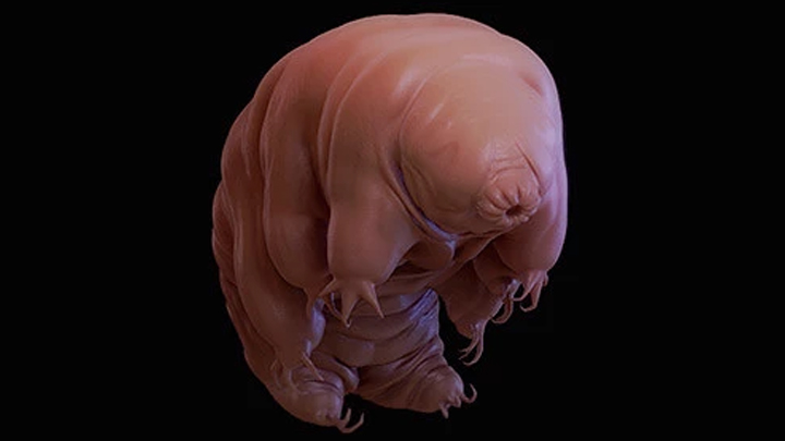
For several years, Traverso and Byrne have been working on developing new ways to prevent radiation damage. In the new study, they were inspired by the extraordinary survival ability of tardigrades. Found all over the world, usually in aquatic environments, these organisms are well known for their resilience to extreme conditions. Scientists have even sent them into space, where they were shown to survive extreme dehydration and cosmic radiation.

One key component of tardigrades’ defence systems is a unique damage suppressor protein called Dsup, which binds to DNA and helps protect it from radiation-induced damage. This protein plays a major role in tardigrades’ ability to survive radiation doses 2000 to 3000 times higher than what a human being can tolerate.

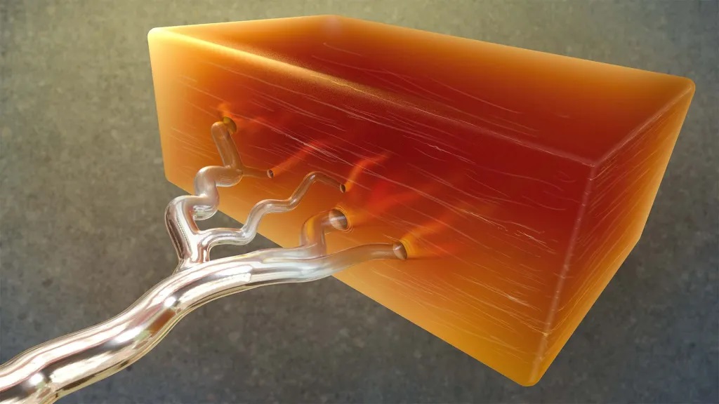
When brainstorming ideas for novel ways to protect cancer patients from radiation, the researchers wondered if they might be able to deliver messenger RNA encoding Dsup to patient tissues before radiation treatment. This mRNA would trigger cells to transiently express the protein, protecting DNA during the treatment. After a few hours, the mRNA and protein would disappear.

For this to work, the researchers needed a way to deliver mRNA that would generate large amounts of protein in the target tissues. They screened libraries of delivery particles containing both polymer and lipid components, which have been used separately to achieve efficient mRNA delivery. From these screens, they identified one polymer-lipid particle that was best-suited for delivery to the colon, and another that was optimized to deliver mRNA to mouth tissue.

“We thought that perhaps by combining these two systems – polymers and lipids – we may be able to get the best of both worlds and get highly potent RNA delivery. And that’s essentially what we saw,” Kirtane says. “One of the strengths of our approach is that we are using a messenger RNA, which just temporarily expresses the protein, so it’s considered far safer than something like DNA, which may be incorporated into the cells’ genome.”

Protection from radiation

After showing that these particles could successfully deliver mRNA to cells grown in the lab, the researchers tested whether this approach could effectively protect tissue from radiation in a mouse model.

They injected the particles into either the cheek or the rectum several hours before giving a dose of radiation similar to what cancer patients would receive. In these mice, the researchers saw a 50 percent reduction in the amount of double-stranded DNA breaks caused by radiation.

The researchers also showed that the protective effect of the Dsup protein did not spread beyond the injection site, which is important because they don’t want to protect the tumour itself from the effects of radiation. To make this treatment more feasible for potential use in humans, the researchers now plan to work on developing a version of the Dsup protein that would not provoke an immune response, as the original tardigrade protein likely would.

If developed for use in humans, this protein could also potentially be used to protect against DNA damage caused by chemotherapy drugs, the researchers say. Another possible application would be to help prevent radiation damage in astronauts in space.

Source: MIT

Using Liquid Metal to Create New Tissue

Artist’s representation of an approach for moulding biological structures in the lab using a metal called gallium. The image shows a metallic branching structure (gallium) and then empty vessels running through a block of organic tissue. Credit: Donny Bliss/NIH

The ability to engineer complex biological tissues, such alveoli or blood vessels, has vast potential to help us unlock fundamental biological insights, test new therapeutics, and one day even build fully functional replacement tissues or whole organs. But researchers have found it challenging to use current technologies such as 3D printing to produce living tissues using natural biological materials that include larger organ structures accurately constructed down to the tiny, cellular level. It has been too complex to recreate the many different tissue architectures of the human body in the lab.

A recent, NIH-supported study reported in Nature suggests a clever solution. The key is taking advantage of the natural properties of a silvery metal known as gallium, which is notable for melting at about 30°C, below body temperature, meaning it can be melted by body temperature. The new study demonstrated the metal’s use as a moulding material for generating soft biological structures complete with hollowed-out internal forms in the wide range of intricate shapes and sizes that would be needed to support the growth of larger, lifelike tissues from cells.

The team behind the new approach called ESCAPE (engineered sacrificial capillary pumps for evacuation), is led by Christopher Chen at Boston University and the Wyss Institute at Harvard University in Boston. The research team realised they needed a single process that could handle fragile biological materials while also building well at both large and extremely tiny scales.

How does ESCAPE achieve this? The strategy is much like traditional metal-casting, which has long been used to make intricate jewelry or sculptures from metals, but in reverse. In this process, a template is made from wax inside a rigid material. When the wax is melted away by molten metal, the desired form is left behind.

The researchers realized that gallium was an ideal material to work with for fashioning scaffolds for use in tissue engineering. Gallium is easy to handle and cast into desired shapes. Its low melting point and biocompatibility also made it especially appealing. In the ESCAPE approach, the researchers start by coming up with a desired shape. They then make a solid metal cast of the shape out of gallium. Next, they form a soft biomaterial around the gallium cast. When the temperature is raised, the gallium can be melted and cleanly removed, leaving behind a perfect scaffold. This works well because gallium also has a high surface tension state, which means that it can be made to readily pump itself out of a confined space. Finally, the researchers add cells to the biomaterial scaffold and grow them to form the desired tissue structure.

To demonstrate the potential of ESCAPE, the researchers chose to create blood vessel networks, including lengths at many different scales. They showed they could make complex, cell-laden vascular networks out of collagen, modelling healthy blood vessel structures, as well as some with dead ends found in disease states such as vascular malformations. The structures included 300 micrometre arterioles, as well as microvasculature ten times smaller than that. For context, the diameter of a human hair is around 75 micrometres.

The team went on to show they could produce distinct and interwoven tissue networks like those in the circulatory system. They also built cavities packed with cardiac cells lined with the blood vessels needed to feed them.

While it’s still early, the researchers say that the ESCAPE moulding approach could pave the way for producing a wide range of tissue architectures that had been previously impossible to make in the lab. They’re continuing to explore the approach with various cell types and shapes found in different organs throughout the body. The hope is that these fabricated tissues could help researchers in numerous ways, including for drug testing, the development of new treatments, and potentially one day with organ replacement.

Source: National Institutes of Health

Hormones may Hold Therapeutic Potential to Prevent Wrinkles and Greying

Photo by Ravi Patel on Unsplash

Hormones may be leveraged to treat and prevent signs of aging such as wrinkles and hair greying, according to a new study published in the Endocrine Society journal Endocrine Reviews.

Until now, only a limited number of hormones, mainly topical retinoids (retinol and tretinoin) and oestrogen which is typically used to treat side effects of menopause, have been used in clinical practice as anti-skin aging compounds. This study reviews a new class of hormones and their anti-aging properties.

“Our paper highlights key hormone players that orchestrate pathways of skin aging such as degradation of connective tissue (leading to wrinkling), stem cell survival and loss of pigment (leading to hair greying),” said lead author Markus Böhm, MD, of the University of Münster in Germany. “Some of the hormones we studied have anti-aging properties and may be used in the future as agents to prevent skin aging.”

The skin is the largest organ and undergoes both intrinsic (chronological) and extrinsic aging which is caused by environmental factors such as sun exposure.

“Skin is not only a target for various hormones that control pathways of skin aging but itself is certainly the largest and richest site for hormone production besides classical endocrine glands,” Böhm said.

To better understand the connection between hormones and skin aging, the researchers studied the pivotal hormones controlling skin aging, including insulin-like growth factor 1, growth hormone, oestrogens, retinoids and melatonin. Melatonin is especially interesting as a potential anti-skin aging substance as it is a small molecule, inexpensive, well-tolerated and a direct and indirect antioxidant as well as a regulator of mitochondrial metabolism. Some of the studied hormones, moreover, have astonishing and unexpected biological effects on skin function and hair aging as highlighted by distinct genetic deficiency syndromes.

They also reviewed the emerging roles of additional endocrine players, including α-melanocyte-stimulating hormone (responsible for skin pigmentation), members of the hypothalamic-pituitary-thyroid axis, oxytocin, endocannabinoids (found in CBD products) and peroxisome proliferator-activated receptor modulators and found they have very promising effects, eg on UV-induced genotoxic stress crucially involved in the development of photoaging and pigment synthesis within skin and hair.

“Further research into these hormones may offer opportunities to develop new therapeutics for treating and preventing skin aging,” Böhm said.

Source: The Endocrine Society

It is a Time for Solutions, Says Prof Tulio de Oliveira in Face of US Funding Cuts

Professor Tulio de Oliveira. (Photo: Supplied)

By Biénne Huisman

Cuts to United States spending on aid and medical research have caused widespread havoc and anxiety in the last month. Professor Tulio de Oliveira sat down with Spotlight’s Biénne Huisman to talk through what it might mean for health research in South Africa.

As the Trump administration moves to freeze foreign aid, halting vital humanitarian health programmes and medical research trials worldwide – leaving patients cut off from lifesaving medicines and scientists in a bind – Professor Tulio de Oliveira argues that the United States stand to lose far more from this move than its 1% government investment in foreign aid.

The non-partisan Pew Research Center recently released figures showing that of the American government’s total 2023 budget, 1.2% or about $71.9 billion was spent on foreign aid. Of this foreign aid budget, 14.7% or about $10.6 billion was earmarked for the “ongoing battle against HIV/AIDS” and 2% or about $1.5 billion for “combatting pandemic influenza and other emerging public health threats”.

Speaking to Spotlight in a boardroom at the Centre for Epidemic Response and Innovation (CERI) at Stellenbosch University, De Oliveira says: “Spending on biosecurity is an investment in the future – I think the United States benefits much more from our research and our work than what we cost them.” Biosecurity refers to measures designed to protect populations against harmful biological or biochemical substances.

During the height of the COVID-19 pandemic, De Oliveira, a professor in bioinformatics, shot to global attention for leading the South African team credited with discovering the Beta and Omicron variants of SARS-CoV-2. Now, in the face of a new global health upheaval, he insists that cross-border scientific collaboration is critical for combating the global spread of disease.

“Pathogens don’t need passports, they don’t care about nationality,” he says, referencing former World Health Organisation Director-General, Dr Margaret Chan, who first used the phrase at the 2007 World Health Assembly.

Professor Tulio de Oliveira. (Photo: Supplied)

De Oliveira is a native Brazilian who speaks accented English. During his interview with Spotlight, his demeanour is calm and his speech unrushed as he expands: “It’s of great interest to America to keep investing – not as a kind of donation, or because we’re entitled to it – but because of how it helps them. We just came out of a pandemic and America actually had much bigger waves of infection than many of the poor countries.”

He lists recent global population health threats: “Like with Covid, now we have influenza; and the virus is mutating, transmitting through multiple animals. We just had an outbreak of Marburg in Rwanda and another one in Kenya. We had an emergence of mpox in central Africa. We had an emergence in Sudan of a strain of Ebola. In Uganda, a growing rate of malaria drug resistance.

“And in the last year, the US saw the biggest number of TB cases ever. So it’s of critical interest that these pathogens get quickly identified, are quickly controlled, that you treat people so that it doesn’t spread to other countries. In the end, it’s the health of the global population, it doesn’t matter which country we live in or how wealthy people are.”

Major funding cuts

Scores of South African research groups (many who provide affiliated public healthcare services) have in the past received funding from United States government entities – including the National Institutes of Health (NIH), the Centers for Disease Control and Prevention (CDC), USAID, and the President’s Emergency Plan for Aids Relief (PEPFAR).

Many of these funding flows have been paused in recent weeks by the Trump administration. As a result, several important clinical trials have been stopped. The impacts are far-reaching – around 28% of the South African Medical Research Council’s (SAMRC) 2025/2026 budget was set to be funded by US government entities. Professor Ntobeko Ntusi, President of the SAMRC, told Spotlight that it would be catastrophic if the funding is cut.

Adding further uncertainty, prominent vaccine sceptic Robert F. Kennedy has been confirmed as the US’s health secretary under the Trump administration. Kennedy has argued that the NIH should reduce its focus on infectious diseases and dedicate more resources to non-communicable diseases like diabetes. The US government has until now been by far the biggest funder of both HIV and TB research.

De Oliveira appears unflustered. At CERI, of which he is the founding director, he says only 7% of funding is from the NIH – “and we have reason to believe that the current NIH grants that we have will not be discontinued”. One such grant was for R40 million over five years awarded in 2023 to CERI’s Professor Frank Tanser for designing HIV prevention strategies.

In fact, De Oliveira says CERI and the KwaZulu-Natal Research Innovation and Sequencing Platform (KRISP) which he also heads, are expanding. Both centres use state-of-the-art genomics – the study of the DNA of organisms – to identify new variants of pathogens and to prevent disease.

“Yes, the opposite, we’re in an expansion phase,” says De Oliveira.

“Just last week, we advertised five post-doctoral fellowship positions. We hope that we can even absorb some of the great talent that may be lost from groups that were unfortunately more reliant on American funding.”

He stresses the importance of having a diversified funding portfolio, saying the work of CERI and KRISP is funded through 46 active grants with another 9 in the offing. “We have multiple grants from multiple funders from multiple countries. So again, I know it’s easily said, but I think it’s something that we should learn going forward, not to grow too reliant on one funder.”

Filling the gap

If the United States pulls back permanently from its leadership role in providing global aid – and medical research funding in particular – who might fill the gap?

The New Yorker quotes Clemence Landers, vice-president of the think tank Centre for Global Development, suggesting that China might come forward.

In response, De Oliveira says: “China could fill the gap. But people don’t realise the biggest foundation in the world at the moment is called the Novo Nordisk Foundation in Denmark which is linked to the company that had the massive breakthrough with Ozempic. They could easily fill the gap if they wanted. There are others as well. I would not be surprised if a completely unexpected foundation came forward to fill the gap.”

Reflecting further, he expresses hope that “people with noble causes step up”.

In 2022, TIME Magazine named De Oliveira one of the world’s 100 most influential people, and in 2024 he cracked the magazine’s top 100 health list. Has this public recognition made it easier for him to attract funding? He shrugs this off.

“We’re really committed to having a global impact that saves lives. And that commitment is not centralised in the director, but in our vision shared across principal investigators. And this is really important for the sustainability of organisations. I get offered good jobs every couple of weeks, and I mean even though I don’t intend on going anywhere, anything could happen. For example, two weeks ago I was skateboarding and cracked my ribs.”

In a moment of levity, he elaborates: “And this is the fifth time I cracked my ribs. Once was while skateboarding, another while snowboarding, surfing, once while mountain biking and another time falling from a children’s tractor.”

De Oliveira moved to South Africa in 1997, as the AIDS crisis was heading toward its peak. He says he feels “eternally grateful” for the boost PEPFAR brought to South Africa’s HIV-programme, adding that today the country might be in a “better position to absorb the loss of the funding than say five, ten years ago”.

He notes that 17% of South Africa’s HIV/AIDS spending was from PEPFAR, but that this does not include the procurement of antiretrovirals. “So yes, I think as South Africans we might be in a position to come up with solutions, as the programme is very well run.”

De Oliveira’s concern is for more vulnerable African countries – he singles out Mozambique – which are reliant on foreign aid for the procurement of medicines like antiretrovirals.

Needless to say, these recent events are a setback in the quest to develop an HIV vaccine. “When you decrease investment in research and science, you keep further away from developing the solutions,” he says. “But in terms of HIV/AIDS, luckily there are antiretroviral therapies that are very efficient.”

As we wrap up the interview, De Oliveira zooms out to the bigger picture: “Unfortunately, we are destroying the environment, there’s increased globalisation and crazy urbanisation, and this is making it easier for infectious diseases to spread.

“This is a challenging time for scientific and medical research. A time to develop solutions.”

Republished from Spotlight under a Creative Commons licence.

Read the original article.

Research Challenges the Understanding of Cancer Predisposition Gene NF-1

Photo by National Cancer Institute on Unsplash

Despite what was previously thought, new research has shown that genetic changes alone cannot explain why and where tumours grow in those with genetic condition neurofibromatosis type 1 (NF-1). Understanding more about the factors involved could, in the future, facilitate early cancer detection in NF-1 patients and even point towards new treatments.

Researchers from the Wellcome Sanger Institute and collaborating institutions, focused on NF-1, a genetic condition that causes specific types of tumours, and investigated how and why these developed.

The study, published in Nature Genetics, reports that the genetic changes thought to cause tumours can be found in normal tissues throughout the body, suggesting that other factors are also necessary for tumour development.

They also uncovered a pattern of changes in the affected gene, NF1, that may explain why the nervous system in particular is a common site for these tumours to develop.

Understanding what other factors are involved in developing these tumours could help inform monitoring programmes for patients with NF-1, who require regular screening to detect tumours early on and could potentially require multiple surgeries and chemotherapy.

In the future, refining our knowledge of why tumours grow in some places and not others may help us identify the patients most likely to need early medical intervention.

This model of tumour development is not unique to NF-1, raising the possibility that similar events occur in related genetic conditions, meaning many more could benefit from tailored management.

NF-1 is a genetic condition that causes brown skin patches, similar to birthmarks, and tumours1. While the tumours are often benign, they can become cancerous over time and may cause a range of symptoms depending on where they are1. For example, NF-1 can cause soft tissue and brain tumours that may restrict movement and vision.

The symptoms and impact of NF-1 can vary greatly from person to person. It is one of the most common inherited genetic conditions, impacting around one in 2500 people. Those with NF-1 have a genetic change that means one copy of the gene encoding the neurofibromin protein, NF1, does not work. It was previously thought that tumours and brown skin patches occurred when the second copy of the gene was lost.

In a new study, researchers from the Sanger Institute, UCL Great Ormond Street Institute of Child Health, Great Ormond Street Hospital, Cambridge University Hospitals NHS Foundation Trust, and their collaborators, studied nearly 500 tissue samples from a child with NF-1 and compared them to tissues from children without the condition.

They found that changes causing a loss of NF1 gene function were not limited to tumours and skin changes but instead can be found throughout other tissues of the child with NF-1 as well. This suggests, whilst advantageous to the affected cells, the mutation is insufficient to cause tumour formation.

For this research, the team applied a new sequencing technology that allowed them to look at genetic changes at a higher resolution than was previously possible and studied additional tissue samples from nine adults with NF-1, showing similar findings.

The team found a pattern of mutations across all patients that showed these were particularly common in tissues of the nervous system. This is a common place for tumours to form in those with NF-1, which can help explain why these tissues are specifically impacted.

“We were astonished to see such extensive genetic changes in the normal tissues of patients with NF-1, seemingly without consequence. This is contrary to our understanding of tumour development in the condition and other related conditions. Additional factors must clearly play a role, perhaps including the cell type and anatomical location affected. Whilst further investigation is needed, I hope this work represents the first step towards developing more personalised care for these patients, such as better identifying who is at greater risk of developing tumours, and adjusting screening to intervene early on and minimise complications.”

Dr Thomas Oliver,co-first author from the Wellcome Sanger Institute and Cambridge University Hospitals NHS Foundation Trust

“NF-1 can have many different impacts on a person’s life. In order to better treat and support those with NF-1, we have to understand more about what is going on at a biological and genetic level, especially in the parts of the body that are most affected, such as the brain and nervous system. Our study showed that these areas of the body have a different pattern of DNA changes, suggesting that if we look further, there could be a potential target for new therapies to help treat or stop tumour development.”

Professor Thomas Jacques,co-senior author from UCL Great Ormond Street Institute of Child Health and Great Ormond Street Hospital

“Loss of the second NF1 gene had always been thought to cause tumours in individuals with NF-1. Our findings fundamentally question this decade-old paradigm and force us to rethink how tumours arise, to pave the way for better screening, prevention, and treatment of cancers.”

Professor Sam Behjati,co-senior author from the Wellcome Sanger Institute and Cambridge University Hospitals NHS Foundation Trust

Source: Wellcome Trust Sanger Institute

Dermatology Researchers Discover New Skin Disease with Innovative Approach

Picture by Macrovector on Freepik

In a recent paper published in Scientific Reports, researchers from the University of Maryland School of Medicine described a new skin disease in a male patient with erythroderma, causing 80% of his skin to be covered with red, exfoliating skin lesions that itched and burned. After undergoing months of treatment with traditional therapies for erythroderma, which included the steroid prednisone, anti-itch creams, and immunosuppressive drugs, the patient experienced little relief.

“We isolated individual circulating blood cells and created a new blood test using flow cytometry to identify specific cytokine signatures,” said study corresponding author Shawn Kwatra, MD, the Joseph W. Burnett Endowed Professor and Chair of Dermatology at UMSOM and Chief of Service Dermatology at the University of Maryland Medical Center (UMMC). The authors received a patent for this new method, involving “peripheral blood flow cytometry-based immunophenotyping enabled us to identify a novel form of a severe and potentially life-threatening skin disease.”

Erythroderma is a rare but severe and potentially life-threatening inflammation that occurs on most of the body’s skin surface. It causes redness and scaling of the skin that spreads over the body and causes the skin to slough off. This can lead to problems with thermoregulation and can cause protein and fluid loss, leading to severe complications.

To determine which of the immune system’s components were driving the inflammatory disease, Dr Kwatra and his team used a new flow cytometry platform technique, for which they received a patent, to immunophenotype skin diseases. They found that two of these cytokines, called interleukin-13 and interleukin-17, were at increased levels in this patient compared to healthy controls as well as when compared to patients with other known causes of erythroderma. Subsequently, targeted treatment with biologic inhibitors of IL-13 and IL-17 reversed the patient’s disease.

“We found a new role for interleukin-13 and interleukin-17 in the blood samples taken from this patient which supported the use of those two particular medications,” said study first author Hannah Cornman, MD, an incoming dermatology resident at the University of North Carolina who conducted the research as a medical student at UMSOM.  “These cytokines appeared to be the key cytokines in defining the disease.”

When the patient was treated with a dual therapy of two monoclonal antibodies, dupilumab and secukinumab, his symptoms dramatically decreased and eventually resolved, essentially curing him of his erythroderma. The authors also identified the cell sources of these pathological cytokines and monitored the decline in immunopathogenic (disease-causing) cell numbers, and the decline of interleukin-13, and interleukin-17 levels in the patient’s blood throughout the treatment course. 

“We created a new diagnostic test to discover a previously undescribed skin disease and initiate appropriate treatment. We are now exploring developing our diagnostic test to a range of other inflammatory skin,” said Dr Kwatra.

Source: University of Maryland School of Medicine

Simple Ways to Improve the Wellbeing of Paediatric Critical Care Staff

Photo by RDNE Stock project

Paediatric critical care (PCC) staff are known to experience high levels of moral distress, symptoms of post-traumatic stress disorder (PTSD) and burnout, but often feel little is offered to help them with their mental health. The SWell team at Aston University, led by Professor Rachel Shaw from the Institute of Health and Neurodevelopment, realised following a literature review that there are no existing, evidence-based interventions specifically designed to improve PCC staff wellbeing. Initial work by SWell identified the ‘active ingredients’ likely to create successful intervention designs.

Together with a team from NHS England, the Aston University researchers set up the SWell Collaborative Project: Interventions for Staff Wellbeing in Paediatric Critical Care, in PCC units across England and Scotland. The aim of the project was to determine the feasibility and acceptability of implementing wellbeing interventions for staff working in PCC in UK hospitals. In total, 14 of the 28 UK PCC units were involved. One hundred and four intervention sessions were run, attended by 573 individuals.

Professor Shaw said: “The significance of healthcare staff wellbeing was brought to the surface during the COVID-19 pandemic, but it’s a problem that has existed far longer than that. As far as we could see researchers had focused on measuring the extent of the problem rather than coming up with possible solutions. The SWell project was initiated to understand the challenges to wellbeing when working in paediatric critical care, to determine what staff in that high-pressure environment need, and what could actually work day-to-day to make a difference. Seeing PCC staff across half the paediatric critical care units in the UK show such enthusiasm and commitment to make the SWell interventions a success has been one of the proudest experiences in my academic career to date.”

The two wellbeing sessions tested are low-resource and low-intensity, and can be delivered by staff for staff without any specialist qualifications.

In the session ‘Wellbeing Images’, a small group of staff is shown images representing wellbeing, with a facilitated discussion using appreciative inquiry – a way of structuring discussions to create positive change in a system or situation by focusing on what works well, rather than what is wrong.

In the ‘Mad-Sad-Glad’ session, another small group reflective session, participants explore what makes them feel mad, sad and glad, and identify positive actions to resolve any issues raised.

The key ingredients in both sessions are social support – providing a psychologically safe space where staff can share their sensitive experiences and emotions without judgement, providing support for each other; self-belief – boosting staff’s self-confidence and ability to identify and express their emotions in response to work; and feedback and monitoring – encouraging staff to monitor what increases their stress, when they experience challenging emotions, and what might help boost their wellbeing in those scenarios.

Feedback from staff both running and participating in the SWell interventions was very positive, with high satisfaction and feasibility ratings. Participants like that the session facilitated open and honest discussions, provided opportunities to connect with colleagues and offered opportunities for generating solutions and support.

One hospital staff member responsible for delivering the sessions said:

“Our staff engaged really well, and it created a buzz around the unit with members of the team asking if they could be ‘swelled’ on shift. A really positive experience and we are keeping it as part of our staff wellbeing package.”

The team concluded that even on busy PCC units, it is feasible to deliver SWell sessions. In addition, following the sessions, staff wellbeing and depression scores improved, indicating their likely positive impact on staff. Further evaluations are needed to determine whether positive changes can be sustained over time following the SWell sessions.

Donna Austin, an advanced critical care practitioner at University Hospital Southampton paediatric intensive care unit, said: “We were relatively new to implementing wellbeing initiatives, but we recognised the need for measures to be put in place for an improvement in staff wellbeing, as staff had described burnout, stress and poor mood. SWell has enabled our unit to become more acutely aware of the needs of the workforce and adapt what we deliver to suit the needs of the staff where possible. Staff morale and retention has been the greatest outcomes from us participating in the SWell study and ongoing SWell related interventions.”

Read the paper about the SWell interventions in the journal Nursing in Critical Care.

Source: Aston University

Scans of Viking Skulls Reveal a Harsh Life of Disease

Photo by Gioele Fazzeri on Unsplash

Sweden’s Viking Age population appears to have suffered from severe oral and maxillofacial disease, sinus and ear infections, osteoarthritis, and much more. This is shown in a study from the University of Gothenburg in which Viking skulls were examined using modern X-ray techniques.

About a year ago saw the publication of research based on the examination of a large number of teeth from the Viking Age population of Varnhem, known for its thousands of ancient graves and excavations of well-preserved skeletons.

Now, odontologists at the University of Gothenburg have taken this research further, looking at not only teeth but also entire skulls, by using modern computed tomography, also known as CT scans.

The results, presented in British Dental Journal Open, suggest that the 15 individuals whose skulls were examined suffered from a broad range of diseases. The CT scans show pathological bone growths in the cranium and jawbone, revealing infections and other conditions.

Detailed image analysis

Several individuals showed signs of having suffered from sinus or ear infections that left traces in the adjacent bone structures. Signs of osteoarthritis and various dental diseases were also found. All the skulls came from adults who died between 20 and 60 years of age.

The study lead, Carolina Bertilsson, is an assistant researcher at the University of Gothenburg and a dentist within Sweden’s Public Dental Service. The study was performed with specialists in dental radiology at the University of Gothenburg and an archaeologist from Västergötlands museum.

About a year ago saw the publication of research based on the examination of a large number of teeth from the Viking Age population of Varnhem in the Swedish province of Västergötland. Varnhem is known for its thousands of ancient graves and excavations of well-preserved skeletons.

Now, odontologists at the University of Gothenburg have taken this research further, looking at not only teeth but also entire skulls, by using modern computed tomography, also known as CT scans.

The results presented in British Dental Journal Open suggest that the 15 individuals whose skulls were examined suffered from a broad range of diseases. The CT scans show pathological bone growths in the cranium and jawbone, revealing infections and other conditions.

Detailed image analysis

Several individuals showed signs of having suffered from sinus or ear infections that left traces in the adjacent bone structures. Signs of osteoarthritis and various dental diseases were also found. All the skulls came from adults who died between 20 and 60 years of age.

The study lead, Carolina Bertilsson, is an assistant researcher at the University of Gothenburg and a dentist within Sweden’s Public Dental Service. The study was performed with specialists in dental radiology at the University of Gothenburg and an archaeologist from Västergötlands museum.

“There was much to look at. We found many signs of disease in these individuals. Exactly why we don’t know. While we can’t study the damage in the soft tissue because it’s no longer there, we can see the traces left in the skeletal structures,” says Carolina Bertilsson, and continues:

“The results of the study provide greater understanding of these people’s health and wellbeing. Everyone knows what it’s like to have pain somewhere, you can get quite desperate for help. But back then, they didn’t have the medical and dental care we do, or the kind of pain relief – and antibiotics – we now have. If you developed an infection, it could stick around for a long time.”

The study is described as a pilot study. One important aspect was to test CT as a method for future and more extensive studies.

“Very many of today’s archaeological methods are invasive, with the need to remove bone or other tissue for analysis. This way, we can keep the remains completely intact yet still extract a great deal of information,” says Carolina Bertilsson.

Source: University of Gothenburg

US Funding Remains Frozen for Many Life-saving Services

Despite waivers, court judgments and assurances from the embassy, USAID funding for projects that provide HIV medication has not resumed

The Ivan Toms Centre for Health building in Green Point, Cape Town. Photo: Jesse Copelyn

By Jesse Copelyn

Numerous South African health projects funded by the US President’s Emergency Plan for AIDS Relief (PEPFAR) remain closed. This is despite a federal court judgment which ordered President Donald Trump’s administration to lift the blanket freeze on global aid.

A waiver on life-saving humanitarian services appears to have had little effect. Funding remains frozen for many projects that provided services explicitly covered by the waiver, such as antiretroviral (ARV) medicines for people with HIV.

A spokesperson for one of these projects said that the United States Agency for International Development Aid (USAID) had not provided any communication regarding the waiver, despite requests for information.

A second organisation said USAID instructed it to provide an adapted budget that only covers services included in the waiver. The organisation submitted it, but it has not yet been approved. The organisation supports orphaned children living with HIV.

CDC funding

PEPFAR is a US initiative that provides billions of dollars a year toward combating HIV in different parts of the world. These funds are primarily distributed through two agencies: USAID and the Centers for Disease Control and Prevention (CDC).

In late January USAID issued stop-work orders to the organisations which it funds. A few days later, the CDC did the same. This was after an executive order by Trump which paused foreign development funding for 90 days pending a review. As a result, US-funded health organisations across South Africa were forced to close their doors. In some cases, HIV patients were left without ARVs.

Last week the CDC issued notifications to its recipient organisations rescinding the stop-work orders. The CDC stated that this was because of a temporary restraining order issued by a federal judge in Rhode Island that halted the Trump administration’s ability to freeze congressional funds. Since then, many South African organisations that get money from the CDC have reopened.

See also: How USAid freeze sent shockwaves through Ethiopia published in The Guardian

But USAID did not send out similar notifications. PEPFAR funds from this agency largely remain frozen.

In a separate judgment on 13 February, a federal judge in Washington DC blocked the implementation of Trump’s executive order to freeze foreign aid. The administration’s lawyers have argued that the US government can continue to freeze aid via other channels unrelated to the executive order.

Dangerous disruption

GroundUp and Spotlight visited three health centres in South Africa funded by USAID, and found all three remained closed. Representatives from a fourth USAID-funded organisation confirmed that its funding has not been restored, and that its partner organisation was in the same boat.

The first centre that we visited is a clinic in Rosebank, Johannesburg, run by OUT LGBT Wellbeing. It provided free HIV testing, ARVs, and the daily HIV-prevention pill (this is referred to as Pre-exposure Prophylaxis or PrEP). It’s one of several US-funded clinics that OUT operates around the country.

Its services are geared toward men who have sex with men. The reason is that rates of HIV are high among this group, and stigma may prevent some from seeking help in general healthcare settings.

When we visited the centre in Rosebank, a note was tied to the gate, stating: “Regrettably our clinic is temporarily closed and consequently no health services are available”. It encouraged patients to go to their nearest health facility.

According to OUT spokesperson Luiz De Barros, the clinics were forced to halt immediately after stop-work orders were issued. This prevented them from making alternative plans, leaving many people without ARVs or PrEP.

He said the centres had a total of 84 staff, who are now “at home without pay”, and about 5000 clients. Without their ARVs, De Barros worries that many clients are at risk of falling ill or developing drug-resistant HIV. Stopping HIV prevention services like PrEP will also “heighten the spread of HIV within communities,” he noted.

De Barros said they had not yet received any communication from USAID about the limited waiver, despite asking for information.

A clause in the waiver says it does not apply to “gender or DEI [diversity, equality and inclusion] ideology programs”. The Trump administration has not spelled out exactly what these terms mean, but it appears that DEI includes any health project which targets particular groups, like LGBTQ people.

GroundUp and Spotlight visited a second health centre in Hillbrow run by the WITS Reproductive Health Institute (RHI). A sign on the gate stated: “USAID has served the WITS RHI Key Populations Programme a notice to pause programme implementation. As of Tuesday, 28 January, we are unable to provide services until further notice.”

WITS RHI’s annual reports suggest that USAID has previously sponsored its projects to treat and prevent HIV, including among high-risk groups like sex workers and transgender people.

The third health facility that we visited is the Ivan Toms Centre for Health, based in Green Point, Cape Town. A temporary closure notification hung from the door. The centre provided HIV and TB testing, ARVs, PrEP, and counselling services – all focused on men who have sex with men.

Representatives from a fourth organisation, NACOSA, told GroundUp and Spotlight that it had been forced to halt all of its USAID-funded services. Subsequently, USAID instructed the organisation to provide a revised budget which only includes activities listed under the waiver. As part of this limited budget, NACOSA proposed retaining a project which helps orphaned and vulnerable children living with HIV in the Western Cape.

Dr Ntlotleng Mabena, a technical specialist at NACOSA, said the project provides these children with psychological support and connects them to health providers. Clinical workers linked to the ANOVA health institute (which is also US-funded) provide the children with ARV treatment, she said.

NACOSA submitted the revised budget with the hope of restarting this service, but they are still awaiting approval. Mabena stated that ANOVA was also waiting for permission to continue. In the meantime, the service remains closed.

The US embassy in South Africa maintains that Trump’s funding cuts do not affect PEPFAR initiatives that provide life-saving services as defined in the limited waiver.

Yet all of the life-saving PEPFAR services that we investigated on Thursday are closed. The only services which have reopened are those funded by the CDC, which is unrelated to the waiver.

Sign outside a Wits RHI clinic in Johannesburg. Photo: Ihsaan Haffejee

Published by GroundUp and Spotlight

Correction on 2025-02-21 12:29

Three paragraphs were removed from the article after publication because of confusion that arose as to whether they were on the record or not.

Republished from GroundUp under a Creative Commons Attribution-NoDerivatives 4.0 International License.

Read the original article.

Ensuring Safe Motherhood: The Need for Quality Maternity Care

Photo by Shvets Production on Pexels

As we observe Pregnancy Awareness Month this February, it is crucial to reflect on the journey of motherhood and the importance of comprehensive maternity care. According to the latest data from 2024, South Africa’s maternal mortality rate stands at approximately 119 deaths per 100 000 live births1. This statistic underscores the urgent need for improved maternity care services across the country.

Pregnancy is a transformative time, and the right support can make all the difference. From prenatal education to quality healthcare services, expectant mothers require tools and support to navigate this journey confidently. Focus must remain on education, enhancing maternity care services, and addressing the unique challenges faced by South African mothers to ensure that we promote healthy pregnancies and safe motherhood.

Empowering Mothers Through Education

“Education is the cornerstone of empowerment,” says Margot Brews, Head of Health Risk Management Strategy at Momentum Health. “By providing expectant mothers with accurate information about prenatal care, nutrition, and the stages of pregnancy, we can help them make informed decisions that benefit their health and the health of their babies.”

Margot Brews, Head of Health Risk Management Strategy at Momentum Health. Photo: Supplied.

Early antenatal care is crucial, as it allows for the early detection and management of potential complications such as hypertension, diabetes, and infections. In South Africa, where maternal and neonatal mortality rates remain a concern, education can be a powerful tool in reducing these numbers.

Enhancing Maternity Care Services

Quality maternity care is essential for ensuring safe pregnancies and healthy births. In South Africa, access to quality healthcare services can be disparate, with rural areas often facing significant challenges.

“Maternity care is critically important as it directly impacts the health and well-being of both mothers and their babies,” Brews emphasises. “Comprehensive maternity care includes not only medical care but also emotional and psychological support for expectant mothers.”

Early and regular antenatal visits are crucial for detecting and managing potential complications, such as hypertension, diabetes, and infections. Additionally, providing mental health support as part of maternity care can help address anxiety and depression, which are common during pregnancy and postpartum.

Efforts to enhance maternity care services must focus on improving infrastructure in healthcare facilities, training healthcare providers, and ensuring the availability of essential medicines and equipment. “By addressing these challenges, we can create a supportive environment that promotes healthy pregnancies and safe motherhood for all women in South Africa,” Brews adds.

Addressing the Challenges Faced by Mothers

South African mothers face a range of challenges, from financial constraints to social stigma. Teenage pregnancies, in particular, pose significant risks to both the mother and the child. Additionally, teenage mothers often face barriers to continuing their education, which can impact their long-term economic prospects.

“To address these challenges, we must create supportive environments which encourage young mothers to seek prenatal care and continue their education. This should start before pregnancy in the form of guidance and support within the context of broader sexual reproductive health. Community-based programs that provide childcare support, financial assistance, and educational opportunities can make a significant difference in the lives of young mothers and their children,” says Brews.

The Role of Partners and Families

Pregnancy is not only a journey for the mother; it involves the entire family. Partners and families play a crucial role in providing support and creating a nurturing environment for the expectant mother. This involvement can help strengthen the family unit and ensure that the mother receives the emotional and practical support she needs.

“Partners and families are integral to the pregnancy journey, specifically within the cultural context of South Africa,” Brews notes. “Their support can make a significant difference in the emotional and physical well-being of the expectant mother.”

Promoting Maternal Mental Health

Mental health is a critical aspect of maternity care that is often overlooked. Pregnancy and childbirth can be emotionally challenging, and many women experience anxiety, depression, or other mental health strains during this time. It is essential to provide mental health support as part of comprehensive maternity care.

“February presents an opportunity to reflect on the importance of empowering mothers and enhancing maternity care in South Africa,” says Brews. “By working together, we can ensure that every mother has the resources and support she needs for a healthy and positive pregnancy journey.”

Momentum Health believes that by prioritising maternity care, a significant difference can be made in the lives of mothers and their babies. Education and advocating for quality maternity care for all is critical and more must be done to improve maternal care, address disparities in healthcare access, and provide comprehensive support to expectant mothers and mothers in general. “Together, we can make a significant difference in the lives of mothers and their babies, creating a healthier and brighter future,” concludes Brews.