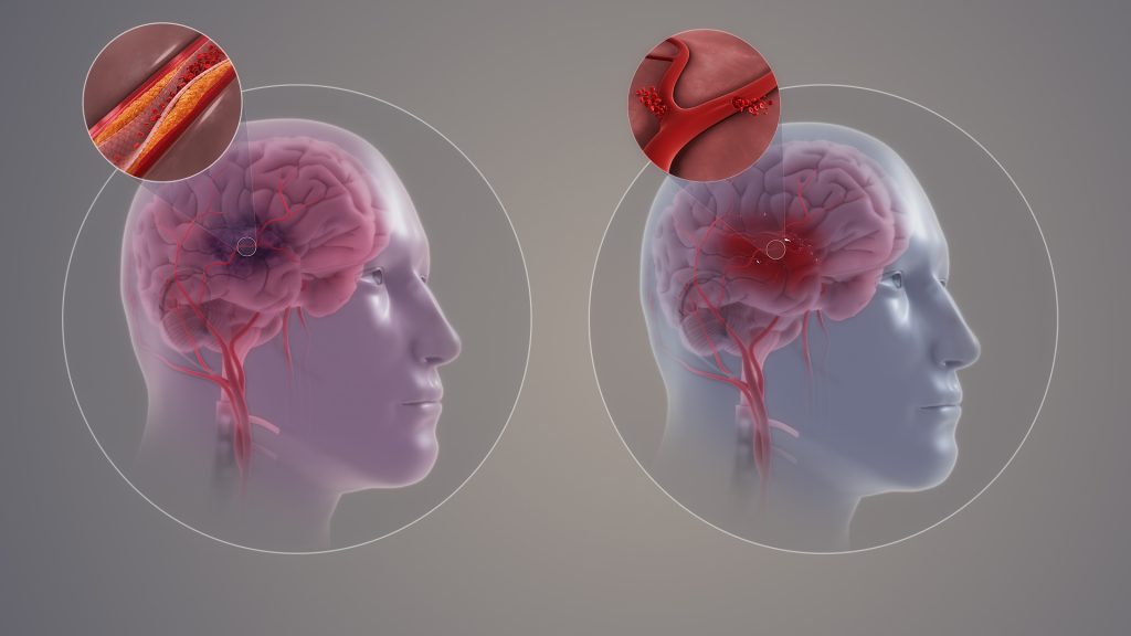
A new study by researchers at the Department of Molecular Medicine at SDU sheds light on one of the most severe consequences of stroke: damage to nerve fibres – the brain’s “cables” – which leads to permanent impairments. The study, which is published in the Journal of Pathology, used unique tissue samples from Denmark’s Brain Bank located at SDU, may pave the way for new treatments that help the brain repair itself.
The brain tries to repair damage
Following an injury, the brain tries to repair the damaged nerve fibres by re-establishing their insulating myelin sheaths. Unfortunately, the repair process often succeeds only partially, meaning many patients experience lasting damage to their physical and mental functions. According to Professor Kate Lykke Lambertsen, one of the study’s lead authors, the brain has the resources to repair itself. “We need to find ways to help the cells complete their work, even under difficult conditions,” Prof Lykke said.
The researchers have thus focused on how inflammatory conditions hinder the rebuilding. The study has identified a particular type of cell in the brain that plays a key role in this process. These cells work to rebuild myelin, but inflammatory conditions often block their efforts.
How researchers used the brain collection
-Using the brain collection, we can precisely map which areas of the brain are most active in the repair process, explains Professor Kate Lykke Lambertsen.
This mapping has enabled researchers to analyse tissue samples from Denmark’s Brain Bank and gain a deeper understanding of the mechanisms that control the brain’s ability to heal itself.
Through advanced staining techniques, known as immunohistochemistry, the researchers have been able to detect specific cells that play a central role in the reconstruction of myelin in the damaged areas of the brain.
The samples were analysed to distinguish between different areas of the brain, including the infarct core (the most damaged area), the peri-infarct area (surrounding tissue where rebuilding is active), and tissue that appears unaffected.
The analysis provided insight into where repair cells accumulate and how their activity varies depending on gender and time since the stroke.
Women and men react differently
An interesting discovery in the study is that women’s and men’s brains react differently to injuries.
-The differences underscore the importance of future treatments being more targeted and taking into account the patient’s gender and individual needs, says Kate Lykke Lambertsen.
In women, it seems that inflammatory conditions can prevent cells from repairing damage, while men have a slightly better ability to initiate the repair process. This difference may explain why women often experience greater difficulties after a stroke.
The brain collection at SDU is key to progress
The researchers behind the study emphasise that the discoveries could not have been made without the Danish Brain Bank at SDU. The collection consists of tissue samples from humans, used to understand brain diseases at a detailed level.
With access to this resource, researchers can investigate the mechanisms behind diseases like stroke and develop new treatment strategies.
Source: University of Southern Denmark Faculty of Health Sciences

