Month: January 2025

New Tech could Cut Epilepsy Misdiagnoses by up to 70% Using Routine EEGs

Source: Pixabay

Doctors could soon reduce epilepsy misdiagnoses by up to 70% using a new tool that turns routine electroencephalogram, or EEG, tests that appear normal into highly accurate epilepsy predictors, a Johns Hopkins University study has found.

By uncovering hidden epilepsy signatures in seemingly normal EEGs, the tool could significantly reduce false positives, seen in around 30% of cases globally, and spare patients from medication side effects, driving restrictions, and other quality-of-life challenges linked to misdiagnoses.

“Even when EEGs appear completely normal, our tool provides insights that make them actionable,” said Sridevi V. Sarma, a Johns Hopkins biomedical engineering professor who led the work. “We can get to the right diagnosis three times faster because patients often need multiple EEGs before abnormalities are detected, even if they have epilepsy. Accurate early diagnosis means a quicker path to effective treatment.”

A report of the study is newly published in Annals of Neurology.

Epilepsy causes recurrent, unprovoked seizures triggered by bursts of abnormal electrical activity in the brain. Standard care involves scalp EEG recordings during initial evaluations. These tests track brainwave patterns using small electrodes placed on the scalp.

Clinicians partly rely on EEGs to diagnose epilepsy and decide whether patients need anti-seizure medications. However, EEGs can be challenging to interpret because they capture noisy signals and because seizures rarely occur during the typical 20 to 40 minutes of an EEG recording. These characteristics makes diagnosing epilepsy subjective and prone to error, even for specialists, Sarma explained.

To improve reliability, Sarma’s team studied what happens in the brains of patients when they are not experiencing seizures. Their tool, called EpiScalp, uses algorithms trained on dynamic network models to map brainwave patterns and identify hidden signs of epilepsy from a single routine EEG.

“If you have epilepsy, why don’t you have seizures all the time? We hypothesized that some brain regions act as natural inhibitors, suppressing seizures. It’s like the brain’s immune response to the disease,” Sarma said.

The new study analyzed 198 epilepsy patients from five major medical centers: Johns Hopkins Hospital, Johns Hopkins Bayview Medical Center, University of Pittsburgh Medical Center, University of Maryland Medical Center, and Thomas Jefferson University Hospital. Out of these 198 patients in the study, 91 patients had epilepsy while the rest had non-epileptic conditions mimicking epilepsy.

When Sarma’s team reanalysed the initial EEGs using EpiScalp, the tool ruled out 96% of those false positives, cutting potential misdiagnoses among these cases from 54% to 17%.

“This is where our tool makes a difference because it can help us uncover markers of epilepsy in EEGs that appear uninformative, reducing the risk of patients being misdiagnosed and treated for a condition they don’t have,” said Khalil Husari, co-senior author and assistant professor of neurology at Johns Hopkins. “These patients experienced side effects of the anti-seizure medication without any benefit because they didn’t have epilepsy. Without the correct diagnosis, we can’t find out what’s actually causing their symptoms.”

In certain cases, misdiagnosis happens due to misinterpretation of EEGs, Husari explained, as doctors may overdiagnose epilepsy to prevent the dangers of a second seizure. But in some cases, patients experience nonepileptic seizures, which mimic epilepsy. These conditions can often be treated with therapies that do not involve epilepsy medication.

In earlier work, the team studied epileptic brain networks using intracranial EEGs to demonstrate that the seizure onset zone is being inhibited by neighboring regions in the brain when patients are not seizing. EpiScalp builds on this research, identifying these patterns from routine scalp EEGs.

Traditional approaches to improve EEG interpretation often focus on individual signals or electrodes. Instead, EpiScalp analyses how different regions of the brain interact and influence one another through a complex network of neural pathways, said Patrick Myers, first author and doctoral student in biomedical engineering at Johns Hopkins.

“If you just look at how nodes are interacting with each other within the brain network, you can find this pattern of independent nodes trying to cause a lot of activity and the suppression from nodes in a second region, and they’re not interacting with the rest of the brain,” Myers said. “We check whether we can see this pattern anywhere. Do we see a region in your EEG that has been decoupled from the rest of the brain’s network? A healthy person shouldn’t have that.”

Source: Johns Hopkins University

Why the Road for New Heart Cell Treatments is so Long

Right side heart failure. Credit: Scientific Animations CC4.0

Pathways to new treatments for heart failure take time – as long as four decades for two now accepted therapies. So, new attempts to repair scar tissue in infarcted hearts using cells or cell products need more time to develop clinical therapies that can reduce risk of death from heart failure after a heart attack.

This message is part of a critical review of cell-based and cell product-based therapies for the treatment of heart failure. The review details 20 years of completed and ongoing clinical trials. While none has gained medical approval, they have proven safe and some have shown beneficial effects.

More importantly, the reviewers note, it took longer, nearly 40 years, to optimise two current therapies to reduce mortality in heart failure: implantable cardioverter–defibrillators and guideline-directed medical therapy.

“The history of the development of life-saving medical therapies for heart failure serves as an important lesson that we should remain hopeful of the promise of cell therapy in heart failure,” Jianyi “Jay” Zhang, MD, PhD, and colleagues write in the review, “Trials and tribulations of cell therapy for heart failure: an update on ongoing trials,” published in Nature Reviews Cardiology. Zhang is professor and chair of the University of Alabama at Birmingham Department of Biomedical Engineering.

Heart failure is responsible for 13% of deaths worldwide. Half of patients with heart failure die within five years. The most common cause of heart failure is blockage of coronary arteries leading to death of the cardiomyocyte heart muscle cells. When that muscle tissue is replaced by dense scar tissue with little blood circulation, the infarcted heart loses contractile power, leading to heart enlargement, progressive loss of pumping ability, increased chance of ventricular arrhythmias and clinical end-stage heart failure.

The problem is that shortly after birth, human heart muscle cells lose their ability to divide, so a damaged infarcted heart cannot repair itself by growing new muscle cells. Thus, the simple idea behind initial cell therapies was to add or inject replacement cells to the scar area to restore muscle tissue.

The two decades since has been a long road, with bumps and turns. The three parts of the Nature Reviews Cardiology paper describe the journey. 

First is a history of the slow development, obstacles, setbacks and scepticism for two current heart failure therapies, implantable cardioverter–defibrillators and guideline-directed medical therapy. The next two sections, and main focus of the review, survey 13 completed clinical trials published in the last 12 years and 10 very recently initiated and ongoing clinical trials that are based on the lessons learned from the past 20 years of research, to assess the safety and efficacy of cell- and cell products-based therapy approaches.

While several randomised, double-blind, multicentre phase II or III trials published in the past 20 years support the concept that even a single dose of cell products has beneficial effects in patients with heart failure on optimal medical therapy, the ongoing trial are taking novel directions, Zhang says. 

These include:

  • New cell types — pluripotent stem cell-derived cardiomyocytes/ spheroids and umbilical cord-derived mesenchymal stem cells
  • Repeated intravenous injections as a noninvasive cell delivery method
  • New cell products, such as engineered epicardial cardiomyocyte patches
  • Novel cell-free products — extracellular vesicle-enriched or exosome-enriched secretomes.

“The results of these trials will continue to define and refine our understanding of cell and cell product therapy as a novel addition in the treatment of patients with heart failure,” Zhang said. 

The review acknowledges scientific criticism during the slow but consistent progress and evolution of cell therapy. Some have questioned the use of public funding to support cell therapy research for heart failure treatment, due to poorly designed or underpowered clinical trials and very modest improvements in cardiac function in preclinical studies that are not always substantiated in large-scale clinical trials.

“These criticisms must be addressed in future trials that are adequately powered and rigorously designed to ensure continued progress of the field,” Zhang said. “Critique is an essential part of science, and the basis for growth, innovation and evolution – this is no less true for the field of cell therapy.” 

Yet Zhang is confident that current research will yield clinical translation. “In the past 20 years, cell therapy has emerged and evolved as a promising avenue for cardiac repair and regeneration,” he said. “Cell therapy has encountered substantial barriers in both preclinical studies and clinical trials, but the field continues to progress and evolve through lessons learned from such research.”

Source: University of Alabama at Birmingham

New Combination Immunotherapy for Melanoma and Breast Cancer

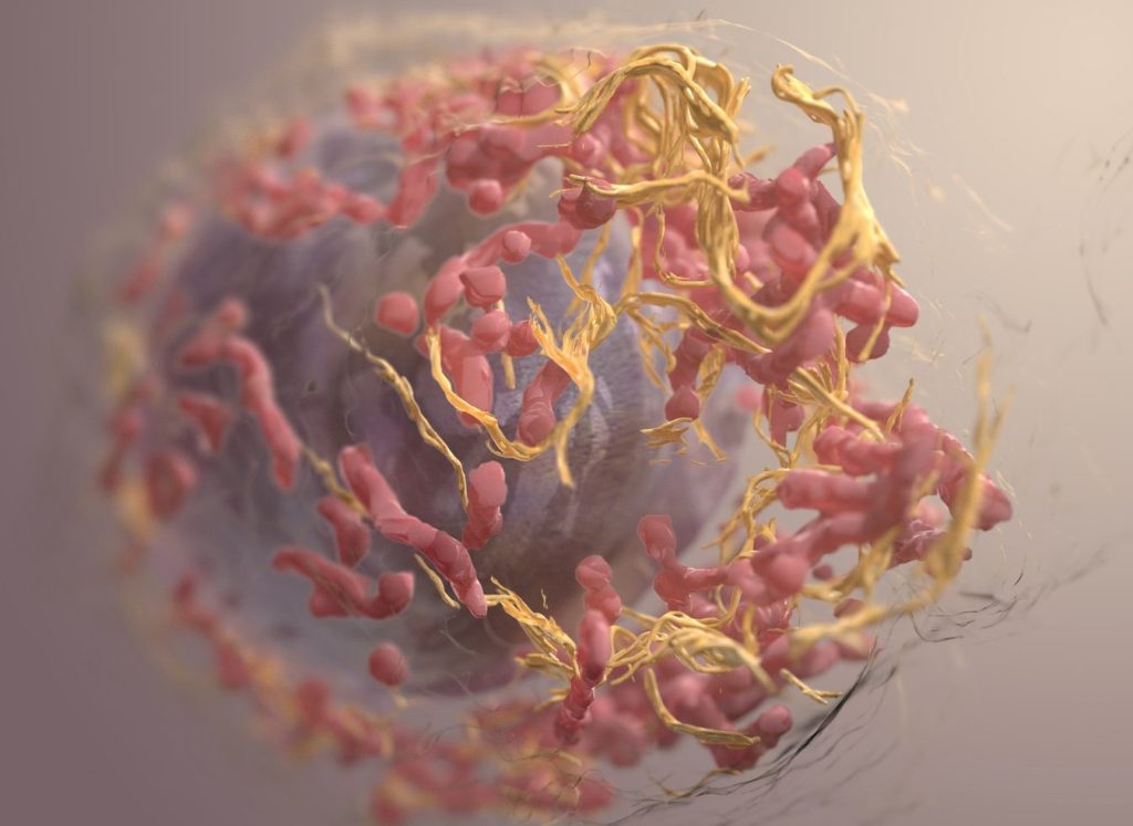
3D structure of a melanoma cell derived by ion abrasion scanning electron microscopy. Credit: Sriram Subramaniam/ National Cancer Institute

A research team at the Medical University of Vienna led by Maria Sibilia has investigated a new combination therapy against cancer. This therapy employs systemic administration of the tissue hormone interferon-I combined with local application of Imiquimod. Promising results were seen in topically accessible tumours like melanoma and breast cancer models: the therapy led to the death of tumour cells at the treated sites and simultaneously activated the adaptive immune system to fight even distant metastases. The findings, published in Nature Cancer, could improve the treatment of superficial tumours such as melanoma and breast cancer.

In recent years, immunotherapies have had significant success in the treatment and cure of a wide range of cancers. However, for some patients, these agents are still not sufficiently effective. As part of a preclinical study, Maria Sibilia, Head of the Center for Cancer Research at the Medical University of Vienna, therefore investigated the effects of a combination immunotherapy consisting of systemic administration of the tissue hormone interferon (IFN)-I and local imiquimod therapy. Imiquimod is an active substance that activates the innate receptors TLR7/8 and used to treat basal cell carcinomas. The researchers employed various preclinical mouse tumour models of melanoma and breast cancer. What both tumours have in common is that they are accessible to local therapy and often form distant metastases.

Effective for local tumours and distant metastases

Immunotherapies use the body’s own immune system to fight cancer cells. Plasmacytoid dendritic cells (pDCs), which are activated by Imiquimod via TLR7/8, play an important role in this process. The study showed that oral imiquimod stimulates pDCs to produce the tissue hormone IFN-I. This sensitised other dendritic cells and macrophages in the tumour environment to topical imiquimod therapy, which inhibited the formation of new blood vessels via the cytokine IL12 leading to the death of tumour cells.
The combination immunotherapy not only had an effect on the treated tumours, but also on distant metastases. It reduced the formation of new metastases thus preventing tumour relapses and increasing the sensitivity of melanomas to checkpoint inhibitors.

“These findings illustrate that the combination of systemic treatment with imiquimod or IFN-I and topical therapy with imiquimod has the potential to expand treatment options for patients and improve therapy outcomes in locally accessible tumors such as melanoma or breast cancer,” emphasizes Maria Sibilia.
“Topical treatment of the primary tumor with imiquimod is essential for this combination therapy with systemic IFN-I to be effective at the treated site and also to clear distant metastases,” adds Philipp Novoszel, MedUni Vienna, one of the first authors of the study.

The results suggest that this therapeutic strategy has the potential to improve treatment outcomes in superficial and thus locally accessible tumors such as melanoma and breast cancer – on the one hand through therapy-associated cancer cell death at the locally treated tumors, but also through the induction of a T cell-induced anti-tumor immune response at distant metastases, which is further enhanced by checkpoint inhibitors.

“Our aim is to continue developing immunotherapeutic strategies in order to improve the long-term prospects for patients who are not yet responding well to these agents,” says Maria Sibilia, who is also Deputy Head of the Comprehensive Cancer Center of MedUni Vienna and University Hospital Vienna.
“As systemic interferon is a well-known cancer therapy and dendritic cells are activated in a similar way to our preclinical models, we believe that the new combination therapy can show an effect in patients,” adds Martina Sanlorenzo, dermato-oncologist at MedUni Vienna and co-first author of the study.

Publication: Nature Cancer
Systemic IFN-I combined with topical TLR7/8 agonists promotes distant tumor-suppression by c-Jun-dependent IL-12 expression in dendritic cells
Sanlorenzo M, Novoszel P, Vujic I, Gastaldi T, Hammer M, Fari O, De Sa Fernandes C, Landau AD, Göcen-Oguz BV, Holcmann M, Monshi B, Rappersberger K, Agnes Csiszar A, Sibilia M
DOI: 10.1038/s43018-024-00889-9; https://www.nature.com/articles/s43018-024-00889-9

Source: Medical University of Vienna

Routine Brain MRI Screening Urged in Asymptomatic Late Stage Breast Cancer

Photo by Anna Shvets on Pexels

A new study led by researchers at Moffitt Cancer Center shows that asymptomatic brain metastasis is more common in stage 4 breast cancer patients than previously believed. The study, published in Neuro-Oncology, suggests that doctors may need to rethink current screening guidelines for detecting brain metastasis in patients without symptoms.

Researchers examined 101 asymptomatic patients diagnosed with stage 4 breast cancer, including triple-negative, HER2-positive and hormone receptor-positive/HER2-negative breast cancer. These patients underwent MRI scans to check for brain metastasis, with a follow-up MRI six months later if the initial scan showed no signs of cancer spread.

Of the patients who completed the initial MRI, 14% had brain metastasis. The rates by subtype were:

  • 18% in triple-negative breast cancer
  • 15% in HER2-positive breast cancer
  • 10% in hormone receptor-positive/HER2-negative breast cancer

After the second MRI, the number of patients with brain metastasis grew to about 25% in each subtype. Following diagnosis, patients went on to receive early treatment for their brain metastases, including changes in systemic therapy and local therapies.

“Our study suggests that asymptomatic brain metastasis is quite common in stage 4 breast cancer,” said Kamran Ahmed, MD, associate member and section chief for Breast Radiation Oncology at Moffitt and principal investigator of the study. “Although larger studies are needed to confirm our findings, given the improvements in systemic and local therapies for breast cancer brain metastasis, the time may be appropriate to reconsider current guidelines that recommend against routine MRI surveillance in late stage breast cancer.”

Source: H. Lee Moffitt Cancer Center & Research Institute

Removing Fallopian Tubes during Other Abdominal Surgeries may Lower Ovarian Cancer Risk

Mathematical modelling study suggests this approach could also reduce total healthcare costs in Germany

Female reproductive system. Credit: Scientific Animations CC4.0 BY-SA

A mathematical modelling study conducted in Germany suggests that ovarian cancer incidence could be reduced and healthcare savings boosted if women who have already completed their families were offered fallopian tube removal during any other suitable abdominal surgeries. Angela Kather and Ingo Runnebaum of Jena University Hospital, Germany, and colleagues present these findings on January 30th in the open-access journal PLOS Medicine.

Some of the most widespread and serious forms of ovarian cancer begin in the fallopian tubes, and removing them may reduce ovarian cancer risk. While women at average risk of ovarian cancer are not recommended to have surgery solely to remove their fallopian tubes, many surgeons offer “opportunistic” tube removal during other gynaecologic surgeries such as hysterectomy or tubal sterilisation. Opportunistic removal may also be feasible during other abdominal surgeries, such as gallbladder removal.

However, the overall potential benefits of opportunistic fallopian tube removal have been unclear. To help clarify, Kather and colleagues developed a mathematical model that incorporates real-world patient statistics to predict population-level risks of ovarian cancer after opportunistic fallopian tube removal, as well as the potential healthcare cost savings.

By applying the model to statistics from Germany, the researchers predicted that opportunistic fallopian tube removal during every hysterectomy and tubal sterilisation could reduce ovarian cancer cases by 5% across the female population of Germany. Removal during every suitable abdominal surgery for women who are done having children could reduce nationwide cancer cases by 15%, the analysis suggests, and it could save more than €10 million in healthcare costs annually.

Ovarian cancer is the third most common gynaecologic cancer in the world and has a mortality rate of 66%. Overall, these findings suggest that opportunistic fallopian tube removal during appropriate abdominal surgeries could not only lower population-level ovarian cancer risks and prevent ovarian cancer deaths, but also provide economic benefits. This study could help inform health policy and insurance costs for the procedure.

The authors add, “We developed a mathematical model to estimate the likelihood of women undergoing surgeries that offer an opportunity for fallopian tube removal and the potential for reducing their ovarian cancer risk. Applying this model to the entire female population of Germany revealed that 15% of ovarian cancer cases could be prevented if fallopian tubes were removed during every suitable abdominal surgery in women who have completed their families. This approach has the potential to extend healthy years of life and significantly save healthcare costs.”

Provided by PLOS

Urine-based Test Detects Aggressive Prostate Cancer

Credit: Darryl Leja National Human Genome Research Institute National Institutes Of Health

Traditional approaches to prostate cancer screening involve blood tests, MRI, and biopsies. Besides being uncomfortable, some of these procedures result in overdiagnosis of low-grade cancers.

In a new study, researchers at the University of Michigan Health Rogel Cancer Center have clinically validated a previously developed urine test, which can potentially bypass these invasive procedures among men who are unlikely to benefit.

Prostate cancers are categorised based on their Gleason Grade or Grade Group. Those with Gleason 3+4=7, or Grade Group 2, or higher are more likely to grow and cause harm in comparison with Gleason 6 or Grade Group 1 prostate cancers, which are considered non-aggressive.

The urine test, called MyProstateScore 2.0, or MPS2, looks at 18 different genes linked to high-grade prostate cancer. 

The researchers had previously demonstrated that the test was effective in identifying GG2 or higher cancers, helping patients avoid unnecessary biopsies. But in that study, urine samples were obtained after a digital rectal examination.

“The process requires the prostate to be compressed, causing the release of cellular debris into a urine sample that the patient provides after the rectal exam,” said Ganesh S. Palapattu, MD, a professor of urology.

Its primary benefit is that the test can accurately predict your probability of developing aggressive prostate cancer, putting both the patient and physician at ease.”

-Ganesh Palapattu, MD

Such an examination may not be practical for many and is associated with some discomfort.

Developing a potential at-home test

In the study, the team modified the urine collection approach so that the MPS2 test could detect markers for prostate cancer, without requiring a prior rectal exam.

Using urine samples from a cohort of 266 men who did not undergo a rectal exam, they found that the test could detect 94% of GG2 or higher cancers and was more sensitive than blood tests.

Further, the team used mathematical models to demonstrate that the use of MPS2 would have avoided up to 53% of unnecessary biopsies.

“These results show that MPS2 has promise as an at-home test,” Palapattu said. 

“Its primary benefit is that the test can accurately predict your probability of developing aggressive prostate cancer, putting both the patient and physician at ease.”

MPS2 can also help patients save on healthcare costs since it is significantly cheaper than an MRI.

The team is interested in repeating the study and corroborating their results with a larger, diverse population of men.

They’re also hoping to study the test’s performance in men as a surveillance screen for low-risk prostate cancer.

“MPS2 could potentially improve the health of our patients by avoiding overdiagnosis and overtreatment and allowing us to focus on those who are most likely to have aggressive cancers,” Palapattu said.

Source: Michigan Medicine – University of Michigan

New Clues to the Mechanism Behind Food Tolerance and Allergies

Photo by Corleto on Unsplash

How the gut decides which food to tolerate and which food to respond to as an allergen has long puzzled scientists. Now, new research identifies specific gut cell types that communicate with T cells – prompting them to tolerate, attack, or simply ignore – and explains how these opposing responses are triggered.

The findings, published in Science, give scientists a new understanding of how the intestinal immune system keeps the gut in balance, and may ultimately shed light on the root causes and mechanisms of food allergies and intestinal diseases.

“The big question is, how do we survive eating?” says lead author Maria C.C. Canesso, a postdoctoral fellow in the laboratories of Daniel Mucida and Gabriel D. Victora. “Why do our bodies normally tolerate food, and what goes wrong when we develop food allergies?”

Gut decisions

The intestinal immune system is complicated machinery. Tolerance to food begins with antigen presenting cells, or APCs, instructing T cells to stand down. This signal gives rise to pTregs, a special type of T cell that calms the immune response to food particles, and kicks off a cascade of activity involving additional immune cells that reinforce the message. But without knowing which specific APCs run the show, it’s difficult to tease out the ins and outs of the body’s eventual tolerance to food and intolerance to pathogens.

“There are so many types of antigen-presenting cells,” Canesso says. “Pinpointing which ones are doing what is a longstanding technical challenge.”

She began exploring this conundrum as a PhD student in the Mucida lab, which focuses on how the intestine balances defense with tolerance. During her postdoc, Canesso also joined the Victora lab, which developed a technology known as LIPSTIC that helps scientists catalogue cell-to-cell interactions, particularly among immune cells.

“The technological advances made by the Victora lab allowed us to understand immune cell dynamics that would not have been possible using existing tools,” says Mucida, head of the Laboratory of Mucosal Immunology.

After optimising LIPSTIC for the task, Canesso and colleagues succeeded in pinpointing those APCs that promote tolerance – a process primarily handled by two types: cDC1s and Rorγt+ APCs. These cells capture dietary antigens from ingested food and present them to T cells, giving rise to the pTregs that ensure food tolerance.

“When we first developed LIPSTIC, we were aiming to specifically measure the interactions between B and T cells that promote antibody responses to vaccines,” says Victora, head of the Laboratory of Lymphocyte Dynamics. “It was to Maria’s credit that she was able to adapt this to settings so different from those it was originally intended for.”

They also uncovered how infections of the intestines can cause interference, demonstrating in mice that the parasitic worm Strongyloides venezuelensis shifts the balance away from tolerance promoting APCs and toward those that promote inflammation. Indeed, mice infected with this worm during a first exposure to a dietary protein display reduced tolerance towards this protein, and signs of allergy when challenged.

Finally, the team characterised the molecular signals underpinning these immune shifts, identifying key cytokines and pathways that influence how APCs present antigens and modulate immune responses. For example, the infection induced a surge in pro-inflammatory cytokines such as IL-6 and IL-12, which have been shown to nudge APC activity toward inflammatory outcomes. This inflammatory environment appears to override the immune system’s tolerance mechanisms. “The worm infection induces this an expansion of non-tolerogenic APCs that help deal with the infection, outnumbering the tolerance-related APCs,” Canesso says.

From food to food allergies

Together, the findings illuminate how the immune system maintains food tolerance and, in the case of parasitic infections, highlights the specific immune mechanisms that can go awry. “It’s important to note that our findings do not suggest that worm infections trigger food allergies,” clarifies Mucida, head of the Laboratory of Mucosal Immunology. “They reduce tolerance mechanisms while the immune response focuses on dealing with the worms.”

While these findings aren’t directly relevant to food allergies, they do lay some groundwork for further investigation into food intolerance. “If food allergies are derived from dysregulation on intestinal APCs inducing tolerance and protective responses to infections, perhaps we could one day modulate those APCs specifically to prevent food allergies,” Canesso says.

Next up, Canesso plans to shift her focus toward early life, exploring how maternal-neonatal interactions shape food intolerance. “Most allergies develop early in life,” she says. “I want to focus on how breast milk and maternal exposure to dietary antigens may influence a baby’s immune system, potentially shaping their risk of developing food allergies.”

Source: Rockefeller University

Moderate Exercise Keeps Appetite at Bay

Photo by Ketut Subiyanto on Unsplash

A recent study involving researchers at Murdoch University’s Health Futures Institute has revealed that moderate-intensity exercise can significantly influence appetite-related hormones and perceptions in males with obesity.

The study, titled “Acute effect of exercise on appetite-related factors in males with obesity,” provides new insights into how exercise can aid appetite control and weight management. 

One of the study authors, Associate Professor Timothy Fairchild from Murdoch’s School of Allied Health, said the study confirms their previous work showing the benefits of incorporating regular exercise into daily routines for individuals looking to manage their weight and improve their overall health. 

“People understand that exercise helps ‘burn energy’. A lot of people assume that exercise also increases hunger and energy intake afterwards,” Associate Professor Timothy Fairchild said.  

“We have previously shown, using high-intensity exercise, that this is not the case. 

“This latest study shows that even moderate-intensity exercise can have immediate and beneficial effects on appetite control in males with obesity.” 

The study not only assessed food intake and appetite, but also measured changes in hormones which help to regulate appetite.  

“Despite a strong focus on weight loss drugs in society at present, this study shows that lifestyle factors still have a strong and relevant role in helping people to live their healthiest life,” Associate Professor Fairchild said. 

“In fact, the hormones which have been shown to increase after exercise, are the same hormones which the most successful weight loss drugs are mimicking.”  

“The added benefit of exercise is that you also receive the additional physical and mental health benefits of exercise”.  

The full study can be found in Physiological Reports journal.  

Source: Murdoch University

Stopping Health Funding in Africa Weakens America

This is an opportunity for President Ramaphosa to lead

Photo by Andy Feliciotti on Unsplash

By Nathan Geffen and Marcus Low

President Donald Trump’s administration took a cruel decision this past week to freeze US foreign aid for health, potentially leaving millions of people in many African countries without their life-saving antiretroviral treatment.

On Wednesday morning, Trump’s secretary of state Marco Rubio backtracked on part of that decision. But if it is not reversed permanently we can expect advances in life expectancy in sub-Saharan Africa of the past two decades to start coming undone. We can also expect HIV infection rates to start picking up again, as people with HIV start getting viral rebound and become more infectious.

The President’s Emergency Plan for AIDS Relief (PEPFAR) was started by the Republican administration led by George W. Bush in 2003. The complexity of world politics is such that the president who perhaps did more than anyone else to unravel confidence in global rules and norms – by invading Iraq – also championed a programme that has saved many millions of lives. Bush described PEPFAR as “compassionate conservatism”.

PEPFAR had bipartisan support. It is one of the greatest contributions the US has made to the world. It is now under threat by people claiming with straight faces – who came to power while the US economy is booming – to make America great again.

About $5-billion went into PEPFAR last year. Although it’s a huge amount of money it’s a tiny fraction of the US budget. It’s not straightforward to measure how many lives PEPFAR has saved but it is in the millions. This is a lot of bang for the buck.

The US government is also the largest contributor to the other major funder of global health: the Global Fund. Its future is also bleak.

Already in South Africa, vital services for extremely vulnerable clients had to pause, such as those provided by the Wits Reproductive Health and HIV Institute clinics in Johannesburg. Hopefully with Rubio’s announcement these can now resume but the situation remains chaotic and the future of this and other US-funded health programmes across Africa is fraught with uncertainty.

Opportunities

America’s abandonment of foreign aid for health relinquishes soft power. There is an opportunity here for the European Union, Canada, Australia, Japan and China to step into the breach and increase their contributions to the Global Fund, or even to directly plug holes left by PEPFAR using bilateral aid – though such funding may come too late for some.

This would not merely be an act of charity. In the post-World War II world, what has made countries great, powerful, prestigious and influential is not nastiness and murder, but investing in projects of solidarity that make the world a better place. US wars in Vietnam, Afghanistan and Iraq degraded US power. Its arming of Israel, especially during the war on Gaza, has shown US concern for universal human rights to be hypocritical and worsened its global standing. By contrast PEPFAR unequivocally enhanced its superpower status.

President Cyril Ramaphosa can display great leadership by meeting with leaders of wealthy countries and convincing them to increase spending to support the health systems of poorer countries.

But perhaps the biggest opportunity is for African countries themselves. Many remain far too dependent on foreign aid to run their health systems. A country like South Africa should be able to pay for every last cent of its health systems. Corruption and mismanagement have had an inordinate role in making this difficult.

For countries like Malawi, Mozambique and others, there is a long way to go before they can pay their own way for HIV treatment. But pressure, from within and out these countries, must be put on their governments to build robust economies capable of delivering tax revenue to spend more on health.

In a very divided world where illiberal nationalist populism is on the rise and African governments are for the most part still weak and corrupt, these opportunities seem unlikely to be seized. But we hope we are proven wrong.

Geffen is the editor of GroundUp. Low is the editor of Spotlight. Both served in the Treatment Action Campaign which successfully campaigned for HIV medicines in South Africa, as well as other countries.

Republished from GroundUp under a Creative Commons Attribution-NoDerivatives 4.0 International License.

Read the original article.

Microbial Therapy Offers New Hope for Vitiligo Patients

Photo by Hanen BOUBAHRI on Unsplash

A natural compound derived from gut-friendly bacteria significantly slows the progression of vitiligo and may restore pigmentation, reports a new Northwestern University pre-clinical study in mice. 

The findings, published in the Journal of Investigative Dermatology, could offer hope to millions affected by the autoimmune disease, which causes visible patches of skin discoloration and carries profound emotional and physical consequences.

“The results in our model were astonishing,” said senior study author I. Caroline Le Poole, a professor of dermatology and microbiology-immunology at Northwestern University Feinberg School of Medicine. 

“We found that administering a microbial compound weekly to vitiligo-prone mice significantly suppressed disease progression. It made a spectacular difference in an aggressive model of the disease.”

What is vitiligo?

Vitiligo affects 0.5% to 2% of the global population and is linked to other health challenges, including higher risks of cardiovascular disease, psychological distress and endocrine disorders.

Discoloration often appears on the face, scalp, hands and arms, as well as around body openings like the mouth and genitals. Many individuals with vitiligo also have at least one other autoimmune condition.

How the study was conducted

Le Poole and her colleagues administered a microbial product weekly to vitiligo-prone mice over an 18-week period. By the end, pigment loss on the mice’s backs was reduced by 74%.

The product reduced killer T cells that attack the skin’s pigment and increased protective regulatory T cells, which are typically scarce in vitiligo patients.

“This simple microbial compound could work as a standalone therapy, or in synergy with existing treatments,” Le Poole said.

Existing therapy has limitations

In 2022, the FDA approved the first re-pigmentation therapy for vitiligo: Opzelura (ruxolitinib) cream. Clinical trials found that only 30% of patients using the cream regained 75% or more skin re-pigmentation on the face.

“Our findings about the effectiveness of microbial therapy could give hope to patients who are not well-served by existing treatments,” Le Poole said.

Vitiligo is more severe in patients with darker skin

Le Poole, who has spent more than 30 years studying vitiligo, said the disease has been found to be more severe in people with darker skin tones. The visibility of the discolouration in people with darker skin can also amplify stigma and emotional distress. 

“Patients often feel powerless as they watch their disease progress, unsure how they will look next month,” Le Poole said. “For many, stabilising the disease would be life changing.” 

While the disease can affect self-esteem – particularly for adolescents navigating social pressures – some people embrace it.

“We’ve seen beautiful models with vitiligo challenging societal norms, and that’s wonderful,” Le Poole added. “But for those who want treatment, it’s important to provide real options that can halt disease progression.”

Next steps

The next challenge is adapting the microbial product for human use. “Weekly injections could work, but we need to explore simpler options, like a food additive or ointment,” said Le Poole. “We also need to understand how long the effects last and the best timing for treatment.”

Le Poole added that the same microbial compound she and her colleagues injected in mice could potentially treat other autoimmune conditions, particularly those involving killer T cells in the skin.  

“Our next step is collaborating with scientists from several institutions to refine the compound, understand its mechanisms and determine whether it works alongside existing treatments for auto-immune disease.”

Source: Northwestern University