Year: 2025

How an SAMRC Study Found that HIV Deaths in SA may be Massively Undercounted

Photo by Sergey Mikheev on Unsplash

By Chris Bateman

It is widely acknowledged among health and demographic experts that relying solely on what is written on death certificates does not paint an accurate picture of what people in South Africa are actually dying of. Now, an SAMRC study has provided evidence that the undercounting of deaths due to HIV might be even greater than previously thought.

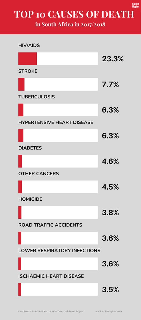
Many in health circles were surprised by a recent South African Medical Research Council (SAMRC) study that found that 23% of deaths in a nationally representative sample drawn from 2017/2018 were due to HIV. By comparison, Stats SA data for roughly the same period puts the figure at only 5.7%.

That Stats SA’s HIV mortality figures differs from other sources is not new and not in itself surprising. This is because Stats SA reports a relatively straight-forward count of what is written on death certificates – where it is known HIV is often not indicated, even if it is the underlying cause of death. By contrast, the new SAMRC study looked at autopsy reports, death certificates, medical records, and interviews with next of kin to come up with its much higher estimate.

The thing that did come as a surprise, is just how much higher the SAMRC figures were than anticipated. Previously, the real number of HIV deaths were thought to be around double the Stats SA number, rather than four times as much. For example, according to Thembisa, the leading model of HIV in South Africa and the basis for UNAIDS’s estimates for the country, around 12% of deaths in the country in 2018 were due to HIV.

“Accurate mortality data are essential for informed public health policies and targeted interventions; however, this study highlights critical gaps in our cause-of-death data, particularly in the underreporting of HIV/AIDS and suicides,” says Professor Debbie Bradshaw, study co-author and Chief Specialist Scientist at the SAMRC Burden of Disease Research Unit, in a media statement. (The study also found substantial under-reporting of suicide on death certificates.)

Multiple data sources

The study was conducted in three phases, examining deaths that were registered in 27 randomly selected health sub-districts between 1 September 2017 and 13 April 2018.

In addition to the examination of autopsy reports, death certificates, and medical records, trained fieldworkers interviewed next of kin to conduct verbal autopsies using a World Health Organization (WHO) questionnaire that had been translated into the country’s nine official languages.

Based on these various sources of data, the cause of each death was categorised into one or more of 44 categories and then compared to the cause of death indicated on the person’s death certificate. (The process for ensuring accuracy, including a review shared by a team of 49 medical doctors, is described in detail in this report.)

The researchers collected data for over 26 000 deaths, although not all types of data were available for each death. Medical records were available for over 17 600 cases, forensic pathology (autopsy) records for 5 700, and about 5 400 verbal autopsies were conducted. In the end, “to save costs”, not all medical records were reviewed.

Overall, for just over 15 000 deaths, the researchers could link and compare their assessment of why a person died to what was written on death certificates.

‘Poor agreement’

The researchers found that “there was poor agreement between the underlying cause of death obtained from the study and the official cause of death data”. The cause of death was the same in only 37% of cases. In addition to the under-reporting of HIV, the researchers also identified “severe under-reporting” of suicide as a cause of death.

A strong link between TB and HIV was observed, with TB responsible for 46% of deaths among people with HIV and 63% of TB deaths occurring in individuals with HIV. Together, these two diseases accounted for almost 30% of deaths.

Some question marks

As noted earlier, the new numbers are substantially higher than estimates from the highly respected Thembisa model. According to their data only 12% of deaths from mid-2017 to mid-2018 were due to HIV-related causes, with a further 9% of deaths occurring in persons with HIV but due to other causes.

Dr Pam Groenewald, a co-author of the new study and also with the SAMRC, describes Thembisa as “an excellent source”. She tells Spotlight they had a long discussion with the Thembisa researchers, “but we weren’t able to fully explain the differences”.

The study authors cite several factors that might contribute to a higher proportion of HIV deaths in their study. Firstly, the weighted national causes of death validation sample aimed to represent the registered deaths in the country, and it was known that deaths in rural areas and child deaths were under-represented. Secondly, deaths that occurred in private sector hospitals were not represented. Groenewald says the HIV-linked deaths in private hospitals are “definitely lower”, but doubts they would have had a significant impact on their findings.

One thing in favour of the study numbers is the fact that the cases they identified with HIV/AIDS as the underlying cause of death were independently reviewed by clinicians. As Groenewald points out, they looked at medical records of people admitted to and who died in hospital, including CD4 cell counts and HIV viral loads. The suggestion is that if someone had a very low CD4 count and a very high HIV viral load at the time of death, then it is very likely HIV played a role in their death, unless of course they died of a clearly non-associated cause like injuries from a car accident.

On the other hand, it might be argued that since HIV is very widely tested for in South Africa, it is more likely to appear on medical records than other less tested for diseases.

Another interesting wrinkle is that the proportion of deaths from HIV/AIDS from this study was higher than anticipated based on observed declines in adult mortality. It is widely accepted that the decline in adult mortality and the increase in life-expectancy over the last two decades was driven by antiretroviral therapy keeping more people with HIV alive. While the new findings do not challenge this narrative, it does suggest the effect may be less pronounced than previously thought.

What to do?

The researchers suggest their study has immediate implications for the country’s response to HIV and TB.

“The study recommends strengthening case finding, follow-up, prevention, and treatment for HIV, AIDS and TB to reduce mortality rates, and underlines the importance of government’s rapid response to counter the recent abrupt withdrawal of Pepfar funding,” Bradshaw comments in the media release.

But more broadly, the findings put the spotlight on major problems in the country’s death certification systems.

“Our findings highlight the need for improved record quality and adherence to testing guidelines within the medical community. Poor record keeping included incomplete documentation of clinical findings and results,” the study authors write.

“A lot of doctors’ report HIV as ‘retroviral disease’, for example, and it’s not coded as HIV,” Groenewald explains to Spotlight.

Urging doctors to record the actual underlying cause of death when writing up death certificates, she also called for improved training in death certification at medical schools.

Doctors’ reluctance to report HIV on death certificates likely has various reasons, including stigma related to HIV and the fact that some medical insurance policies used to exclude HIV, though policies now treat HIV like any other chronic condition.

Overall, Groenewald says, we need to step back and probe the rationale of compiling underlying cause of death statistics.

“The public health aim of the medical certificate of cause of death, (MCCD), is to prevent premature deaths. We therefore need to record the cascade of events or causal sequence of medical conditions leading to death and target our interventions at the underlying cause of death. The coding rules focus on the underlying cause of death, (UCOD), to compile the mortality statistics,” she says.

Groenewald stresses that the law requires doctors to provide accurate information on death causation. The Health Professions Council of SA’s ethical rules also recognised that a statute requiring disclosure about a deceased person’s health must be complied with and is not considered unethical. Contrary to common physician misconception, Groenewald says all this combined to show “it is completely ethical to disclose on a death certificate that a person has died from an AIDS related illness”.

In the meantime, routine mortality data from Stats SA should clearly be taken with a pinch of salt. As Groenewald points out, vital registration data should not be accepted at face value but should be interrogated and cross-checked with other data sources to get coherent and consistent estimates that fit within an envelope of all causes of mortality.

– Additional reporting by Marcus Low.

Republished from Spotlight under a Creative Commons licence.

Read the original article

New Biomarker Discovered in CSF for Parkinson’s Disease

A misfolded protein facilitates reliable diagnosis even in the early stages of Parkinson’s disease in body fluids.

Source: CC0

Parkinson’s disease is a neurodegenerative disorder that is usually diagnosed in its late stage on the basis of clinical symptoms, mainly motor disorders. By this point, however, the brain is already severely and irreparably damaged. Moreover, diagnosis is difficult and often incorrect because the disease takes many forms and symptoms overlap with other disorders. Researchers from the PRODI Center for Protein Diagnostics at Ruhr University Bochum, Germany, and the biotech company betaSENSE have now discovered a biomarker in the spinal fluid that facilitates a reliable diagnosis at an early stage and can shed light on the progression of the disease and the effect of a therapy. They report their findings in the journal EMBO Molecular Medicine.

Parkinson’s disease – an unstoppable condition

Parkinson’s disease is characterised by the loss of dopaminergic nerve cells in the brain, typically leading to increasing motor impairments as the symptoms progress. Dopamine supplements can compensate for the loss and temporarily alleviate the symptoms. The misfolding of the key protein alpha-synuclein (αSyn) from α-helical structures to β-sheet-rich structures plays a crucial role in the development of Parkinson’s disease. “These misfoldings make the protein sticky, leading to the formation of larger complexes, so-called oligomers. The oligomers then produce long fibrillar filaments and cause the aggregation of these filaments into macroscopically large Lewy bodies in the brain,” explains Professor Klaus Gerwert, founding and managing director at PRODI and CEO of betaSENSE.

Advanced platform technology

In two independent clinical cohorts with a total of 134 participants, the Bochum-based researchers showed that, with a sensitivity and specificity of well over 90 percent, this misfolding of αSyn in body-fluids is a viable biomarker for the diagnosis of Parkinson’s disease. The research was conducted using cerebrospinal fluid samples from patients at the Parkinson’s centers in Bochum (St. Josef Hospital, Professor Lars Tönges, Professor Ralf Gold) and Kassel (Paracelsus-Elena-Klinik, Dr. Sandrina Weber, Professor Brit Mollenhauer). The measurements were carried out using the patented iRS (immuno-infrared sensor) technology from betaSENSE GmbH.

betaSENSE has already successfully implemented the iRS technology for diagnosing Alzheimer’s disease. In this case, it was shown that the misfolding of the biomarker Aβ can indicate the risk of Alzheimer’s dementia at a later stage with high accuracy up to 17 years before clinical diagnosis. “We have now transferred this approach to Parkinson’s for the misfolding of αSyn,” stresses Klaus Gerwert.

Development of Parkinson’s drugs

In addition to diagnostic applications, the technology can also help to develop new active substances and prove their efficacy in clinical trials.

Source: Ruhr-University Bochum

The Rise in Dry Eye Disease Among Young Adults

Photo by Steinar Engeland on Unsplash

Researchers at Aston University have called for more advice to be given to young people about preventing dry eye disease, after a study carried out in conjunction with Oslo University Hospital and Sørlandet Hospital Trust in Norway found that 90% of participants had at least one sign of the condition in their eyes.

Dry eye disease occurs when the eyes do not make enough tears, or make poor-quality tears without sufficient lipid or mucus levels which leads to poor tear film stability and rapid evaporation. Sufferers may have gritty feeling eyes, itching or stinging in the eyes, red eyes, sensitivity to light and blurry vision. There are several risk factors for dry eye disease, including stress and wearing contact lenses. It is also more prevalent in females. In the 18-25 age group, a major risk factor is screen use.

The research, following 50 18-25-year-olds over time, was led by Dr Rachel Casemore at Aston University School of Optometry and is the first of its kind. It was published in The Ocular Surface. The researchers looked for symptoms of dry eye disease in the participants, studied lifestyle factors, and followed up with participants one year on to find out if there had been any progression of the condition.

The initial study showed that 56% of participants had dry eye disease, while 90% had at least one symptom of the condition. Around half of the participants in the study had lost at least 25% of a type of gland in the eye called the meibomian gland. These glands produce the outer lipid layer of the eye’s tear film, which is responsible for preventing evaporation of tears, and therefore keeps the tear film stable and the eye moist. One year on, the researchers found that there had been significant progression of dry eye disease in the study participants.

Additionally, the researchers found correlation found between how long the study group used screens and signs of dryness on the eye surface. The average screen use of participants was eight hours per day.

The researchers concluded that the evidence of dry eye disease symptoms and progression in the young adults in their study shows the need for early detection of potential signs, and the identification of those who may go on to develop dry eye disease. These individuals can then be advised on managing the condition before progression.

The progression and development of dry eye disease can be slowed by various methods. Dr Casemore says that the simplest ways are to take regular screen breaks, to carry out blink exercises to ensure the release of oils from the meibomian glands and to keep hydrated. A healthy, balanced diet, including sources of omega-3 fatty acids, such as oily fish, is also important, as is regular sleep patterns.

Dr Casemore suggests that those with irregular sleep patterns, such as those caused by sleep disorders or anxiety, should seek advice. People who wear contact lenses need to ensure they get regular check-ups to ensure optimum fitting, and that they adhere to their replacement schedule, wearing time schedule, cleaning regimes and safety advice, such as no sleeping, showering or swimming in contact lenses.

Dr Casemore said:

“It is concerning to note the increasing prevalence of dry eye disease signs and symptoms in young adults, which has been referred to as a ‘lifestyle epidemic’ by some researchers. Eye care practitioners are well placed to identify the clinical indicators of dry eye disease and counsel young adults around modifiable risk factors, such as screen use habits, sleeping habits, contact lens use, diet, blinking patterns, and management of stress levels.

“Our future research aims to continue investigation of the potential tear and meibomian gland oil biomarkers which were identified during the study and further explore the effect of diet on dry eye disease development.”

Source: Aston University

Preventing Unnecessary Pancreatic Cancer Surgery

Pancreatic cancer. Credit: Scientific Animations CC BY-SA 4.0

Pancreatic cysts are fluid-filled sacs that can form in the pancreas. Some remain benign, while others have the potential to develop into pancreatic cancer. A recent study, which followed 257 patients in Japan for an average of five years, showed that the presence or absence of invasive nodules in pancreatic cysts is key to assessing whether these cysts are benign or cancerous.

The findings, published in the journal Annals of Surgery, may help patients diagnosed with a high risk of pancreatic cancer to avoid unnecessary surgery.

Pancreatic cancer is one of the most life-threatening and rapidly growing cancers. Pancreatic cysts, known as pancreatic intraductal papillary mucinous neoplasms (IPMNs), are gaining attention as one of the precursors of the cancer that can be identified by radiological imaging. In this context, patients diagnosed with pancreatic cysts are referred for further evaluation, and if they meet the criteria for being at particularly high risk of developing cancer, called high-risk stigmata, they are often recommended for surgery.

However, it was not clear whether all patients who met the criteria would need to undergo surgery. “In fact, among patients who underwent surgery, there were a number of cases where pathological examination results showed that their IPMNs were still benign and had not progressed to cancer,” explained Ryohei Kumano from Nagoya University, the first author of the study. “Pancreatic surgery is a significant burden for patients, so we wanted to find a more accurate way to diagnose whether their IPMNs are benign or cancerous in order to avoid unnecessary surgery.”

A research group consisting of Professor Hiroki Kawashima and Dr Kumano from Nagoya University Graduate School of Medicine, Professor Eizaburo Ohno from Fujita Health University, and their colleagues focused on the presence or absence of invasive nodules in 257 IPMN patients with high-risk stigmata. The researchers evaluated the prognosis of the patients with and without these nodules.

Invasive nodules, solid growths within cysts that have begun to invade surrounding tissues, are difficult to detect with a conventional method that uses a CAT scan. Therefore, the researchers instead used contrast-enhanced endoscopic ultrasound, which is thought to detect invasive nodules more accurately.

To track the prognosis of patients with and without invasive nodules between surgical and non-surgical groups, the researchers followed them for an average of about five years (ranging from 6 months to 24 years, depending on the patient).

The results showed that the presence or absence of invasive nodules had a significant impact on their survival. For patients with invasive nodules, undergoing surgery had a positive effect on improving their survival. On the other hand, most patients without invasive nodules had a favorable outcome even without surgery. 

Endoscopic ultrasound (EUS) enables differentiation between non-invasive and invasive nodules within IPMN, providing crucial information for surgical decision making. (Credit: Ryohei Kumano) 

In this study, a total of 21 patients who did not have invasive nodules opted for clinical monitoring instead of surgery. Notably, their five-year survival rates were 84.7% for overall survival and 100% for disease-specific survival.

In addition, in patients at higher risk for surgery, such as the elderly, there was little difference in survival rates between patients who underwent surgery and those who did not, if they had no invasive nodules. “Avoiding surgery, especially in such patients, seems to be a reasonable treatment strategy, given the fact that pancreatic surgery is highly invasive, carries a high risk of complications, and requires a long recovery period,” Kumano said.  

“We expect that our findings will contribute to future clinical guidelines for IPMNs, leading to more accurate cancer diagnosis and optimised treatment selection.”

Source: Nagoya University

Study Links Teen Girls’ Screen Time to Sleep Disruptions and Depression

The association between screen time and depression may be mediated by poor sleep, the new findings suggest.

Photo by Steinar Engeland on Unsplash

Excessive screen time among adolescents negatively impacts multiple aspects of sleep, which in turn increases the risk of depressive symptoms – particularly among girls. That is the conclusion of a new study published in the open-access journal PLOS Global Public Health by Sebastian Hökby of Karolinska Institutet, Sweden, and colleagues.

Recently, the Swedish Public Health Agency published recommendations that adolescents use no more than two-to-three hours of daily leisure screen time, partly to promote better sleep. Previous studies have suggested associations between screen time, sleep disruptions, and depression in teens. However, sleep problems and depression often coincide, and the direction of these associations has been unclear.

In the new study, researchers tracked 4,810 Swedish students aged 12-16, collecting data on sleep quality and quantity, depressive symptoms, and screen usage at three timepoints over the course of a year.  

The researchers found that increased screen time led to deteriorated sleep within three months, impacting both the duration and quality of sleep. Screen time was also found to postpone sleep times towards later hours – disrupting multiple aspects of the human sleep-wake cycle at once. Among boys, screen time had a direct adverse effect on depression after twelve months, while among girls the depressive effect was mediated through sleep disturbances. Sleep could explain about half (38%-57%) of the association between screen time and depression in girls. Boys who spent more time on screens also experienced sleep disruptions, but these were not strongly associated to later depression.

The authors summarize: “In this study, we found that adolescents who reported longer screen times also developed poorer sleep habits over time. In turn, this led to increased depression levels, especially among girls.”

They add: “Our results do suggest that less[…] screen time seems healthier, in line with previous World Health Organization statements…if screen times were somehow reduced, for example through public health policies, our results imply that the high burden of depressive states among young Swedish women, and maybe young men, would likely decrease.”

Provided by PLOS

Cinnamon Could Affect Drug Metabolism in the Body

Photo by Rens D on Unsplash

Cinnamon is one of the oldest and most commonly used spices in the world – but a new study from the University of Mississippi indicates a compound in it could interfere with some prescription medications.

In a recent study published in Food Chemistry: Molecular Sciences, the researchers found that cinnamaldehyde, a primary component of cinnamon, activates receptors that control the metabolic clearance of medication from the body, meaning consuming large amounts of cinnamon could reduce the effects of drugs.

“Health concerns could arise if excessive amounts of supplements are consumed without the knowledge of health care provider or prescriber of the medications,” said Shabana Khan, a principal scientist in the natural products centre. “Overconsumption of supplements could lead to a rapid clearance of the prescription medicine from the body, and that could result in making the medicine less effective.”

Aside from its culinary uses, cinnamon has a long history of being used in traditional medicine and can help manage blood sugar and heart health and reduce inflammation. But how the product actually functions in the body remains unclear.

Sprinkling cinnamon on your morning coffee is unlikely to cause an issue, but using highly concentrated cinnamon as a dietary supplement might.

“Despite its vast uses, very few reports were available to describe the fate of its major component – cinnamaldehyde,” Khan said. “Understanding its bioaccessibility, metabolism and interaction with xenobiotic receptors was important to evaluate how excess intake of cinnamon would affect the prescription drugs if taken at the same time.”

Not all cinnamon is equal. Cinnamon oil – which is commonly used topically as an antifungal or antibacterial and as a flavouring agent in food and drinks – presents almost no risk of herb-drug interactions, said Amar Chittiboyina, the center’s associate director.

But cinnamon bark – especially Cassia cinnamon, a cheaper variety of cinnamon that originates in southern China – contains high levels of coumarin, a blood thinner, compared to other cinnamon varieties. Ground Cassia cinnamon bark is what is normally found in grocery stores.

“In contrast, true cinnamon from Sri Lanka carries a lower risk due to its reduced coumarin content,” he said. “Coumarin’s anticoagulant properties can be hazardous for individuals on blood thinners.”

More research is needed to fully understand the role that cinnamon plays in the body and what potential herb-drug interactions may occur, said Bill Gurley, a principal scientist in the Ole Miss center and co-author of the study.

“We know there’s a potential for cinnamaldehyde to activate these receptors that can pose a risk for drug interactions,” he said. “That’s what could happen, but we won’t know exactly what will happen until we do a clinical study.”

Until those studies are complete, the researchers recommend anyone interested in using cinnamon as a dietary supplement to check with their doctor first.

“People who suffer from chronic diseases – like hypertension, diabetes, cancer, arthritis, asthma, obesity, HIV, AIDS or depression – should be cautious when using cinnamon or any other supplements,” Khan said. “Our best advice is to talk to a health care provider before using any supplements along with the prescription medicine.

“By definition, supplements are not meant to treat, cure or mitigate any disease.”

Source: University of Mississippi

Roman-era Skeleton from Britain is Rare Evidence of Human–animal Gladiator Combat

The bones show evidence of bite marks from a large cat such as a lion, used in some gladiator shows

Although most Roman-era gladiators are believed to have mostly fought animal as well as human opponents, to date there has been little archaelogical evidence of this. Photo by David Cruz asenjo

A skeleton from Roman-era England has bite marks consistent with those of a large cat like a lion, suggesting that this individual may have died as part of a gladiator show or execution, according to a study published April 23, 2025 in the open-access journal PLOS One by Tim Thompson from Maynooth University, Ireland, and colleagues.

Records of gladiator combat in the Roman Empire have been well-documented, with evidence of both human-human conflicts and fights between humans and animals such as lions and bears. But actual gladiator remains are relatively scarce in the archaeological record – and in Britain specifically, which was occupied by the Romans from the first through fifth centuries, there has so far been no confirmed evidence of human-animal combat.

Puncture injuries by large felid scavenging on both sides of bone. Credit: Thompson et al., 2025, PLOS One, CC-BY 4.0

The skeleton described in the new paper was likely buried sometime between 200-300 CE, near the Roman city of Eboracum, which is now York. This site contains the remains of mostly younger men, often with evidence of trauma, which has led to speculation that it could be a gladiator burial site. This specific skeleton has a series of depressions on the pelvis, which had previously been suggested as possible evidence of carnivore bites. By creating a three-dimensional scan of these marks, the researchers on this new study could compare these marks to bites from a variety of different animals.

They determined that these marks were likely bite marks from a large cat, possibly a lion. Since they were on the pelvis, they note it’s possible that these bites came as a result of the lion scavenging on the body around the time of death.

This skeleton is the first direct, physical evidence of human-animal combat from Europe during the Roman Empire. By demonstrating the possibility of gladiatorial combat or similar spectacles in modern York, this finding also gives archaeologists and historians new insight into the life and history of Roman-era England.

Lead author Prof Tim Thompson, of Maynooth University, adds: “The implications of our multidisciplinary study are huge. Here we have physical evidence for the spectacle of the Roman Empire and the dangerous gladiatorial combat on show. This provides new evidence to support our understanding of the past.”

Co-author Dr John Pearce, of King’s College London, adds: “As tangible witnesses to spectacles in Britain’s Roman amphitheatres, the bitemarks help us appreciate these spaces as settings for brutal demonstrations of power. They make an important contribution to desanitising our Roman past.”   

David Jennings, CEO of York Archaeology, adds: “One of the wonderful things about archaeology is that we continue to make discoveries even years after a dig has concluded, as research methods and technology enable us to explore the past in more detail; it is now 20 years since we unearthed 80 burials at Driffield Terrace. This latest research gives us a remarkable insight into the life – and death – of this particular individual, and adds to both previous and ongoing genome research into the origins of some of the men buried in this particular Roman cemetery. We may never know what brought this man to the arena where we believe he may have been fighting for the entertainment of others, but it is remarkable that the first osteo-archaeological evidence for this kind of gladiatorial combat has been found so far from the Colosseum of Rome, which would have been the classical world’s Wembley Stadium of combat.”

Provided by PLOS

Funding Cuts Risk the Resurgence of Preventable Diseases, WHO Warns

Photo by Mufid Majnun on Unsplash

The World Health Organization warns that global health funding cuts are paving the way for a resurgence of diseases that had been brought to the brink by vaccination.

One example of prior success is Africa’s “meningitis belt”, spanning parts of sub-Saharan Africa, where vaccination campaigns had successfully eliminated meningitis A. Likewise, yellow fever and related deaths were drastically cut by improved routine immunisation and emergency vaccine stockpiles.

The WHO says that this hard-won progress is now threatened. “Funding cuts to global health have put these hard-won gains in jeopardy,” warned Tedros Adhanom Ghebreyesus, WHO Director-General.

Outbreaks on the rise

In 2023, measles cases were estimated at more than 10.3 million – a 20% year-on-year increase. In a statement marking the beginning of World Immunization Week, the WHO, UN Children’s Fund UNICEF and their partners warned that this upward trend is expected to continue into 2025.

After years of declining cases in Africa thanks to improved vaccine access, yellow fever is also making a return. The start of 2025 has already seen a rise in outbreaks across the continent, with cases also confirmed in the Americas.

The threat of vaccine misinformation

Vaccination efforts are increasingly under pressure due to a combination of misinformation, population growth, humanitarian crises, and funding cuts.

Earlier this month, a WHO review across 108 countries found that nearly half are experiencing moderate to severe disruptions to vaccination campaigns, routine immunisations, and supply chains due to falling donor support.

“The global funding crisis is severely limiting our ability to vaccinate over 15 million vulnerable children in fragile and conflict-affected countries against measles,” said Catherine Russell, Executive Director of UNICEF.

High healthcare returns on vaccination

Vaccines save around 4.2 million lives each year, protecting against 14 different diseases. Almost half of those lives are saved in Africa.

Despite this, falling investment now risks the re-emergence of diseases once thought to be under control.

Health experts emphasise that immunisation is one of the most cost-effective health interventions. Every $1 invested in vaccines brings an estimated return of $54 through better health and economic productivity.

UNICEF, WHO, and their partners are calling on parents, the public, and political leaders to support immunisation programmes and ensure long-term investment in vaccines and public health systems.

Source: WHO

Statin Use May Improve Survival in Patients for Some Blood Cancers

Photo by Towfiqu Barbhuiya on Unsplash

Patients with chronic lymphocytic leukaemia (CLL) or small lymphocytic lymphoma (SLL) who were taking cholesterol-lowering statin medications at the start of their cancer treatment had a 61% lower risk of dying from their cancer compared to similar patients who were not taking statins, according to a study published today in the journal Blood Advances.

“This is the first systematic evaluation of the association of statin use with survival outcomes in patients with CLL or SLL who have been treated with contemporary targeted agents such as ibrutinib,” said the study’s principal investigator, Ahmad Abuhelwa, PhD, an assistant professor of pharmacy practice and pharmacotherapeutics at the University of Sharjah in the United Arab Emirates. “Our results highlight a strong link between statin use and improved survival in this patient population.”

CLL is a slow-growing cancer that starts in the blood-forming cells of the bone marrow and is the most common form of leukaemia in adults in the United States. SLL, also a slow-growing cancer, affects the same type of cells as CLL but starts in lymphoid tissues such as the spleen instead of in the blood-forming cells.

Statins are among the most widely prescribed medications. It’s estimated that over 90 million adults in the United States take a statin drug to reduce their cholesterol levels and lower their risk for heart disease, which can lead to heart attacks or strokes. Previous studies have linked statin use to reduced death rates from several cancers, including CLL, said Dr Abuhelwa. However, those studies did not evaluate the effects of statin use in patients who were treated with newer cancer therapies such as the targeted drug ibrutinib, he said.

In the current study, Dr Abuhelwa and his colleagues analysed data from 1467 patients with CLL or SLL who participated in four international clinical trials conducted between 2012 and 2019. In these trials, patients were randomly assigned to treatment with ibrutinib either alone or in combination with other anti-cancer drugs, or to a drug regimen that did not include ibrutinib. A total of 424 patients (29%) were taking a statin at the time they started treatment across the four clinical trials. The median patient age was 65, and 66% were men; 92% had CLL, which was either newly diagnosed, had come back, or had not responded to prior treatment.

The study’s primary endpoints were cancer-specific survival (how long patients lived after starting treatment before dying specifically from their cancer), overall survival (how long patients lived after starting treatment, regardless of the cause of death), and progression-free survival (how long patients lived after starting treatment before their cancer worsened or they died from any cause). The secondary endpoint was the proportion of patients who experienced severe or life-threatening adverse events. The median follow-up time for all patients enrolled in the four trials was five years for overall survival and 22 months for progression-free survival.

To account for potential confounding factors, the investigators adjusted their analysis for variables including each patient’s diagnosis, age, sex, weight, physical functioning (as assessed by doctors), disease severity, length of time since their diagnosis, number of co-existing illnesses, use of other medications for heart conditions or high blood pressure, and the specific anti-cancer treatment regimen received.

Results showed that, regardless of any of these factors, patients who took a statin had, on average, a 61% reduced risk of dying from their cancer, a 38% reduced risk of death from any cause, and a 26% reduced risk of disease progression. Importantly, statin use did not increase the likelihood of severe or life-threatening adverse events.

“These findings don’t allow us to say for certain that statins directly improve cancer outcomes,” said Dr. Abuhelwa. “However, the fact that this association remained strong even after accounting for multiple factors makes it an important area for future research.” As next steps, he recommended conducting laboratory studies to better understand how statins may influence cancer biology, as well as prospective clinical trials in which patients with CLL or SLL are randomly assigned to take a statin or not.

The study has several limitations given its observational nature. For example, patients enrolled in clinical trials tend to be monitored more closely than those who receive treatment outside of a clinical trial, so the study findings may not be generalizable to patients treated in non-clinical trial settings. Additionally, because patients used various statins at different doses, the study could not determine the effects of specific statin types, doses, or duration of use on patients’ survival.

“While our results are very promising, we can’t recommend starting statins for CLL/SLL treatment based on this study alone,” Dr Abuhelwa said. “Future clinical trials are needed to determine definitively whether statins have a direct benefit on cancer survival.”

Source: American Society of Hematology

Adolescents Who Sleep Longer Perform Better at Cognitive Tasks

Photo by Eren Li

Adolescents who sleep for longer – and from an earlier bedtime – than their peers tend to have improved brain function and perform better at cognitive tests, researchers from the UK and China have shown.

But the study of adolescents in the US also showed that even those with better sleeping habits were not reaching the amount of sleep recommended for their age group.

Sleep plays an important role in helping our bodies function. It is thought that while we are asleep, toxins that have built up in our brains are cleared out, and brain connections are consolidated and pruned, enhancing memory, learning, and problem-solving skills. Sleep has also been shown to boost our immune systems and improve our mental health.

During adolescence, our sleep patterns change. We tend to start going to bed later and sleeping less, which affects our body clocks. All of this coincides with a period of important development in our brain function and cognitive development. The American Academy of Sleep Medicine says that the ideal amount of sleep during this period is between eight- and 10-hours’ sleep.

Professor Barbara Sahakian from the Department of Psychiatry at the University of Cambridge said: “Regularly getting a good night’s sleep is important in helping us function properly, but while we know a lot about sleep in adulthood and later life, we know surprisingly little about sleep in adolescence, even though this is a crucial time in our development. How long do young people sleep for, for example, and what impact does this have on their brain function and cognitive performance?”

Studies looking at how much sleep adolescents get usually rely on self-reporting, which can be inaccurate. To get around this, a team led by researchers at Fudan University, Shanghai, and the University of Cambridge turned to data from the Adolescent Brain Cognitive Development (ABCD) Study, the largest long-term study of brain development and child health in the United States.

As part of the ABCD Study, more than 3200 adolescents aged 11-12 years old had been given FitBits, allowing the researchers to look at objective data on their sleep patterns and to compare it against brain scans and results from cognitive tests. The team double-checked their results against two additional groups of 13-14 years old, totalling around 1190 participants. The results are published today in Cell Reports.

The team found that the adolescents could be divided broadly into one of three groups:

Group One, accounting for around 39% of participants, slept an average (mean) of 7 hours 10 mins. They tended to go to bed and fall asleep the latest and wake up the earliest.

Group Two, accounting for 24% of participants, slept an average of 7 hours 21 mins. They had average levels across all sleep characteristics.

Group Three, accounting for 37% of participants, slept an average of 7 hours 25 mins. They tended to go to bed and fall asleep the earliest and had lower heart rates during sleep.

Although the researchers found no significant differences in school achievement between the groups, when it came to cognitive tests looking at aspects such as vocabulary, reading, problem solving and focus, Group Three performed better than Group Two, which in turn performed better than Group One.

Group Three also had the largest brain volume and best brain functions, with Group One the smallest volume and poorest brain functions.

Professor Sahakian said: “Even though the differences in the amount of sleep that each group got was relatively small, at just over a quarter-of-an-hour between the best and worst sleepers, we could still see differences in brain structure and activity and in how well they did at tasks. This drives home to us just how important it is to have a good night’s sleep at this important time in life.”

First author Dr Qing Ma from Fudan University said: “Although our study can’t answer conclusively whether young people have better brain function and perform better at tests because they sleep better, there are a number of studies that would support this idea. For example, research has shown the benefits of sleep on memory, especially on memory consolidation, which is important for learning.”

The researchers also assessed the participants’ heart rates, finding that Group Three had the lowest heart rates across the sleep states and Group One the highest. Lower heart rates are usually a sign of better health, whereas higher rates often accompany poor sleep quality like restless sleep, frequent awakenings and excessive daytime sleepiness.

Because the ABCD Study is a longitudinal study – that is, one that follows its participants over time – the team was able to show that the differences in sleep patterns, brain structure and function, and cognitive performance, tended be present two years before and two years after the snapshot that they looked at.

Senior author Dr Wei Cheng from Fudan University added: “Given the importance of sleep, we now need to look at why some children go to bed later and sleep less than others. Is it because of playing videogames or smartphones, for example, or is it just that their body clocks do not tell them it’s time to sleep until later?”

The research was supported by the National Key R&D Program of China, National Natural Science Foundation of China, National Postdoctoral Foundation of China and Shanghai Postdoctoral Excellence Program. The ABCD Study is supported by the National Institutes of Health.

Reference

Ma, Q et al. Neural correlates of device-based sleep characteristics in adolescents. Cell Reports; 22 Apr 2025; DOI: 10.1016/j.celrep.2025.115565


Republished under a Creative Commons Attribution-NonCommercial-ShareAlike 4.0 International License.

Read the original article.