Month: November 2024

Bilateral Magnetic Stimulation of the Brain Improves Symptoms of Depression
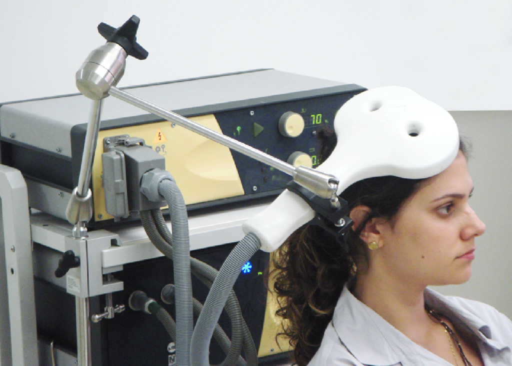
By BaburovOwn work, CC BY-SA 4.0, Link

A type of therapy that involves applying a magnetic field to both sides of the brain has been shown to be effective at rapidly treating depression in patients for whom standard treatments have been ineffective.

Our accelerated approach means we can do all of the sessions in just five days, rapidly reducing an individual’s symptoms of depression

Valerie Voon

The treatment – known as repetitive transcranial magnetic stimulation (TMS) – involves placing an electromagnetic coil against the scalp to relay a high-frequency magnetic field to the brain.

Around one in 20 adults is estimated to suffer from depression. Although treatments exist, such as anti-depressant medication and cognitive behavioural therapy (‘talking therapy’), they are ineffective for just under one in three patients.

One of the key characteristics of depression is under-activity of some regions (such as the dorsolateral prefrontal cortex) and over-activity of others (such as the orbitofrontal cortex (OFC)).

Repetitive transcranial magnetic stimulation applied to the left side of the dorsolateral prefrontal cortex (an area at the upper front area of the brain) is approved for treatment of depression in the UK by NICE and in the US by the FDA. It has previously been shown to lead to considerable improvements among patients after a course of 20 sessions, but because the sessions usually take place over 20-30 days, the treatment is not ideal for everyone, particularly in acute cases or where a person is suicidal.

In research published in Psychological Medicine, scientists from Cambridge, UK, and Guiyang, China, tested how effective an accelerated form of TMS is. In this approach, the treatment is given over 20 sessions, but with four sessions per day over a period of five consecutive days.

The researchers also tested a ‘dual’ approach, whereby a magnetic field was additionally applied to the right-hand side of the OFC (which sits below the dorsolateral prefrontal cortex).

Seventy-five patients were recruited to the trial from the Second People’s Hospital of Guizhou Province in China. The severity of their depression was measured on a scale known as the Hamilton Rating Scale of Depression.

Participants were split randomly into three groups: a ‘dual’ group receiving TMS applied first to the right- and then to the left-hand sides of the brain; a ‘single’ group receiving sham TMS to the right-side followed by active TMS applied to the left-side; and a control group receiving a sham treatment to both sides. Each session lasted in total 22 minutes.

There was a significant improvement in scores assessed immediately after the final treatment in the dual treatment group compared to the other two groups. When the researchers looked for clinically-relevant responses – that is, where an individual’s score fell by at least 50% – they found that almost half (48%) of the patients in the dual treatment group saw such a reduction, compared to just under one in five (18%) in the single treatment group and fewer than one in 20 (4%) in the control group.

Four weeks later, around six in 10 participants in both the dual and single treatment groups (61% and 59% respectively) showed clinically relevant responses, compared to just over one in five (22%) in the control group.

Professor Valerie Voon from the Department of Psychiatry at the University of Cambridge, who led the UK side of the study, said: “Our accelerated approach means we can do all of the sessions in just five days, rapidly reducing an individual’s symptoms of depression. This means it could be particularly useful in severe cases of depression, including when someone is experiencing suicidal thoughts. It may also help people be discharged from hospital more rapidly or even avoid admission in the first place.

“The treatment works faster because, by targeting two areas of the brain implicated in depression, we’re effectively correcting imbalances in two import processes, getting brain regions ‘talking’ to each other correctly.”

The treatment was most effective in those patients who at the start of the trial showed greater connectivity between the OFC and the thalamus (an area in the middle of the brain responsible for, among other things, regulation of consciousness, sleep, and alertness). The OFC is important for helping us make decisions, particularly in choosing rewards and avoiding punishment. Its over-activity in depression, particularly in relation to its role in anti-reward or punishment, might help explain why people with depression show a bias towards negative expectations and ruminations.

Dr Yanping Shu from the Guizhou Mental Health Centre, Guiyang, China, said: “This new treatment has demonstrated a more pronounced – and faster – improvement in response rates for patients with major depressive disorder. It represents a significant step forward in improving outcomes, enabling rapid discharge from hospitals for individuals with treatment-resistant depression, and we are hopeful it will lead to new possibilities in mental health care.”

Dr Hailun Cui from Fudan University, a PhD student in Professor Voon’s lab at the time of the study, added: “The management of treatment-resistant depression remains one of the most challenging areas in mental health care. These patients often fail to respond to standard treatments, including medication and psychotherapy, leaving them in a prolonged state of severe distress, functional impairment, and increased risk of suicide.

“This new TMS approach offers a beacon of hope in this difficult landscape. Patients frequently reported experiencing ‘lighter and brighter’ feelings as early as the second day of treatment. The rapid improvements, coupled with a higher response rate that could benefit a broader depressed population, mark a significant breakthrough in the field.”

Just under a half (48%) of participants in the dual treatment group reported local pain where the dual treatment was applied, compared to just under one in 10 (9%) of participants in the single treatment group. However, despite this, there were no dropouts.

For some individuals, this treatment may be sufficient, but for others ‘maintenance therapy’ may be necessary, with an additional day session if their symptoms appear to be worsening over time. It may also be possible to re-administer standard therapy as patients can then become more able to engage in psychotherapy. Other options include using transcranial direct current stimulation, a non-invasive form of stimulation using weak electrical impulses that can be delivered at home.

The researchers are now exploring exactly which part of the orbitofrontal cortex is most effective to target and for which types of depression.

The research was supported by in the UK by the Medical Research Council and by the National Institute for Health and Care Research Cambridge Biomedical Research Centre.*

Reference
Cui, H, Ding, H & Hu, L et al. A novel dual-site OFC-dlPFC accelerated repetitive transcranial magnetic stimulation for depression: a pilot randomized controlled study. Psychological Medicine; 23 Oct 2024; DOI: 10.1017/S0033291724002289

*A full list of funders is available in the journal paper.

Source: University of Cambridge. The original text of this story is licensed under a Creative Commons Attribution-NonCommercial-ShareAlike 4.0 International LicenseNote: Content may be edited for style and length.

SARS-CoV-2 Hijacks Three Key Proteins in the Complement System

SARS-CoV-2 viruses (yellow) infecting a human cell (blue). Photo by CDC on Pexels

Researchers at the Medical University of Vienna and the Medical University of Innsbruck discovered that SARS-CoV-2 hijacks three important host proteins that dampen the activity of the complement system, a key component of early antiviral immunity. This significantly impairs viral clearance which may affect the course of both acute COVID infections and post-COVID sequelae. The study was recently published in the journal Emerging Microbes & Infections.

An early and effective immune response is crucial for resolving viral infections and preventing post-infectious complications. The complement system, a pivotal element of antiviral immunity, is a cascade of proteins found in the bloodstream and at mucosal sites, such as the respiratory tract. Activated through three different pathways, complement facilitates the clearance of virus particles by directly inducing their destruction (lysis). To prevent bystander damage to host cells, complement is rapidly inactivated by a set of host molecules referred to as complement regulatory proteins. The new study led by Anna Ohradanova-Repic and colleagues from the Center for Pathophysiology, Infectiology and Immunology at the Medical University of Vienna in collaboration with the team of Heribert Stoiber from the Institute of Virology at the Medical University of Innsbruck shows that SARS-CoV-2 hijacks three of these regulatory proteins, CD55, CD59 and Factor H, and thereby successfully shields itself from complement-mediated lysis.

Hijacking host proteins for effective complement resistance

By propagating SARS-CoV-2 in human cells the researchers discovered that the virus particles acquire the cellular proteins CD55 and CD59. Further experiments showed that SARS-CoV-2 also binds to Factor H, another complement regulatory protein that is primarily found in the bloodstream. Confronting the virus particles with active complement revealed that they are partially resistant to complement-mediated lysis. By removing CD55, CD59 and Factor H from the virus surface or inhibiting their biological functions, the researchers could successfully restore complement-mediated clearance of SARS-CoV-2.

“Through hijacking these three proteins, SARS-CoV-2 can evade all three complement pathways, resulting in reduced or delayed viral clearance by the infected host,” Anna Ohradanova-Repic, the leader of the study explains. Because complement is intricately linked with other components of the immune system, this not only affects virus elimination but can also cause significant inflammation, a core feature of both severe COVID-19 and Long COVID. “Uncovering immune evasion mechanisms that allow the virus to linger within the host for longer, deepen our understanding of the acute and long-term impacts of SARS-CoV-2 infection,” says first author Laura Gebetsberger.

Source: Medical University of Vienna

An Unexpected Effect Unlocks New Treatment Option for Prostate Cancer

Credit: Darryl Leja National Human Genome Research Institute National Institutes Of Health

An international research team led by MedUni Vienna may have found a new cancer treatment strategy – by activating a pathway which normally promotes cancer. Unexpectedly, this turned out to not only slow tumour growth, but also stimulates the immune system to combat tumour cells. The results of the study have just been published in Molecular Cancer.

The scientific team focused its investigations on the GP130 signalling pathway, which researchers expect to have a major potential in the fight against cancer. The signalling pathway, which is mediated by the protein GP130, plays a central role in cell communication and influences the activity of the transcription factor STAT3, which in turn is associated with the development and spread of tumours. Accordingly, blocking the GP130 signalling pathway is currently seen as a great hope in cancer medicine. Yet the current study proves the opposite: tumour growth can be slowed down not by inhibiting but by activating the GP130 signalling pathway in prostate cells.
 
New hope, especially for aggressive tumours

To achieve these new findings, the researchers investigated genetically modified mice in which GP130 was specifically activated in the prostate. “This allowed us to directly observe the reduction in tumour growth in the cell,” reports Lukas Kenner (Clinical Department of Pathology, MedUni Vienna), who led the study together with Stefan Rose-John (Biochemical Institute, University of Kiel). The results were further backed up by analyses of tissue samples from prostate cancer patients. This showed that high GP130 values correlate with a better survival rate. At the same time, extensive molecular analyses were carried out, including gene expression profiling.

“Our research provides exciting new evidence that the activation of GP130 in prostate cells not only slows tumour growth, but also stimulates the immune system to actively fight the cancer cells,” says Lukas Kenner, summarising the significance of the results, which will now be confirmed in further studies. The research work opens up a promising new therapeutic option, particularly for aggressive prostate cancer, which is still difficult to treat.

Source: Medical University of Vienna

Memories are Stored in Cells Outside the Brain, Too

It’s common knowledge that the neurons in the brain store memories. But a team of scientists has discovered that cells from other parts of the body also perform a memory function, opening new pathways for understanding how memory works and creating the potential to enhance learning and to treat memory-related afflictions. 

“Learning and memory are generally associated with brains and brain cells alone, but our study shows that other cells in the body can learn and form memories, too,” explains New York University’s Nikolay V. Kukushkin, the lead author of the study in Nature Communications

The research sought to better understand if non-brain cells help with memory by borrowing from a long-established neurological property – the massed-spaced effect – which shows that we tend to retain information better when studied in spaced intervals rather than in a single, intensive session – better known as cramming for a test.

In the Nature Communications research, the scientists replicated learning over time by studying two types of non-brain human cells in a laboratory (one from nerve tissue and one from kidney tissue) and exposing them to different patterns of chemical signals – just like brain cells are exposed to patterns of neurotransmitters when we learn new information. In response, the non-brain cells turned on a “memory gene” – the same gene that brain cells turn on when they detect a pattern in the information and restructure their connections in order to form memories.

“Learning and memory are generally associated with brains and brain cells alone, but our study shows that other cells in the body can learn and form memories, too.”

NYU’s Nikolay Kukushkin 

To monitor the memory and learning process, the scientists engineered these non-brain cells to make a glowing protein, which indicated when the memory gene was on and when it was off.

The results showed that these cells could determine when the chemical pulses, which imitated bursts of neurotransmitter in the brain, were repeated rather than simply prolonged – just as neurons in our brain can register when we learn with breaks rather than cramming all the material in one sitting. Specifically, when the pulses were delivered in spaced-out intervals, they turned on the “memory gene” more strongly, and for a longer time, than when the same treatment was delivered all at once.

“This reflects the massed-space effect in action,” says Kukushkin, a clinical associate professor of life science at NYU Liberal Studies and a research fellow at NYU’s Center for Neural Science. “It shows that the ability to learn from spaced repetition isn’t unique to brain cells, but, in fact, might be a fundamental property of all cells.”

The researchers add that the findings not only offer new ways to study memory, but also point to potential health-related gains.

“This discovery opens new doors for understanding how memory works and could lead to better ways to enhance learning and treat memory problems,” observes Kukushkin. “At the same time, it suggests that in the future, we will need to treat our body more like the brain – for example, consider what our pancreas remembers about the pattern of our past meals to maintain healthy levels of blood glucose or consider what a cancer cell remembers about the pattern of chemotherapy.”

Source: New York University

South Africa Amended its Research Guidelines to Allow for Heritable Human Genome Editing

Source: Pixabay CC0

Françoise Baylis, Dalhousie University

A little-noticed change to South Africa’s national health research guidelines, published in May of this year, has put the country on an ethical precipice. The newly added language appears to position the country as the first to explicitly permit the use of genome editing to create genetically modified children.

Heritable human genome editing has long been hotly contested, in large part because of its societal and eugenic implications. As experts on the global policy landscape who have observed the high stakes and ongoing controversies over this technology — one from an academic standpoint (Françoise Baylis) and one from public interest advocacy (Katie Hasson) — we find it surprising that South Africa plans to facilitate this type of research.

In November 2018, the media reported on a Chinese scientist who had created the world’s first gene-edited babies using CRISPR technology. He said his goal was to provide children with resistance to HIV, the virus that causes AIDS. When his experiment became public knowledge, twin girls had already been born and a third child was born the following year.

The fate of these three children, and whether they have experienced any negative long-term consequences from the embryonic genome editing, remains a closely guarded secret.

Controversial research

Considerable criticism followed the original birth announcement. Some argued that genetically modifying embryos to alter the traits of future children and generations should never be done.

Many pointed out that the rationale in this case was medically unconvincing – and indeed that safe reproductive procedures to avoid transmitting genetic diseases are already in widespread use, belying the justification typically given for heritable human genome editing. Others condemned his secretive approach, as well as the absence of any robust public consultation, considered a prerequisite for embarking on such a socially consequential path.

In the immediate aftermath of the 2018 revelation, the organizing committee of the Second International Summit on Human Genome Editing joined the global uproar with a statement condemning this research.

At the same time, however, the committee called for a “responsible translational pathway” toward clinical research. Safety thresholds and “additional criteria” would have to be met, including: “independent oversight, a compelling medical need, an absence of reasonable alternatives, a plan for long-term follow-up, and attention to societal effects.”

Notably, the additional criteria no longer included the earlier standard of “broad societal consensus.” https://www.youtube.com/embed/XAhFoaT6Kik?wmode=transparent&start=0 Nobel laureate David Baltimore, chair of the organizing committee for the Second International Summit on Human Genome Editing, talks about the importance of public global dialogue on gene editing.

New criteria

Now, it appears that South Africa has amended its Ethics in Health Research Guidelines to explicitly envisage research that would result in the birth of gene-edited babies.

Section 4.3.2 of the guidelines on “Heritable Human Genome Editing” includes a few brief and rather vague paragraphs enumerating the following criteria: (a) scientific and medical justification; (b) transparency and informed consent; (c) stringent ethical oversight; (d) ongoing ethical evaluation and adaptation; (e) safety and efficacy; (f) long-term monitoring; and (g) legal compliance.

While these criteria seem to be in line with those laid out in the 2018 summit statement, they are far less stringent than the frameworks put forth in subsequent reports. This includes, for example, the World Health Organization’s report Human Genome Editing: Framework for Governance (co-authored by Françoise Baylis).

Alignment with the law

Further, there is a significant problem with the seemingly permissive stance on heritable human genome editing entrenched in these research guidelines. The guidelines clearly require the research to comply with all laws governing heritable human genome research. Yet, the law and the research guidelines in South Africa are not aligned, which entails a significant inhibition on any possible research.

This is because of a stipulation in section 57(1) of the South African National Health Act 2004 on the “Prohibition of reproductive cloning of human beings.” This stipulates that a “person may not manipulate any genetic material, including genetic material of human gametes, zygotes, or embryos… for the purpose of the reproductive cloning of a human being.”

When this act came into force in 2004, it was not yet possible to genetically modify human embryos and so it’s not surprising there’s no specific reference to this technology. Yet the statutory language is clearly wide enough to encompass it. The objection to the manipulation of human genetic material is therefore clear, and imports a prohibition on heritable human genome editing.

Ethical concerns

Photo by Tingey Injury Law Firm on Unsplash

The question that concerns us is: why are South Africa’s ethical guidelines on research apparently pushing the envelope with heritable human genome editing?

In 2020, we published alongside our colleagues a global review of policies on research involving heritable human genome editing. At the time, we identified policy documents — legislation, regulations, guidelines, codes and international treaties — prohibiting heritable genome editing in more than 70 countries. We found no policy documents that explicitly permitted heritable human genome editing.

It’s easy to understand why some of South Africa’s ethicists might be disposed to clear the way for somatic human genome editing research. Recently, an effective treatment for sickle cell disease has been developed using genome editing technology. Many children die of this disease before the age of five and somatic genome editing — which does not involve the genetic modification of embryos — promises a cure.

Implications on future research

But that’s not what this is about. So, what is the interest in forging a path for research on heritable human genome editing, which involves the genetic modification of embryos and has implications for subsequent generations? And why the seemingly quiet modification of the guidelines?

How many people in South Africa are aware that they’ve just become the only country in the world with research guidelines that envisage accommodating a highly contested technology? Has careful attention been given to the myriad potential harms associated with this use of CRISPR technology, including harms to women, prospective parents, children, society and the gene pool?

Is it plausible that scientists from other countries, who are interested in this area of research, are patiently waiting in the wings to see whether the law in South Africa prohibiting the manipulation of human genetic material will be an insufficient impediment to creating genetically modified children? Should the research guidelines be amended to accord with the 2004 statutory prohibition?

Or if, instead, the law is brought into line with the guidelines, would the result be a wave of scientific tourism with labs moving to South Africa to take advantage of permissive research guidelines and laws?

We hope the questions we ask are alarmist, as now is the time to ask and answer these questions.

Katie Hasson, Associate Director at the Center for Genetics and Society, co-authored this article.

Françoise Baylis, Distinguished Research Professor, Emerita, Dalhousie University

This article is republished from The Conversation under a Creative Commons license. Read the original article.

Eight Reasons Why ADHD Diagnoses are Increasing

Photo by Annie Spratt on Unsplash

Sven Bölte, Karolinska Institutet

For a long time it was assumed that somewhere between 5 and 6% of children have attention-deficit hyperactivity disorder (ADHD). But the rates, in practice, are often higher. The American Centers for Disease Control and Prevention put the prevalence at 11.4% in children in 2022.

The Swedish Board of Health and Welfare reports that in 2022 10.5% of boys and 6% of girls received an ADHD diagnosis, which is 50% more than in 2019. And the board forecast that the rates will eventually plateau at 15% for boys and 11% for girls.

So, what might be the reasons behind the startling rise? Here are eight possible causes, many of which overlap and interact with each other.

1. Multiple diagnoses made in the same person

Previously, doctors were recommended by diagnostic manuals and trained to limit diagnoses in an individual to the most prominent one, and not to make certain combinations of diagnoses at all – for example, autism and ADHD. Today, it is recommended and common practice in the mental health sector to make as many diagnoses needed to meaningfully describe and cover the symptoms and challenges of a person.

2. Increased knowledge and awareness by professionals

Today, there is a new generation of professionals working in services with higher awareness and knowledge of ADHD. This has led to earlier detection and to ADHD being diagnosed in groups that were previously neglected, particularly girls and women – but also in adults, generally

3. Reduced stigma

In many societies, ADHD is far less stigmatised than previously. Doctors have fewer doubts about making the diagnosis, and those receiving it feel less stigmatised. For more and more people, ADHD has fewer negative connotations and is becoming a natural part of people’s identities .

4. Modern society places higher demands on cognitive skills

ADHD is not a disease but a malfunctioning composition of cognitive traits that exist on more functional levels even in the general population, such as “attention control” (concentration) and organisational and self-regulation skills. Modern societies are fast and complex, placing high demands on these cognitive traits. So people with lower than average skills in these key cognitive areas begin struggling to cope with everyday demands and might receive an ADHD diagnosis.

5. Higher expectations on health and performance

People’s expectations of their own and others’ performance and health are rising. The so-called “social baseline” of average health and performance is higher today. Therefore, people may express concerns about their own and others’ functioning earlier and more often, and may presume that ADHD could be an explanation.

6. Changes in schools have led to more students struggling

Schools have gone through substantial changes in how they teach, such as digitisation and introducing more project- and group-based learning, as well as much more self-guided education.

These changes have led to a less clear learning environment, including increased demands on students’ motivation and their cognitive skills, factors that can make it harder for students with even just a few traits of ADHD to succeed. It has also caused schools to refer more students whom they suspect of having ADHD for assessment.

7. Policymakers prioritise assessment

Politicians in many countries have tried to address the rising diagnosis rates predominantly by making diagnostic assessments more accessible so that people don’t have to wait a long time to receive a diagnosis.

While this is understandable, it fuels the number of diagnoses made and does not focus on avoiding diagnoses, such as by improving how children are taught, improving workplaces to make them more neurodivergent friendly, and offering support without requiring that a person have a diagnosis.

8. Diagnosis guarantees access to support and resources

In most societies, services are constructed as such that only a clinical diagnosis guarantees access to support and resources. It is often the only way for people and their families to get support.

Generally, not a lot is done for people without a diagnosis as service providers do not get reimbursed and are therefore less obliged to take action. So people in need of support are more likely to actively seek a diagnosis. And professionals are more inclined to assist them by giving a diagnosis, even if the person doesn’t quite meet the diagnostic criteria for ADHD – a phenomenon called “diagnostic upgrading”.

Sven Bölte, Professor of Child and Adolescent Psychiatric Science, Karolinska Institutet

This article is republished from The Conversation under a Creative Commons license. Read the original article.

Opinion Piece: Business Continuity and Data Management – a Life-or-death Situation in Healthcare

Photo by Nahel Abdul on Unsplash

By Hemant Harie, Group CTO at DMP SA / Gabsten Technologies

Ransomware attacks are a growing concern for healthcare facilities worldwide, with attacks wreaking havoc, including encrypting complex patient records, cancelling appointments, delaying life-saving surgeries, and even rerouting ambulances. The critical nature of healthcare services, combined with the sensitive personal and medical data they handle, makes hospitals and healthcare providers a prime target for cybercriminals.

When these systems are compromised, the impact is severe, jeopardising patient safety, disrupting service delivery and causing financial strain. It has become imperative for healthcare facilities to adopt more robust cybersecurity measures, including effective data management strategies as part of an overall business continuity approach. Partnering with an expert third-party service provider can assist healthcare facilities in ensuring continuity of care and business operations even in the face of cyberattacks.

Attractive targets with unique vulnerabilities

Digital transformation within the healthcare space, while vital for improving patient care,  can also introduce significant cybersecurity risks. Many hospitals and healthcare facilities are at different stages in their digital transformation , and legacy infrastructure is a common challenge, alongside immature cybersecurity posture and processes, making them more susceptible to attacks.

Cybercriminals often target these systems because they handle vast amounts of sensitive data, including Personal Health Information (PHI), which is highly valuable on the black market. In addition, these facilities often lack the dedicated IT and cybersecurity specialists they need to adequately defend against or recover from ransomware incidents.

The nature of information housed within healthcare and the consequences of a breach mean the stakes are high. This, combined with the fact that healthcare facilities are legally bound by regulations such as the Protection of Personal Information Act (PoPIA), Health Insurance Portability and Accountability Act (HIPAA) and General Data Protection Regulation (GDPR) to protect this information, means potential breaches could have catastrophic consequences.

The impact of ransomware on healthcare

Ransomware attacks can have devastating effects on healthcare organisations, leading to significant downtime that directly threatens patient care. Operations may be postponed or cancelled, disrupting treatment schedules and putting patients’ lives at risk. Additionally, the exposure of PHI can result in severe legal and ethical repercussions, including costly regulatory fines and lawsuits. Financial losses also extend to ransom payments, the cost of recovery, and reputational damage, all of which can linger long after the attack is resolved.

Moreover, a ransomware attack on one healthcare facility can damage the reputation of the entire network, as trust is critical in healthcare. Patients may be less likely to seek care from a hospital they perceive as insecure, leading to long-term financial and operational challenges.

Data management mitigates ransomware risks

To effectively combat ransomware, healthcare organisations must prioritise data management and cyber resilience. This starts with classifying and understanding the types of data being processed and stored , such as medical records, surgical files, and other critical patient information. Once this data is properly categorised, healthcare facilities can implement security controls that ensure the integrity and availability of the data.

Regular, automated backups stored offline are essential for mitigating ransomware risks. These backups allow facilities to restore their systems quickly without paying a ransom, minimising downtime and ensuring continuity of care. In addition to regular backups, hospitals should adopt advanced security measures such as multi-factor authentication, firewalls, and intrusion detection systems to safeguard against unauthorised access.

An expert partner enhances data management and security

Third-party service providers offer critical expertise and comprehensive solutions that healthcare organisations may lack in-house. These providers specialise in data management, backup, and disaster recovery, ensuring that hospitals have access to the latest technologies and best practices for defending against cyber threats. These experts bring valuable experience from handling multiple cyber incidents across various sectors, which can inform and improve the healthcare facility’s own data management practices. In addition to providing technical expertise, third-party providers can offer ongoing education, helping healthcare staff stay informed about the latest cybersecurity threats and recovery processes.

One of the key services offered by third-party providers is automated backup and disaster recovery solutions. These services typically include offsite storage, secure cloud options, and regular backups, all of which are vital for restoring data and reducing downtime during a ransomware attack. Offsite storage and cloud solutions also protect data from physical threats like floods or fires, adding an extra layer of security. In addition to traditional backup services, advanced tools can enhance data protection by providing early warning systems and simulating real-time production environments, which allow healthcare facilities to detect and respond to potential threats before they can cause damage. For example, scanning tools can identify which versions of data are clean and free from malware, enabling faster and more effective recovery.

Partnering with a third-party provider ensures that healthcare organisations have access to continuous support and the latest innovations in data protection. These providers not only help mitigate ransomware risks but also assist in compliance with industry regulations and offer scalable solutions to meet the growing needs of healthcare facilities.

As ransomware threats continue to rise, healthcare organisations must take proactive steps to safeguard their systems and protect patient data. Effective data management, including regular backups and disaster recovery plans, is essential for mitigating these risks. By partnering with third-party service providers, healthcare facilities can leverage specialised expertise and advanced technologies to enhance their cybersecurity defences and maintain continuity of care, even in the face of growing cyber threats.

A New Era of Treating Neurological Diseases at the Blood-brain-immune Interface

This is a pseudo-colored image of high-resolution gradient-echo MRI scan of a fixed cerebral hemisphere from a person with multiple sclerosis. Credit: Govind Bhagavatheeshwaran, Daniel Reich, National Institute of Neurological Disorders and Stroke, National Institutes of Health

The question of what causes complex neurological diseases such as Alzheimer’s or multiple sclerosis continues to confound scientists and doctors, with the unknowns standing in the way of early diagnoses and effective treatments.

Even among identical twins who share the same genetic risk factors, one may develop a particular neurological disease while the other does not.

That’s because unlike diseases such as cystic fibrosis or sickle-cell anaemia, which are caused by a single gene, most neurological disorders are associated with many, sometimes hundreds, of rare genetic variants. And on their own, these variants can’t predict who will develop disease, as neurological conditions are also strongly influenced by environmental factors and vascular risks such as high blood pressure, aging, heart disease, or obesity.

But there’s one often-overlooked thread that connects most neurological diseases, says Katerina Akassoglou, PhD, a senior investigator at Gladstone Institutes: They’re marked by a toxic immune reaction caused by blood that leaks into the brain through damaged blood vessels.

“Interactions between the brain, blood vessels, and the immune system are a common thread in the development and progression of many neurological diseases that have been traditionally viewed as very different conditions,” says Akassoglou. “Knowing that leaked blood is a key driver of brain inflammation, we can now approach these diseases from a different angle.”

She and her collaborators share their insights on this topic in a commentary article published in Cell’s 50th anniversary “Focus on Neuroscience” issue. 

Neutralising the Culprit

Akassoglou and her lab have long investigated how blood that leaks into the brain triggers neurologic diseases, essentially by hijacking the brain’s immune system and setting off a cascade of harmful often-irreversible effects that result in damaged neurons.

One blood protein in particular, fibrin, normally involved in blood coagulation, is responsible for setting off this detrimental cascade. The process has been observed in conditions as diverse as Alzheimer’s, traumatic brain injury, multiple sclerosis, premature birth, and even COVID-19. However, Akassoglou and her team found that the process could be prevented or interrupted by “neutralising” fibrin to deactivate its toxic properties – an approach that appears to protect against many neurological diseases when tested in animal models.

“As a first step, we know that neutralizing fibrin reduces the burden posed by vascular dysfunction,” Akassoglou says. Regardless of what initially caused the blood leaks, be it a head injury, autoimmunity, genetic mutations, brain amyloid or infection, neutralizing fibrin appears to be protective in multiple animal models of disease.

The scientists previously developed a drug, a therapeutic monoclonal antibody, that specifically targets fibrin’s inflammatory properties without affecting its essential role in blood coagulation. This fibrin-targeting immunotherapy has shown, in mice, to protect from multiple sclerosis and Alzheimer’s, and to treat neurological effects of COVID-19. A humanized version of this first-in-class fibrin immunotherapy is already in Phase 1 safety clinical trials by Therini Bio, a biotech company launched to advance discoveries from Akassoglou’s lab.

A New Era of Brain Research

In the Cell commentary, Akassoglou and her colleagues make the case that seemingly disparate neurological diseases must be viewed differently in light of new research on the blood-brain-immune interface.

They say that in the coming decade, scientific breakthroughs will emerge from collaborative networks of immunologists, neuroscientists, haematologists, geneticists, computer scientists, physicists, bioengineers, drug developers, and clinical researchers. These partnerships, forged across academia, industry, and foundations, will catalyse innovation in drug discovery and transform medical practice for neurological diseases.

“This is a new opportunity for drug discovery that goes beyond addressing genes alone or environmental factors alone,” Akassoglou says. “To usher in this new era, we must leverage new technologies and embrace an interdisciplinary approach that accounts for the important roles of immune and vascular systems in neurodegeneration.”

Source: Gladstone Institutes

Extra Year of Education does Not Protect the Brain

Photo by Andrea Piacquadio on Pexels

Thanks to a ‘natural experiment’ involving 30 000 people, researchers at Radboud university medical centre were able to very precisely determine the effect of an extra year of education to the brain in the long term. To their surprise, they found no effect on brain structure and no protective benefit of additional education against brain ageing. Their findings appear in eLife.

It is well-known that education has many positive effects. People who spend more time in school are generally healthier, smarter, and have better jobs and higher incomes than those with less education. However, whether prolonged education actually causes changes in brain structure over the long term and protects against brain ageing, was still unknown.

It is challenging to study this, because alongside education, many other factors influence brain structure, such as the conditions under which someone grows up, DNA traits, and environmental pollution. Nonetheless, researchers Rogier Kievit (PI of the Lifespan Cognitive Dynamics lab) and Nicholas Judd from Radboudumc and the Donders Institute found a unique opportunity to very precisely examine the effects of an extra year of education.

Ageing

In 1972, a change in the law in the UK raised the number of mandatory school years from 15 to 16, while all other circumstances remained constant. This created an interesting ‘natural experiment’, an event not under the control of researchers which divides people into an exposed and unexposed group. Data from approximately 30 000 people who attended school around that time, including MRI scans taken much later (46 years after), is available. This dataset is the world’s largest collection of brain imaging data.

The researchers examined the MRI scans for the structure of various brain regions, but they found no differences between those who attended school longer and those who did not. ‘This surprised us’, says Judd. ‘We know that education is beneficial, and we had expected education to provide protection against brain aging. Aging shows up in all of our MRI measures, for instance we see a decline in total volume, surface area, cortical thickness, and worse water diffusion in the brain. However, the extra year of education appears to have no effect here.’

Brain structure

It’s possible that the brain looked different immediately after the extra year of education, but that wasn’t measured. “Maybe education temporarily increases brain size, but it returns to normal later. After all, it has to fit in your head,” explains Kievit. “It could be like sports: if you train hard for a year at sixteen, you’ll see a positive effect on your muscles, but fifty years later, that effect is gone.” It’s also possible that extra education only produces microscopic changes in the brain, which are not visible with MRI.

Both in this study and in other, smaller studies, links have been found between more education and brain benefits. For example, people who receive more education have stronger cognitive abilities, better health, and a higher likelihood of employment. However, this is not visible in brain structure via MRI. Kievit notes: “Our study shows that one should be cautious about assigning causation when only a correlation is observed. Although we also see correlations between education and the brain, we see no evidence of this in brain structure.”

Source: Radboud University Medical Centre

Defibrillation Using 1/1000th the Energy could be Possible

Photo by Mikhail Nilov

Researchers from Sergio Arboleda University in Colombia and the Georgia Institute of Technology in the US used an electrophysiological computer model of the heart’s electrical circuits to examine the effect of the applied voltage field in multiple fibrillation-defibrillation scenarios. Their research, published in the interdisciplinary journal Chaos, discovered that far less energy is needed than is currently used in state-of-the-art defibrillation techniques.

“The results were not at all what we expected. We learned the mechanism for ultra-low-energy defibrillation is not related to synchronisation of the excitation waves like we thought, but is instead related to whether the waves manage to propagate across regions of the tissue which have not had the time to fully recover from a previous excitation,” author Roman Grigoriev said. “Our focus was on finding the optimal variation in time of the applied electric field over an extended time interval. Since the length of the time interval is not known a priori, it was incremented until a defibrillating protocol was found.”

The authors applied an adjoint optimization method, which aims to achieve a desired result, defibrillation in this case, by solving the electrophysiologic model for a given voltage input and looping backward through time to determine the correction to the voltage profile that will successfully defibrillate irregular heart activity while reducing the energy the most.

Energy reduction in defibrillation devices is an active area of research. While defibrillators are often successful at ending dangerous arrhythmias in patients, they are painful and cause damage to the cardiac tissue.

“Existing low-energy defibrillation protocols yield only a moderate reduction in tissue damage and pain,” Grigoriev said. “Our study shows these can be completely eliminated. Conventional protocols require substantial power for implantable defibrillators-cardioverters (ICDs), and replacement surgeries carry substantial health risks.”

In a normal rhythm, electrochemical waves triggered by pacemaker cells at the top of the atria propagate through the heart, causing synchronised contractions. During arrhythmias, such as fibrillation, the excitation waves start to quickly rotate instead of propagating through and leaving the tissue, as in normal rhythm.

“Under some conditions, an excitation wave may or may not be able to propagate through the tissue. This is called the ‘vulnerable window,’” Grigoriev said. “The outcome depends on very small changes in the timing of the excitation wave or very small external perturbations.

“The mechanism of ultra-low-energy defibrillation we uncovered exploits this sensitivity. Varying the electrical field profile over a relatively long time interval allows blocking the propagation of the rotating excitation waves through the ‘sensitive’ regions of tissue, successfully terminating the irregular electric activity in the heart.”

Source: American Institute of Physics