Month: November 2024

CPR with Breaths Essential for Cardiac Arrest after Drowning

Photo by Kampus Production

Updated guidance reaffirms the recommendation for cardiopulmonary resuscitation (CPR) and highlights the importance of compressions with rescue breaths as a first step in responding to cardiac arrest following drowning, according to a new, focused update to Special Circumstances Guidelines from the American Heart Association and the American Academy of Pediatrics. The recommendations were published simultaneously in Circulation (focusing on adults) and Pediatrics (focusing on children).

Drowning is the third-leading cause of death from unintentional injury worldwide. The World Health Organization estimates there are about 236 000 deaths due to drowning each year globally. According to the CDC, it’s the number one cause of death for children ages 1-4 years old in the US.

“The focused update on drowning contains the most up-to-date, evidence-based recommendations on how to resuscitate someone who has drowned, offering practical guidance for health care professionals, trained rescuers, caregivers and families,” said writing group Co-Chair Tracy E. McCallin, M.D., FAAP, associate professor of paediatrics in the division of paediatric emergency medicine at Rainbow Babies and Children’s Hospital in Cleveland. “While we work on a daily basis to lower risks of drowning through education and community outreach on drowning prevention, we still need emergency preparedness training that can be used in tragic circumstances if a drowning occurs.”  

Detailed in the new guideline update:

  • Anyone removed from the water without showing signs of normal breathing or consciousness should be presumed to be in cardiac arrest.
  • Rescuers should immediately initiate CPR that includes rescue breathing in addition to chest compressions. Multiple large studies over time show more people with cardiac arrest from non-cardiac causes such as drowning survive when CPR includes rescue breaths compared to Hands-Only CPR (calling 911 [10111 in South Africa] and pushing hard and fast in the centre of the chest).

Drowning generally progresses quickly from initial respiratory arrest (when a person is unable to breathe) to cardiac arrest, meaning that the heart stops beating. As a result, blood cannot circulate properly throughout the body, and it is starved of oxygen.

“CPR for cardiac arrest due to drowning must focus on restoring breathing as well as restoring blood circulation,” said writing group Co-Chair Cameron Dezfulian, MD, FAHA, FAAP, senior faculty in paediatrics and critical care at Baylor College of Medicine in Houston.

“Cardiac arrest following drowning is most often due to severe hypoxia, or low blood oxygen levels,“ Dezfulian said. ”This differs from sudden cardiac arrest from a cardiac cause where the individual generally collapses with fully oxygenated blood.”

The updated guidance advises untrained rescuers and the public to:

  • Provide CPR with breaths and compressions to all people who have a cardiac arrest after drowning. If a person is untrained, unwilling, or unable to give breaths, they can provide chest compressions only until help arrives. 
  • In-water rescue breathing should be given only by rescuers trained in this special skill if it doesn’t compromise their own safety. Trained rescuers should also provide supplemental oxygen if available.
  • The initiation of CPR should always be prioritised and begin as soon as possible as early lay responder CPR has been shown to improve outcomes from drowning.
  • The writing group recommends an automated external defibrillator (AED) should be placed in public facilities where aquatic activities are present such as swimming pools or beaches. They can be used once the person is removed from the water, if available, yet should not delay initiation of CPR. If available, the AED should be connected to the patient to assess for shockable rhythms once CPR is ongoing. Although most cases of cardiac arrest following drowning do not have shockable rhythms, if a primary cardiac event such as a heart attack occurs while in the water, the best outcomes are when defibrillation is done quickly. AED use is safe and feasible in aquatic environments.
  • All individuals requiring any level of resuscitation following drowning, including those who only need rescue breaths, should be transported to a hospital for evaluation, monitoring and treatment.

In addition to the recommendations on drowning resuscitation, the guideline update also highlights the Drowning Chain of Survival, which includes the steps needed to improve chances of survival: preventionrecognition and safe rescue.

Prevention

It has been estimated that more than 90% of all drownings are preventable. Research has found most infants drown in bathtubs, and the majority of preschool-aged children drown in swimming pools. The American Heart Association and the American Academy of Pediatrics recommend being water aware and practicing water safety. See: Prevention of Drowning and other guidelines.  

Recognition

Recognition of drowning may be challenging because someone who is drowning may not be able to verbalise distress or signal for help. Drowning happens quickly. People in distress will rapidly submerge, lose consciousness and may be hidden from anyone not actively seeking them.

Safe Rescue and Removal

The guideline update recommends that appropriately trained rescuers, such as lifeguards, swim instructors or first responders, should provide in-water rescue breathing to an unresponsive person who has drowned if it does not compromise their own safety. Previous studies have proven this leads to more favourable survival outcomes. A drowning person who is unconscious and likely in cardiac arrest should be removed from the water in a near-horizontal position, with the head maintained above body level and airway open. If the drowning individual is conscious, a more vertical position may be preferable to reduce the risk of vomiting.

In summary, “These updated guidelines are based on the latest available evidence and are designed to inform trained rescuers and the public how to proceed in resuscitating people who have drowned. Drowning can be fatal. Our recommendations maximise balancing the need for rapid rescue and resuscitation, while prioritising rescuer safety,” Dezfulian said.

Source: American Heart Association

More than a Quarter of New Mothers have Fallen Asleep while Breastfeeding

Photo by Wendy Wei

More than a quarter of new mothers have fallen asleep recently while feeding their babies, putting the infants at increased risk of sudden infant death syndrome (SIDS), research published in Pediatrics reveals.

More than 80% had not intended to fall asleep, and many had chosen to feed in chairs or on sofas rather than in a bed. Unfortunately, the cushions and confines of those locations can be very unsafe for babies, raising the risk of death by 49 to 67 times.

The researchers, with UVA Health and UVA Health Children’s, are urging care providers to provide additional guidance for new parents on safe feeding practices, such as informing new moms that a hormone naturally released during breastfeeding will make them feel sleepy.

“While falling asleep while feeding young infants is not in itself too surprising, what is very alarming is that the majority of mothers did not plan to fall asleep, so the sleep space was potentially unsafe for the baby while both slept,” said researcher Fern Hauck, MD, MS, a safe-sleep expert at UVA Health and the UVA School of Medicine. “This highlights the need for parents to be educated about the potential risk of falling asleep while feeding and to plan for that possibility by making the space around the baby as safe as possible. That would include removing pillows and blankets to ensure an open airway for the baby.”

Safe infant feeding

Hauck and her collaborators, including UVA’s Ann Kellams, MD, and Rachel Moon, MD, analysed survey results collected from more than 1250 new mothers as part of the Social Media and Risk-reduction Training (SMART) study conducted at 16 US hospitals in 2015 and 2016. Most respondents completed the survey when their infant was between 2 and 3 months of age.

Among the respondents, more than 28% said they had “usually” or “sometimes” fallen asleep during feeding in the prior two weeks. Of those, a whopping 83.4% said falling asleep was unplanned.

Women who fed in bed were more likely to fall asleep (33.6%) than those who fed on a chair or couch (16.8%). The American Academy of Pediatrics (AAP) recommends mothers at risk of falling asleep while breastfeeding should do so in an adult bed rather than a chair or couch.

Many of the women who fell asleep on chairs or sofas said they chose those locations specifically to avoid falling asleep, to avoid locations (such as a bed) they had been told were unsafe or to avoid disturbing someone else. (The AAP warns moms against sharing a bed or other sleep space with an infant because of the risk the parent might accidentally roll over and smother the child, or that the child could become tangled in bedding. But the group also says that beds are safer than chairs and sofas if falling asleep while feeding is a possibility.)

“We need to meet families where they are and come up with a nighttime plan for sleeping and feeding their baby that works for them and is as safe as possible,” said Kellams, a paediatrician and breastfeeding and lactation medicine specialist at UVA Health Children’s. “Our data suggest that too many of these falling asleep incidents are not planned, so discussions about how to plan for feeding your baby when you are very tired are important.”

The researchers note that providing parents with information about safe sleep and feeding has been shown to reduce risk of unexpected death significantly. But this educational outreach needs to be expanded, they say. Care providers should acknowledge that moms face a very real risk of falling asleep while feeding, even if they are trying not to, and provide practical advice on how to reduce that risk. Further, the researchers are urging additional studies to find ways to assist parents in both safe-sleep practices and breastfeeding.

“We hope that parents of young infants will think proactively about what might happen in the middle of the night,” said Moon, a paediatrician and safe-sleep expert at UVA Health Children’s. “Feeding your baby in your bed is safer than feeding on a couch or armchair if you might fall asleep.”

Source: University of Virginia Health System

Objective Study Shows that Cannabinol does in Fact Increase Sleep

Photo by Cottonbro on Pexels

Research by scientists at the University of Sydney has identified cannabinol (CBN), a constituent in the cannabis plant that improves sleep. Their report is the first to use objective measures to show that (CBN), while not intoxicating, does increase sleep in rats. The study, which has been published in the leading journal Neuropsychopharmacology, found that CBN was comparable in efficacy to zolpidem.

“For decades, cannabis folklore has suggested that aged cannabis makes consumers sleepy via the build-up of CBN, however there was no convincing evidence for this,” said lead author Professor Jonathon Arnold, Director of Preclinical Research, at the Lambert Initiative for Cannabinoid Therapeutics and the Sydney Pharmacy School.

“Our study provides the first objective evidence that CBN increases sleep, at least in rats, by modifying the architecture of sleep in a beneficial way.”

CBN is an end-product of the main intoxicating constituent of cannabis, delta9-tetrahydrocannabinol (THC). THC in cannabis is slowly converted to CBN over time, which means older cannabis contains higher levels of this compound. It has been suggested that the consumption of older cannabis is associated with a sleepier cannabis “high”. 

In the United States, highly purified CBN products are being sold as sleep aids, but there has been little high-quality scientific evidence to support this application.

The research team at the Lambert Initiative for Cannabinoid Therapeutics tested the effects of purified CBN on sleep in rats. Using high-tech monitoring, the experiments provided insights into the rats’ sleep patterns including the amount of non-rapid eye movement (NREM) and rapid eye movement (REM) sleep. 

NREM is deep sleep that promotes physical recovery and strengthens memories, while REM sleep is associated with dreaming and processing of emotions. 

Professor Arnold said: “CBN was found to increase both NREM and REM sleep, leading to increased total sleep time, with a comparable effect to the known sleep drug zolpidem.”

Non-intoxicating

Unlike its parent molecule THC, CBN did not appear to intoxicate rats. THC intoxicates by activating CB1 cannabinoid receptors, which are present in the brain. The study showed that unlike THC, CBN only weakly activates these receptors. To their surprise, the researchers found that a metabolite of CBN had significant effects on cannabinoid CB1 receptors. 

A metabolite is a chemical produced via the metabolism of a larger molecule in the body.

They also found that the 11-OH CBN metabolite had some impact on sleep architecture, which might contribute to the overall effects of CBN on sleep.

“This provides the first evidence that CBN indeed increases sleep using objective sleep measures. It was a surprise that CBN metabolism in the body can yield a much greater effect on cannabinoid CB1 receptors than the parent molecule CBN, which has much more limited activity,” Professor Arnold said.

“At this stage our results are confined to testing in rats. Further research is needed to see if this translates to humans.”  

Further study

In a parallel study, yet to be published, Professor Iain McGregor, Director of Clinical Research at the Lambert Initiative, initiated a placebo-controlled randomised human clinical trial in insomnia patients. This was led by PhD student Isobel Lavender with leading sleep researcher Dr Camilla Hoyos from the Woolcock Institute of Medical Research. The trial has now been completed with very promising results that were recently announced at the International Cannabinoid Research Society and Sleep DownUnder scientific conferences.

“Our research encourages further basic and clinical research on CBN as a new treatment strategy for sleep disorders, including insomnia. Our clinical study only administered CBN on a single occasion. A trial on a larger scale, that includes repeated dosing, is the logical next step,” Professor McGregor said.

Professor Arnold said: “The team has now commenced a preclinical drug discovery program around CBN, as well as observing whether the pro-sleep effects of CBN can be further amplified by other molecules found in cannabis, or by conventional sleep aids, such as melatonin.”

Source: University of Sydney

A Way to Make Glioblastoma Cells Visible to Immune Cells

MRI scan showing brain cancer. Credit: Michelle Monje, MD, PhD, Stanford University

Patients with glioblastoma typically survive less than two years after diagnosis, even with cutting-edge therapies. The latest immunotherapies have been unsuccessful, likely because glioblastoma cells have few, if any, natural targets for the immune system to attack.

In a cell-based study, scientists at Washington University School of Medicine have forced glioblastoma cells to display immune system targets, potentially making them visible to immune cells and newly vulnerable to immunotherapies. The strategy involves a combination of two drugs, each already FDA-approved to treat different cancers.

The study is online in the journal Nature Genetics.

“For patients whose tumours do not naturally produce targets for immunotherapy, we showed there is a way to induce their generation,” said co-senior author Ting Wang, PhD, professor of medicine and Department of Genetics head at WashU Medicine. “In other words, when there is no target, we can create one. This is a very new way of designing targeted and precision therapies for cancer. We are hopeful that in the near future we will be able to move into clinical trials, where immunotherapy can be combined with this strategy to provide new therapeutic approaches for patients with very hard-to-treat cancers.”

To create immune targets on cancer cells, Wang has focused on stretches of DNA in the genome known as transposable elements. In recent years, transposable elements have emerged as a double-edged sword in cancer, according to Wang. His work has shown that transposable elements play a role in causing tumours to develop even as they present vulnerabilities that could be exploited to create new cancer treatment strategies.

For this study, Wang’s team took advantage of the fact that transposable elements naturally can cause a tumour to churn out random proteins that are unique to the tumour and not present in normal cells. Called tumour antigens or neoantigens, these unusual proteins could be the targets for immunotherapies, such as checkpoint inhibitors, antibodies, vaccines and genetically engineered T cell therapies.

Even so, some tumours, including glioblastoma, have few immune targets produced naturally by transposable elements. To address this, Wang and his colleagues, including co-senior author Albert H. Kim, MD, PhD, neurosurgery professor, have demonstrated how to purposely force transposable elements to produce immune system targets on glioblastoma cells that normally lack them.

The researchers used a combination of two drugs that influence the epigenome, which controls gene activation. When treated with the two epigenetic therapy drugs, the tightly packed DNA molecules of the glioblastoma cells unfurled, triggering transposable elements to begin making the unusual proteins that could be used to target the cancer cells. The two drugs were decitabine, which is approved to treat myelodysplastic syndromes, a group of blood cancers; and panobinostat, which is approved for multiple myeloma, a cancer of white blood cells.

Before investigating this strategy in people, the researchers are seeking ways to target the epigenetic therapy so that only the tumour cells are induced to make neoantigens. In the new study, the researchers cautioned that normal cells also produced targets when exposed to the two drugs. Even though normal cells didn’t produce as many neoantigens as the glioblastoma cells did, Wang and Kim said there is a risk of unwanted side effects if normal cells create these targets as well.

In ongoing work, Wang and Kim are investigating how to use CRISPR molecular editing technology to induce specific parts of the genome in cancer cells to produce the same neoantigens from transposable elements that are common across the human population. Such a strategy could give many patients’ tumours – even different cancer types – the same targets that could respond to the same immunotherapy, while sparing healthy cells. There are then multiple possible ways to go after such a shared target, including checkpoint inhibitors, vaccines, engineered antibodies and engineered T cells.

Source: Washington University School of Medicine

How Organisations Can Support Mobile Workforces with Diabetes – From Prevention to Management

Photo by Nataliya Vaitkevich on Pexels

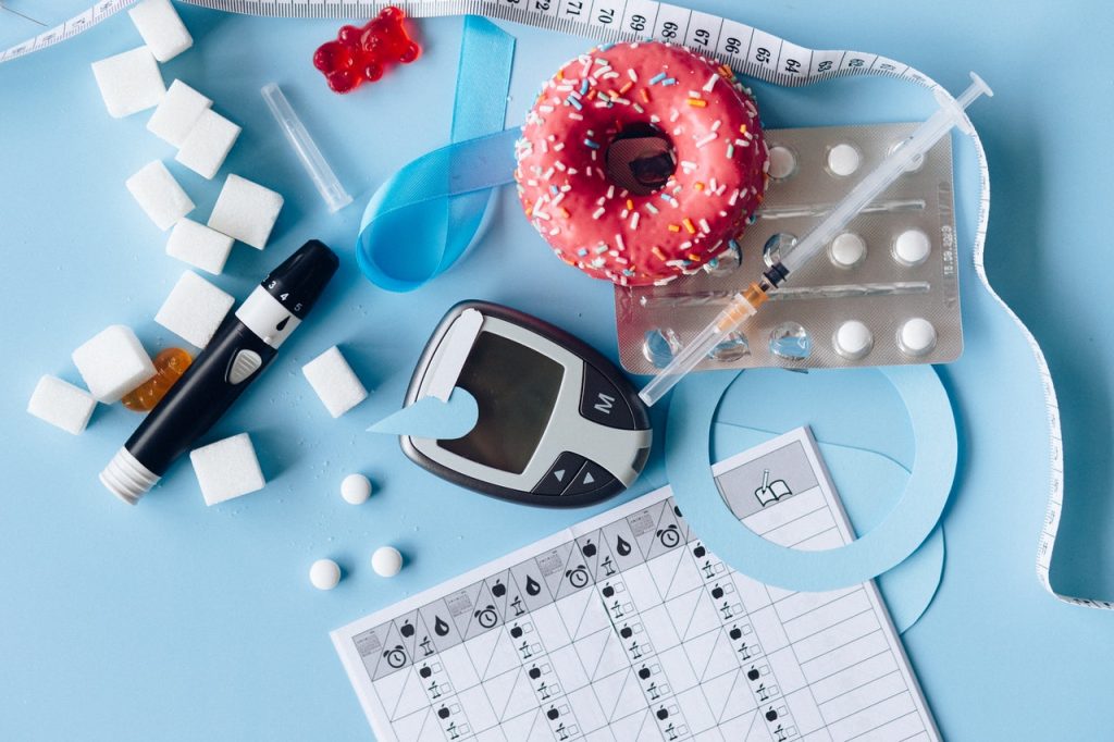
As global mobility surges, managing chronic conditions like diabetes during travel has become a significant concern. Diabetes remains one of the fastest-growing global public health issues1,affecting approximately 422 million people worldwide and causing 1.5 million deaths annually.2 International SOS, the world’s leading health and security services company, has reported a significant year-on-year increase in diabetes-related assistance cases over the past three years, with a 28% increase in 2022 and a 32% increase in 2023.

Meanwhile, year-to-date 2024 data indicates a further uptick.3 With World Diabetes Day approaching on 14 November, organisations are urged to support diabetes prevention and management strategies. This year’s theme, ‘Breaking Barriers, Bridging Gaps’4 highlights the need for equitable, comprehensive and affordable diabetes care.

Dr Katherine O’Reilly, Regional Medical Director at International SOS, emphasises the importance of comprehensive health strategies: “It is important for organisations to understand the unique challenges that employees with diabetes face, particularly when travelling. By recognising these specific needs, companies can provide the necessary support and resources to help their employees manage their condition effectively. This ensures that employees can maintain their health and productivity, even when they are on the go. With thoughtful planning and the right resources, organisations can help their employees navigate the complexities of diabetes, fostering a supportive and inclusive work environment.”

People with diabetes face a double burden: a higher risk of life-threatening conditions like heart attack, stroke, and kidney failure, compounded by the psychological toll of diabetes distress. Individuals with diabetes are two to three times more likely to experience depression compared to those without the condition.5 These challenges can significantly impact employee wellbeing, leading to increased absenteeism, reduced productivity, and higher healthcare costs for employers.

According to The International Diabetes Federation (IDF), the global healthcare costs for individuals living with diabetes are expected to exceed $1054 billion by 2045.6 Furthermore, the prevalence of diabetes is projected to rise, with 643 million people affected by 2030, and 783 million by 2045.With this rising prevalence, it is crucial for organisations to implement strategies that help their workforce manage and prevent this chronic condition. Minor adjustments can reduce absenteeism, increase productivity, concentration and energy levels, and reduce the chance of on-the-job injury.

Dr Katherine O’Reilly continues, “Early diagnosis is crucial. Raising awareness about diabetes symptoms can prompt people to get screened, enabling early detection and intervention to prevent or delay its onset.  This proactive approach can prevent undiagnosed diabetes from causing severe health complications, affecting various organ systems, including eye damage, heart and kidney disease, nerve damage and poor wound healing. By prioritising employee health, organisations can enhance productivity and foster a more engaged and resilient workforce. This approach also promotes a positive work environment and supports overall employee wellbeing.”


International SOS offers five tips for organisations to support employees in managing and preventing diabetes:

  1. Education and awareness: Increase awareness about diabetes symptoms to encourage early diagnosis and effective management, thereby preventing severe health complications.
  2. Provide comprehensive health solutions: Offer resources such as dietary guidelines, exercise programmes and regular health screenings to help employees manage their diabetes.
  3. Supportive culture and policies: Develop and implement policies allowing for flexible work schedules and access to medical care while travelling. Foster a culture that prioritises health and wellbeing by accommodating regular meals and exercise, and ensuring employees have time to rest and recover from travel.
  4. Promote a healthy lifestyle: Offer guidance on maintaining a healthy diet and regular exercise. Provide resources such as a list of healthy meal options and tips for finding nutritious food in different locations.
  5. Facilitate health monitoring and provide adjustments: Ensure employees have scheduled breaks to take medication, check blood sugar levels and eat regular meals. Provide a private space for insulin administration and other medical needs.

  1. Hossain, J., Al-Mamun, Islam, R. Health Science Reports | Diabetes mellitus, the fastest growing global public health concern: Early detection should be focused (2024)
  2. World Health Organization | Diabetes
  3. International SOS Assistance Tracker Data (2021 – 2024)
  4. World Health Organization | World Diabetes Day 2024
  5. CDC | Diabetes and Mental Health (2024)
  6. International Diabetes Federation | Diabetes Atlas Report 2021
  7. Parker ED, Lin J, Mahoney T, Ume N, Yang G, Gabbay RA, ElSayed NA, Bannuru RR. | Economic Costs of Diabetes in the U.S. in 2022. Diabetes Care (2024)

Bystander CPR up to 10 Minutes after Cardiac Arrest may Protect Brain Function

Photo by www.testen.no on Unsplash

The sooner a lay rescuer (bystander) starts cardiopulmonary resuscitation (CPR) on a person having a cardiac arrest at home or in public, up to 10 minutes after the arrest, the better the chances of saving the person’s life and protecting their brain function, according to preliminary research to be presented at the American Heart Association’s Resuscitation Science Symposium 2024.

Cardiac arrest, which occurs when the heart malfunctions and abruptly stops beating, is often fatal without quick medical attention such as CPR to increase blood flow to the heart and brain. More than 357 000 out-of-hospital cardiac arrests happen each year in the US, with a 9.3% survival rate. “Our findings reinforce that every second counts when starting bystander CPR and even a few minutes delay can make a big difference,” said first author Evan O’Keefe, MD, a cardiovascular fellow at Saint Luke’s Mid America Heart Institute and the University of Missouri-Kansas City. “If you see someone in need of CPR, don’t dwell on how long they’ve been down, your quick actions could save their life.”

The study analysed nearly 200 000 cases of witnessed out-of-hospital cardiac arrest to determine whether initiating CPR within different time windows, compared to outcomes with no bystander CPR administered, made a difference in survival and brain function after hospital discharge.

“We found that people who received bystander CPR within the first few minutes of their cardiac arrest were much more likely to survive and have better brain function than those who didn’t,” O’Keefe said. “The longer it took for CPR to start, the less survival benefit one received. However, even when CPR was started up to 10 minutes after cardiac arrest, there was still a significant survival benefit compared to individuals who did not receive CPR from a bystander.”

Results also found: 

  • People who received CPR within two minutes of out-of-hospital cardiac arrest had an 81% higher rate of survival to release from the hospital and 95% higher rate of surviving without significant brain damage compared to people who did not receive bystander CPR.
  • Even people who received bystander CPR up to 10 minutes after cardiac arrest were 19% more likely to survive to hospital discharge and 22% more likely to have a favorable neurological outcome than those who did not receive bystander CPR at all.
  • For those who did not receive bystander CPR, about 12% survived to be released from the hospital, and more than 9% survived without significant brain damage or major disabilities. When bystander CPR was initiated more than 10 minutes after cardiac arrest, bystander CPR, compared to not receiving the lifesaving assistance, was no longer associated with improved survival.

“These results highlight the critical importance of quick action in emergencies. It suggests that we need to focus on teaching more people how to perform CPR, and we also need to emphasise ways to get help to those suffering cardiac arrest faster,” O’Keefe said. “This might include more widespread CPR training programs, as well as better public access to automated external defibrillators (AEDs) and improved dispatch systems.”

O’Keefe noted that future research could explore how technology (like apps that alert nearby trained bystanders or alert dispatchers to likely cardiac arrest) may help to reduce the time to first intervention, information that could be important for emergency dispatchers and policymakers in the development of public interventions for cardiac arrest.

“This study highlights the need for prompt recognition and treatment of cardiac arrest by bystanders. Time is of the essence when a cardiac arrest occurs, and late interventions can be as ineffective as no intervention. Community education and empowerment are critical for us to save lives,” said American Heart Association volunteer expert Anezi Uzendu, MD, an interventional cardiologist at the University of Texas Southwestern Medical Center in Dallas and a cardiac arrest survivor.

A limitation of the study includes that the average time of arrival for emergency medical technicians (EMTs) to the person having cardiac arrest was roughly 10 minutes. This means that in this study, the people who received bystander CPR 10 minutes after their cardiac arrest were likely being compared to a group receiving professional medical attention.

Study details and background:

  • The study identified 160 822 witnessed out-of-hospital cardiac arrests that occurred from 2013-2022.  Among the people whose data was analysed, the average age was 64 years old and about 34% were women.
  • Researchers used data from the Cardiac Arrest Registry to Enhance Survival (CARES), a national, web-based health registry focused on helping communities improve care for and survival of out-of-hospital cardiac arrest.
  • The research categorized time to initiation of bystander CPR in two-minute intervals and analysed the link between each time interval, compared to the group who did not receive CPR, with survival to hospital discharge and favourable neurological survival, or surviving with minor disabilities.

Source: American Heart Association

Dogs and Owners Match Their Heart Rate Variability

Photo by Pauline Loroy on Unsplash

A study at the University of Jyväskylä showed that the heart rate variability of a dog and its owner adapt to each other during interaction. High heart rate variability is associated with relaxation, while low heart rate variability indicates stimulation. The study, published in Scientific Reports, deepens our understanding of the emotional connection between a dog and its owner, including its underlying physiological mechanisms.

Emotional connection enhances interaction in human relationships. Emotional synchronisation in the interaction between a child and a parent is essential for affective attachment. The relationship between a dog and its owner is also based on attachment, but little is known about its physiological mechanisms.

The heart rate variability of a dog and its owner adapt to each other

In a study conducted at the University of Jyväskylä, at the Department of Psychology and Jyväskylä Centre for Interdisciplinary Brain Research, it was found that the heart rate variability of a dog and its owner are interconnected during interaction. Heart rate variability, in other words, the variation in the heartbeat intervals, indicates the state of the autonomic nervous system. High heart rate variability is associated with a state of relaxation and recovery, while low heart rate variability indicates stimulation or strain, such as stress during an exam or sport performance. In this study, the owner’s high heart rate variability was connected to the dog’s high heart rate variability, and vice versa. In addition, the physical activity levels of a dog and its owner mutually adapted to each other during the study. 

Different connections for activity and heart rate variability

The connections of heart rate variability and activity levels between a dog and its owner were monitored during specific interaction tasks. Both heart rate and activity level were interconnected between dogs and their owners, but at different times. During free-form resting periods, the owner’s high heart rate variability was connected to the dog’s high heart rate variability. In other words, when the owner was relaxed the dog was also relaxed. Moreover, the owner’s and dog’s activity levels were similar during the given tasks, such as playing. Although it is known that physical activity has an impact on heart rate, the strongest connections of these variables between dog and owner were found in different situations and do not fully explain each other. This suggests that the connection in heart rate variability reflects the synchrony of emotional state rather than of activity levels.

“The interconnection in heart rate variability between the dog and its owner during resting periods may be explained by the fact that in those instances there were no external tasks, but the counterparts could react more to each other’s state in a natural way,” says Doctoral Researcher Aija Koskela. 

Owner’s temperament is connected to the dog’s heart rate variability

The study also investigated various background factors for the interconnection of the heart rate variability of the dog and its owner. Bigger dogs had higher heart rate variability. In addition, the dog’s high heart rate variability was also explained by the owner’s negative affectivity, a temperament trait that reflects the person’s tendency to become easily concerned about negative things. This type of owner tends to develop a strong emotional bond with the dog, and therefore the shepherd dogs of this study possibly had a higher sense of safety with these owners. 

The dog also influences the owner

A surprising finding in the study was that the owner’s heart rate variability was best explained by the dog’s heart rate variability, even though also the owner’s activity level and body mass index, which are known to impact heart rate, were taken into account in the analysis.

“We exceptionally investigated both a dog’s and its owner’s heart rate and activity level simultaneously, whereas previous studies have commonly focused either on the human’s or the dog’s perspective,” says the leader of the study, Academy Research Fellow Miiamaaria Kujala. “The challenging research setting gives a better opportunity to investigate interactive aspects.”

This study indicates that the emotional states of dogs and their owners as well as the reactions of their nervous system become partially adapted to each other during interaction. The same mechanisms that strengthen human affective attachment also seem to support the relationship between a dog and its owner. This study deepens our understanding about interaction between species and about the meaning of emotional connection between dogs and humans. 

Interaction research involved dogs bred for cooperation

The study was funded mainly by the Research Council of Finland and the Agria & Svenska Kennelklubben Research Fund. It involved altogether 30 voluntary dog owners with their dogs. The dogs represented breeds refined for cooperating with humans, such as sheep dogs and retrievers. The research findings are in line with previous studies, which have suggested that breeds selected for cooperation are particularly sensitive to react to their owners’ behaviour and personality traits. Next, the project will seek to shed light more specifically on the influential mechanisms involved in this phenomenon.

Source: University of Jyväskylä

Beta Blockers may Also Cause Depression for Cardiac Patients

Photo by Towfiqu Barbhuiya on Unsplash

Patients who have had a heart attack are typically treated using beta blockers. According to a Swedish study conducted earlier this year, this drug is unlikely to be needed for those heart patients who have a normal pumping ability. Now a sub-study at Uppsala University shows that there is also a risk that these patients will become depressed by the treatment.

“We found that beta blockers led to slightly higher levels of depression symptoms in patients who had had a heart attack but were not suffering from heart failure. At the same time, beta blockers have no life-sustaining function for this group of patients,” says Philip Leissner, a doctoral student in cardiac psychology and the study’s first author. The study was published in European Heart Journal Acute Cardiovascular Care.

Beta blockers are drugs that block the effects of adrenaline on the heart and have been used for decades as a basic treatment for all heart attack patients. In recent years, their importance has started to be questioned as new, successful treatments have begun to be developed. This is mainly the case for heart attack patients who do not suffer from heart failure.

The researchers wanted to look at the side effects of beta blockers, that is, whether they affect anxiety and depression levels. This is because older research and clinical experience suggests that beta blockers are linked to negative side effects such as depression, difficulty sleeping and nightmares.

Earlier this year, a major national study was conducted in Sweden, which found that those who received beta-blocking drugs were not protected from relapse or death compared to those who did not receive the drug. Leissner and his colleagues based their research on these findings and conducted a sub-study. It ran from 2018 to 2023 and involved 806 patients who had had a heart attack but no problems with heart failure. Half were given beta blockers and the other half were not. About 100 of the patients receiving beta blockers had been taking them since before the study, and the researchers observed more severe symptoms of depression in them.

“Most doctors used to give beta blockers even to patients without heart failure, but as the evidence in favour of doing so is no longer so strong, this should be reconsidered. We could see that some of these patients appear to be more at risk of depression. If the drug doesn’t make a difference to their heart, then they are taking it unnecessarily and at risk of becoming depressed,” adds Leissner.

Source: Uppsala University

Study Identifies Hip Implant Materials with Lowest Risk of Revision

Photo by DanR on Flickr

Hip implants with a delta ceramic or oxidised zirconium head and highly crosslinked polyethylene liner or cup had the lowest risk of revision during the 15 years after surgery, a new University of Bristol-led study has found. The research could help hospitals, surgeons and patients to choose what hip implant to use for replacement surgery.

The aim of the study, which appears in PLOS Medicine, was to establish hip implant materials at risk of revision. This would help orthopaedic surgeons, and patients, and to improve shared decision making before surgery by identifying hip implants with the lowest risk of revision.

The researchers analysed the UK’s National Joint Registry (NJR) data from 1 026 481 hip replacement patients carried out in the NHS and private sectors in England and Wales for up to 15 years after initial hip replacement operations (between 2003 to 2019). 

After reviewing hip implants from the NJR data, the research team found the risk of revision following a hip replacement is influenced by the type of material used in the bearing surface. Bearing surfaces are the moving parts of an artificial hip joint that glide against each other during activity.

The data indicated that hip implants with a delta ceramic or oxidised zirconium head and highly crosslinked polyethylene liner or cup had the lowest risk of revision throughout the 15 years following hip replacement surgery.

These findings were confirmed when the research team investigated the specific reasons for revision hip replacements being performed.  The data also showed 20 869 (2%) of hip replacement patients had to undergo revision after the initial surgery.

Senior author Dr Erik Lenguerrand, Senior Lecturer in Medical Statistics and Quantitative Epidemiologist in the Bristol Medical School: Translational Health Sciences (THS), said: “Our research has found the risk of hip replacement revision depends on the hip implant materials used in the original surgery.  The lowest risk of revision are from implants with delta ceramic or oxidised zirconium head and a highly crosslinked polyethylene (HCLPE) liner or cup.

“Further research is needed to find out the association of implant materials with the risk of rehospitalisation, re-operation other than revision, mortality and the cost-effectiveness of these materials.”

Michael Whitehouse, Professor of Trauma and Orthopaedics at Bristol Medical School: THS, and senior clinical lead for the paper, explained: “Our study has used data from one of the largest registries in the world that includes all public and private health care sectors in England and Wales. This means that the data is more generally applicable than that available previously, which was limited by broad groupings of implant types or much smaller study size. It highlights the importance of considering the whole structure that is created when implants are put together to make up a hip replacement rather than focusing on individual components.

“Our findings will help hospitals, surgeons and patients to choose hip implants and combinations of them with the lowest risk of revision following an initial hip replacement operation.”

Tim Wilton, Medical Director of the National Joint Registry (NJR), added: “We are always delighted when the data from the NJR can be used by researchers to produce important research of this kind which gives meaningful analysis to guide surgeons and patients in their decisions. An important value of the NJR data is that it allows researchers a unique insight to assess the long-term performance of different hip implant materials.

“By tracking the combinations of materials used and subsequent revision rates, this research highlights the role of implant material choice in surgical outcomes. This ensures that the materials used can be optimised for longevity and patient health. Surgeons would be well advised to study these findings carefully in relation to the implant choices they make, and to use the information in pre-operative discussions with their patients. As the demand for joint replacements continues to rise, this insight can be invaluable in reducing revision surgery.”

The research was not a randomised controlled trial and therefore it was not possible to control all factors that can influence the risk of revision.

The categorisation of hip implants used as part of hip replacements is often broad in national joint replacement surgery registries and does not fully show differences in revision risks associated within the different types of implant materials grouped together.

Source: University of Bristol

Gut Health Signals could Transform Arthritis Treatment

Gut Microbiome. Credit Darryl Leja National Human Genome Research Institute National Institutes Of Health

Changes in the gut microbiome before rheumatoid arthritis is developed could provide a window of opportunity for preventative treatments, new research suggests.

Bacteria associated with inflammation is found in the gut in higher amounts roughly 10 months before patients develop clinical rheumatoid arthritis, according to a longitudinal study by researchers at the University of Leeds. 

This new research might give us a major opportunity to act sooner to prevent rheumatoid arthritis.

Dr Christopher Rooney, Leeds Institute of Medical Research

Previous research has linked rheumatoid arthritis to the gut microbiome, which is the ecosystem of microbes in your intestines. But this new study, published in the Annals of the Rheumatic Diseases, reveals a potential intervention point. 

Lead researcher Dr Christopher Rooney, NIHR Academic Clinical Lecturer at the University of Leeds and Leeds Teaching Hospitals NHS Trust, said: “Patients at risk of rheumatoid arthritis are already experiencing symptoms such as fatigue and joint pain, and they may know someone in their family who has developed the disease. As there is no known cure, at-risk patients often feel a sense of hopelessness, or even avoid getting tested.  

“This new research might give us a major opportunity to act sooner to prevent rheumatoid arthritis.” 

Major opportunity for treatment

Funded by Versus Arthritis, the longitudinal study was conducted on 19 patients at risk of rheumatoid arthritis, with samples taken five times during a 15-month period.  

Five of these patients progressed to clinical arthritis, and the research showed they had gut instability with higher amounts of bacteria including Prevotella, which is associated with rheumatoid arthritis, about ten months before progression. The remaining 14, whose disease didn’t progress, had largely stable amounts of bacteria in their gut. 

Potential treatments that the researchers want to test at the 10-month window include changes to diet like eating more fibre, taking prebiotics or probiotics, and improving dental hygiene to keep harmful bacteria from periodontal disease away from the gut. 

The exact relationship between gut inflammation and rheumatoid arthritis development remains unclear. In a small number of patients within the study, the gut changes occurred before there were any changes to the joints observed by a rheumatologist, but more research is needed to determine whether these influence each other. 

Although bacteria is associated with rheumatoid arthritis, the researchers want to make it clear that there is no evidence this is contagious. 

Lucy Donaldson, director for research and health intelligence at Versus Arthritis, said: “At Versus Arthritis, we welcome the findings of this study which could give the clinicians of the future a crucial window of opportunity to delay – or even prevent – the onset of rheumatoid arthritis. This success is testament to the dedication of UK researchers who are working to personalise treatment and prevent chronic conditions that have significant impacts on a person’s ability to work, raise families and live independently.” 

The study initially took data from 124 individuals who had high levels of CCP+, an antibody that attacks healthy cells in the blood, which indicates risk of developing rheumatoid arthritis. The researchers compared their samples to 22 healthy individuals and seven people who had a new rheumatoid arthritis diagnosis.  

The findings from this larger group showed that the gut microbiome was less diverse in the at-risk group, compared to the healthy control group. 

The longitudinal study, which took samples from 19 patients over 15 months, revealed the changes in bacteria at ten months before progression to rheumatoid arthritis. 

The Leeds research team will now carry out an analysis of treatments that have already been trialled, to inform future testing of treatments at this potential 10-month intervention point. 

Source: University of Leeds