Day: November 19, 2024

Genetically Tailored Diets for IBS may Soon be Possible

Irritable bowel syndrome. Credit: Scientific Animations CC4.0

An international study has found that genetic variations in human carbohydrate-active enzymes may affect how people with irritable bowel syndrome (IBS) respond to a carbohydrate-reduced diet.

The research, which is published in Clinical Gastroenterology & Hepatologyshows that IBS patients with genetic defects in carbohydrate digestion had a better response to certain dietary interventions. This could lead to tailored treatments for IBS, using genetic markers to predict which patients benefit from specific diets.

Irritable bowel syndrome (IBS) is a digestive disorder affecting up to 10% of the global population. It is characterised by abdominal pain, bloating, diarrhoea, or constipation. Despite its prevalence, treating IBS remains a challenge as symptoms and responses to dietary or pharmacological interventions vary significantly.

Patients often connect their symptoms to eating certain foods, especially carbohydrates, and dietary elimination or reduction has emerged as an effective treatment option, though not all patients experience the same benefits.

Nutrigenetics (the science investigating the combined action of our genes and nutrition on human health) has highlighted how changes in the DNA can affect the way we process food. A well-known example is lactose intolerance, where the loss of function in the lactase enzyme hinders the digestion of dairy products.

Now, this pioneering new study suggests that genetic variations in human carbohydrate-active enzymes (hCAZymes) may similarly affect how IBS patients respond to a carbohydrate-reduced (low-FODMAP) diet.

The team have now revealed that individuals with hypomorphic (defective) variants in hCAZyme genes are more likely to benefit from a carbohydrate-reduced diet.

The study, involving 250 IBS patients, compared two treatments: a diet low in fermentable carbohydrates (FODMAPs) and the antispasmodic medication otilonium bromide. Strikingly, of the 196 patients on the diet, those carrying defective hCAZyme genes showed marked improvement compared to non-carriers, and the effect was particularly pronounced in patients with diarrhoea-predominant IBS (IBS-D), who were six times more likely to respond to the diet. In contrast, this difference was not observed in patients receiving medication, underscoring the specificity of genetic predisposition in dietary treatment efficacy.

These findings suggest that genetic variations in hCAZyme enzymes, which play a key role in digesting carbohydrates, could become critical markers for designing personalised dietary treatments for IBS. The ability to predict which patients respond best to a carbohydrate-reduced diet has the potential to strongly impact IBS management, leading to better adherence and improved outcomes.

Study leader Dr D’Amato, Gastrointestinal Genetics Research group at CIC bioGUNE and the Department of Medicine and Surgery at LUM University in in Italy.

In the future, incorporating knowledge of hCAZyme genotype into clinical practice could enable clinicians to identify in advance which patients are most likely to benefit from specific dietary interventions. This would not only avoid unnecessary restrictive diets for those unlikely to benefit but also open the door to personalised medicine in IBS.

Source: University of Nottingham

New Minimally Invasive Neural Interface is a Revolutionary Development

The researchers’ experiments showed that the catheter electrodes could be successfully delivered and guided into the ventricular spaces and brain surface for electrical stimulation. Image courtesy of Rice University.

A team of researchers has developed a technique for diagnosing, managing and treating neurological disorders with minimal surgical risks. The team’s findings were published in Nature Biomedical Engineering.

While traditional approaches for interfacing with the nervous system often require creating a hole in the skull to interface with the brain, the researchers have developed an innovative method known as endocisternal interfaces (ECI), allowing for electrical recording and stimulation of neural structures, including the brain and spinal cord, through cerebral spinal fluid (CSF).

“Using ECI, we can access multiple brain and spinal cord structures simultaneously without ever opening up the skull, reducing the risk of complications associated with traditional surgical techniques,” said study leader Robinson Jacob Robinson, professor of electrical and computer engineering and bioengineering at Rice University.

ECI uses CSF, which surrounds the nervous system, as a pathway to deliver targeted devices. By performing a simple lumbar puncture in the lower back, researchers can navigate a flexible catheter to access the brain and spinal cord.

Using miniature magnetoelectric-powered bioelectronics, the entire wireless system can be deployed through a small percutaneous procedure. The flexible catheter electrodes can be navigated freely from the spinal subarachnoid space to the brain ventricles.

“This is the first reported technique that enables a neural interface to simultaneously access the brain and spinal cord through a simple and minimally invasive lumbar puncture,” said University of Texas Medical Branch’s Peter Kan, professor and Chair of Neurosurgery, who also led the study. “It introduces new possibilities for therapies in stroke rehabilitation, epilepsy monitoring and other neurological applications.”

To test the hypothesis, the research team characterised the endocisternal space and measured the width of the subarachnoid, or fluid-filled space, in human patients using MRI. The researchers then conducted experiments in large animal models, specifically sheep, to validate the feasibility of the new neural interface.

Their experiments showed that the catheter electrodes could be successfully delivered and guided into the ventricular spaces and brain surface for electrical stimulation. By using the magnetoelectric implant, the researchers were able to record electrophysiologic signals such as muscle activation and spinal cord potentials.

Preliminary safety results showed that the ECI remained functional with minimal damage up to 30 days after the electronic device was implanted chronically into the brain.

Moreover, the study revealed that unlike endovascular neural interfaces that require antithrombotic medication and are limited by the small size and location of blood vessels, ECI offers broader access to neural targets without the medication.

“This technology creates a new paradigm for minimally invasive neural interfaces and could lower the risk of implantable neurotechnologies, enabling access to wider patient populations,” said Josh Chen, lead author of the study.

Source: Rice University

Adding Vitamin C to Chemotherapy Doubles Pancreatic Cancer Survival Time

Pancreatic cancer. Credit: Scientific Animations CC BY-SA 4.0

Results from a randomised, phase 2 clinical trial show that adding high-dose, intravenous (IV) vitamin C to chemotherapy doubles the overall survival of patients with late-stage metastatic pancreatic cancer from eight months to 16 months. 

“This is a deadly disease with very poor outcomes for patients. The median survival is eight months with treatment, probably less without treatment, and the five-year survival is tiny,” says Joe Cullen, MD, University of Iowa professor of surgery, and radiation oncology, and senior author of the study. “When we started the trial, we thought it would be a success if we got to 12 months survival, but we doubled overall survival to 16 months. The results were so strong in showing the benefit of this therapy for patient survival that we were able to stop the trial early.” 

The findings, published in Redox Biology, mark another success for high-dose, intravenous vitamin C, which has overcome many hurdles in the almost 20 years UI researchers have persevered to demonstrate its benefit for cancer patients. 

“We’ve had ups and downs of course, but this is a culmination of a lot of people’s hard work,” says Cullen who also is a member of UI Health Care Holden Comprehensive Cancer Center. “It’s really a positive thing for patients and for the University of Iowa.”

Increased survival, improved quality of life

In the study, 34 patients with stage 4 metastatic pancreatic cancer were randomized to receive either standard chemotherapy (gemcitabine and nab-paclitaxel), or the chemotherapy plus infusions of high-dose vitamin C. The results showed that average overall survival was 16 months for the patients receiving the chemotherapy plus vitamin C, compared to eight months for the patients getting just chemotherapy. In addition, progression free survival was extended from four months to six months. 

“Not only does it increase overall survival, but the patients seem to feel better with the treatment,” Cullen says. “They have less side effects, and appear to be able to tolerate more treatment, and we’ve seen that in other trials, too.” 

The new study is not the only evidence of the benefit of including IV vitamin C as part of cancer treatment. Earlier this year, the results of another UI phase 2 clinical trial in patients with glioblastoma, a deadly form of brain cancer, were published. That study also showed a significant increase in survival when high-dose, IV vitamin C was added to standard of care chemotherapy and radiation. Cullen was also part of that trial along with his colleague Bryan Allen, MD, PhD, UI professor and head of radiation oncology. 

A third phase 2 trial in non-small cell lung cancer is still underway, with results expected within the year. All three trials were funded by a 2018 grant from the National Cancer Institute (NCI)

“This NCI funding was incredibly important for us to conduct these phase 2 trials and obtain these really encouraging results. Our aim is to show that adding high-dose, IV vitamin C, which is very inexpensive and very well tolerated, can improve treatment for these cancers that are among the deadliest affecting the U.S. population,” Cullen adds. 

A long journey to clinical trials

Cullen, Allen, and their colleagues at UI Health Care have been researching the anti-cancer effect of high-dose, IV vitamin C for decades. Their work revealed a critical difference between intravenous and oral vitamin C. Intravenous vitamin C administration produces very high levels in the blood, which cannot be achieved with oral delivery. These high concentrations result in unique chemical reactions within cancer cells that render the cell more vulnerable to chemo- and radiation therapies. 

Cullen notes that despite scepticism towards vitamin C as a cancer therapy, the results he and his colleagues have obtained, from basic science findings to understand the biological mechanisms at work, through the various clinical trials, have been highly encouraging and robust. 

“Through every step of the process, it continued to improve. We did it in cells, it worked great. We did it in mice, it worked great. Then our phase one trials looked very promising. So, the progression has just been phenomenal, really,” Cullen says. “For example, in one of our phase 1 trials for pancreatic cancer, where we combine high-dose, IV vitamin C with radiation, we still have three long-term survivors. They’re out nine years at this point, which is far beyond the typical survival range.” 

Source: University of Iowa Health Care

A Multiple Sclerosis Drug may Help with Poor Working Memory

This is a pseudo-coloured image of high-resolution gradient-echo MRI scan of a fixed cerebral hemisphere from a person with multiple sclerosis. Credit: Govind Bhagavatheeshwaran, Daniel Reich, National Institute of Neurological Disorders and Stroke, National Institutes of Health

Fampridine is currently used to improve walking ability in multiple sclerosis. A new study shows that it could also help individuals with reduced working memory, as seen in mental health conditions like schizophrenia or depression.

Working memory allows a memory to be actively retained for a few seconds, for cognitive tasks such as remembering an email address to save it, or participating in a conversation. Certain conditions, such as schizophrenia or depression, as well as ADHD, impair working memory. Those affected lose track in conversations and struggle to organise their thoughts.

Fampridine is a drug that could help in such cases, as shown in a study led by Professors Andreas Papassotiropoulos and Dominique de Quervain at the University of Basel. The team has reported their findings in the journal Molecular Psychiatry.

Effective only if working memory is poor

In their study, the researchers tested the effectiveness of fampridine on working memory in 43 healthy adults. It was in those participants whose baseline working memory was at a low level that fampridine showed a more pronounced effect: after taking the active substance for three days, they scored better in the relevant tests than those who took the placebo. In contrast, in people who already had good baseline working memory, the drug showed no effect.

The researchers also observed that fampridine increased brain excitability in all participants, thus enabling faster processing of stimuli. The study was randomized and double-blind.

Established drug, new application

“Fampridine doesn’t improve working memory in everyone. But it could be a treatment option for those with reduced working memory,” explains Andreas Papassotiropoulos. Dominique de Quervain adds: “That’s why, together with researchers from the University Psychiatric Clinics Basel (UPK), we’re planning studies to test the efficacy of fampridine in schizophrenia and depression.”

The drug is currently used to improve walking ability in multiple sclerosis (MS). Particularly in capsule form, which releases the active ingredient slowly in the body, fampridine has shown effects on cognitive performance in MS patients: for some, it alleviates the mental fatigue that can accompany MS.

The researchers did not select the drug at random: this study followed comprehensive analyses of genome data in order to find starting points for repurposing established drugs. Fampridine acts on specific ion channels in nerve cells that, according to the researchers’ analyses, also play a role in mental disorders such as schizophrenia.

Source: University of Basel

A Link between Heart Shape and Cardiovascular Disease Risk

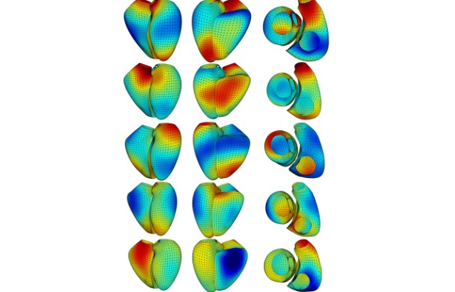
Researchers from Queen Mary University of London and other universities have for the first time examined the genetic basis of the heart’s left and right ventricles using advanced 3D imaging and machine learning.

Prior research primarily focused on the heart’s size and volume and specific chambers. By studying both ventricles together, the team was able to capture the more intricate, multi-dimensional aspects of the heart shape.

This new approach of exploring shape has led to the discovery of new heart-associated genes and provided a better understanding of the biological pathways linking heart shape to cardiovascular disease.

Cardiovascular disease is among the leading causes of death in the UK and globally. The findings of this study could change how cardiac disease risk is evaluated. Genetic information related to heart shape can provide a risk score for heart disease, offering potentially early and more tailored assessment in clinical settings.

“This study provides new information on how we think about heart disease risk,” said Patricia B. Munroe, Professor of Molecular Medicine at Queen Mary and co-author of the study. “We’ve long known that size and volume of the heart matter, but by examining shape, we’re uncovering new insights into genetic risks. This discovery could provide valuable additional tools for clinicians to predict disease earlier and with more precision.”

The team used cardiovascular MRI images from over 40 000 individuals from the UK Biobank to create 3D models of the ventricles. Through statistical analysis, they identified 11 shape dimensions that describe the primary variations in heart shape.

Subsequent genetic analysis found 45 specific areas in the human genome linked to different heart shapes. Fourteen of these areas had not been previously known to influence heart traits.

“This study sets an important foundation for the exploration of genetics in both ventricles”, said Dr Richard Burns, Statistical Geneticist at Queen Mary. “The study confirms that combined cardiac shape is influenced by genetics, and demonstrates the usefulness of cardiac shape analysis in both ventricles for predicting individual risk of cardiometabolic diseases alongside established clinical measures.”

This research marks an exciting new chapter in understanding how genetics influence the heart and opens the doors to further studies on how these findings could be integrated into clinical practice, ultimately benefiting millions at risk of heart disease.

Source: Queen Mary University of London