Day: October 24, 2024

Aerobic Exercise may Help Prevent the Brain Fog from Chemotherapy

Clinical trial reveals improved self-reported cognitive function in women with breast cancer who started an exercise program when initiating chemotherapy.

Photo by Ketut Subiyanto on Pexels

Many women who receive chemotherapy experience a decreased ability to remember, concentrate, and/or think – commonly referred to as “chemo-brain” or “brain fog” – both short- and long-term. In a recent clinical trial of women initiating chemotherapy for breast cancer, those who simultaneously started an aerobic exercise program self-reported greater improvements in cognitive function and quality of life compared with those receiving standard care. The findings are published by Wiley online in CANCER, a peer-reviewed journal of the American Cancer Society.

The study, called the Aerobic exercise and CogniTIVe functioning in women with breAsT cancEr (ACTIVATE) trial, included 57 Canadian women in Ottawa and Vancouver who were diagnosed with stage I–III breast cancer and beginning chemotherapy. All women participated in 12–24 weeks of aerobic exercise: 28 started this exercise when initiating chemotherapy and 29 started after chemotherapy completion. Cognitive function assessments were conducted before chemotherapy initiation and after chemotherapy completion (therefore, before the latter group started the exercise program).

Women who participated in the aerobic exercise program during chemotherapy self-reported better cognitive functioning and felt their mental abilities improved compared with those who received standard care without exercise. Neuropsychological testing – a performance-based method used to measure a range of mental functions – revealed similar cognitive performance in the two groups after chemotherapy completion, however.

“Our findings strengthen the case for making exercise assessment, recommendation, and referral a routine part of cancer care; this may help empower women living with and beyond cancer to actively manage both their physical and mental health during and after treatment,” said lead author Jennifer Brunet, PhD, of the University of Ottawa.

Dr Brunet noted that many women undergoing chemotherapy for breast cancer remain insufficiently active, and there are limited exercise programs tailored to their needs. “To address this, we advocate for collaboration across various sectors – academic, healthcare, fitness, and community – to develop exercise programs specifically designed for women with breast cancer,” she said. “These programs should be easy to adopt and implement widely, helping to make the benefits of exercise more accessible to all women facing the challenges of cancer treatment and recovery.”

Source: Wiley

How Muscle Energy Production is Impaired in Type 2 Diabetes

Mitochondria (red) are organelles found in most cells. They generate a cell’s chemical energy. Credit: NICHD/U. Manor

A new study from Karolinska Institutet, published in Science Translational Medicine, shows that people with type 2 diabetes have lower levels of the protein that breaks down and converts creatine in the muscles. This leads to impaired function of the mitochondria, the ‘powerhouses’ of the cell.

Creatine is a popular supplement for improving exercise performance as it can make muscles work harder and longer before they become fatigued. Previous studies however showed a possible link between high blood creatine levels and increased type 2 diabetes risk. This has raised questions about whether creatine supplementation may contribute to that risk.

New research based on studies in both humans and mice shows that people with type 2 diabetes have lower protein levels in their muscles that metabolises and converts creatine – a protein called creatine kinase. 

“This reduced protein level leads to impaired creatine metabolism in the muscle. This may explain why people with type 2 diabetes accumulate creatine in their blood,” says principal investigator Anna Krook, Professor at the Department of Physiology and Pharmacology at Karolinska Institutet.

Scientists don’t know exactly what high creatine levels in the blood mean for the body, but it is known that it does have an effect outside the cells. 

“The findings indicate that impaired creatine metabolism is a consequence of type 2 diabetes, rather than a cause of the disease,” says Anna Krook. 

Impairs mitochondrial function

The study also shows that low levels of creatine kinase are not only linked to higher creatine levels in the blood, but also impair the function of mitochondria in the muscle. Mitochondria, which convert nutrients into energy, function less well in muscle cells with reduced creatine kinase, leading to both lower energy production and increased cell stress.

“This is quite consistent with the fact that people with type 2 diabetes have poorer energy metabolism. In the future, one possibility could be to regulate creatine kinase as part of the treatment of metabolic diseases such as obesity and diabetes,” says Anna Krook.

An unexpected finding of the study was that changes in creatine kinase levels affected the appearance of mitochondria and also their ability to produce energy, regardless of the amount of creatine available. 

“This suggests that although the main role of creatine kinase is to process creatine, it affects mitochondrial function in other ways,” explains David Rizo-Roca, the study’s first author.

“Our next step is to find the molecular mechanisms behind these effects,” he says. 

Source: Karolinska Institutet

Immunotherapy Blocks Scarring, Improves Cardiac Function in Heart Failure

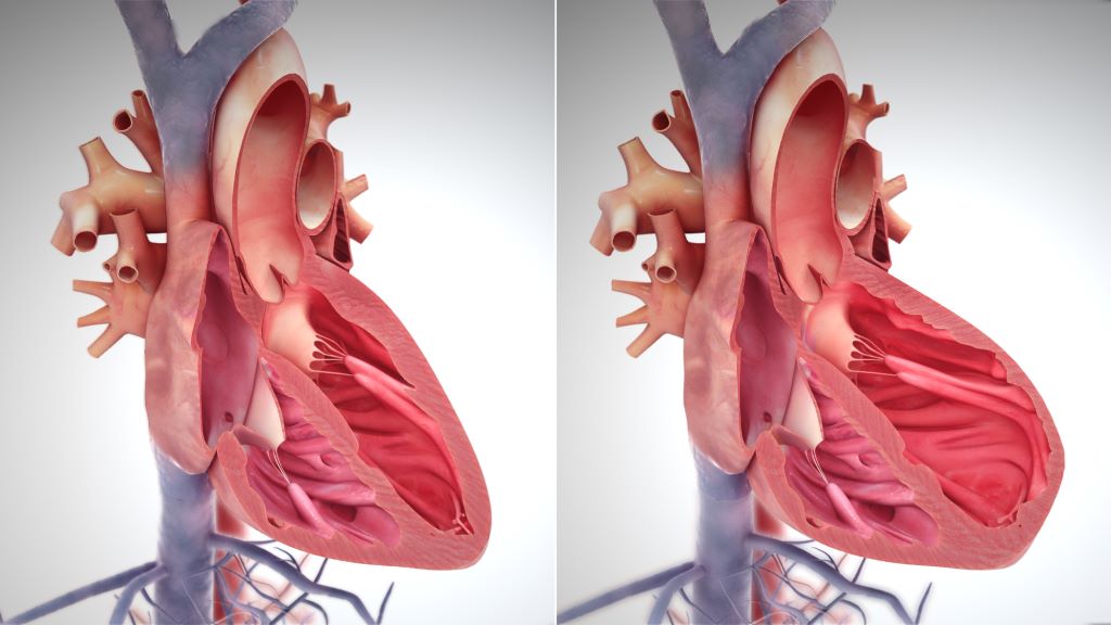
Right side heart failure. Credit: Scientific Animations CC4.0

A new study from Washington University School of Medicine in St. Louis suggests that a type of immunotherapy also may be an effective treatment strategy for heart failure by using an FDA-approved drug to block the signalling protein IL-1 beta. The study is published in Nature.

After a heart attack, viral infection or other injury to the heart, scar tissue often forms in the heart muscle, where it interferes with the heart’s normal contractions and plays a leading role in heart failure, a chronic condition which can only be slowed, not cured.

Studying human tissue samples as part of the new study, the researchers identified a type of fibroblast cell in the heart as the main culprit responsible for the formation of scar tissue in heart failure. To see if they could prevent scar formation, the scientists turned to mouse models of heart failure that have the very same type of fibroblasts. They used a therapeutic monoclonal antibody that blocks the formation of this harmful type of fibroblast, and succeeded in reducing the formation of scar tissue and improving heart function in the mice.

“After scar tissue forms in the heart, its ability to recover is dramatically impaired or impossible,” said cardiologist and senior author Kory Lavine, MD, PhD, a professor of medicine in the Cardiovascular Division at WashU Medicine. “Heart failure is a growing problem in the US and globally, affecting millions of people. Current treatments can help relieve symptoms and slow the progression, but there is a tremendous need for better therapies that actually stop the disease process and prevent the formation of new scar tissue that causes a loss of heart function. We are hopeful our study will lead to clinical trials investigating this immunotherapy strategy in heart failure patients.”

Fibroblasts have many roles in the heart, and parsing out the differences between various populations of these cells has been challenging. Some types of fibroblasts support the heart’s structural integrity and maintain good blood flow through the heart’s blood vessels, while others are responsible for driving inflammation and the development of scar tissue. Only recently, with the wide availability of the most advanced single cell sequencing technologies, could scientists peg which groups of cells are which.

“These various types of fibroblasts highlight newly recognised opportunities to craft treatment strategies that specifically block the type of fibroblasts that promote scarring and protect fibroblasts that maintain the structure of the heart, so the heart doesn’t rupture,” Lavine said. “Our research suggests that the fibroblasts that promote scarring in the injured heart are very similar to fibroblasts associated with cancer and other inflammatory processes. This opens the door to immunotherapies that potentially can stop the inflammation and resulting scar tissue.”

The research team, co-led by Junedh Amrute, a graduate student in Lavine’s lab, used genetic methods to demonstrate that a signaling molecule called IL-1 beta was important in a chain of events driving fibroblasts to create scar tissue in heart failure. With that in mind, they tested a mouse monoclonal antibody that blocks IL-1 beta and found beneficial effects in the mouse hearts. The mouse monoclonal antibody was provided by Amgen, whose scientists were also co-authors of the study. Monoclonal antibodies are proteins manufactured in the lab that modulate the immune system. The treatment reduced the formation of scar tissue and improved the pumping capacity of the mouse hearts, as measured on an echocardiogram.

At least two FDA-approved monoclonal antibodies, canakinumab and rilonacept, can block IL-1 signalling. These immunotherapies are approved to treat inflammatory disorders such as juvenile idiopathic arthritis and recurrent pericarditis, which is inflammation of the sac surrounding the heart.

One of these antibodies also has been evaluated in a clinical trial for atherosclerosis, a buildup of plaque that hardens the arteries. The trial, called CANTOS (Canakinumab Anti-inflammatory Thrombosis Outcome Study), showed a benefit for study participants with atherosclerosis.

“Even though this trial was not designed to test this treatment in heart failure, there are hints in the data that the monoclonal antibody might be beneficial for patients with heart failure,” Lavine said. “Secondary analyses of the data from this trial showed that the treatment was associated with a sizable reduction in heart failure admissions compared with standard care. Our new study may help explain why.”

Even so, the IL-1 antibody used in the CANTOS study had some side effects, such as increased risk of infection, that could perhaps be reduced with a more targeted antibody that specifically blocks IL-1 signaling in cardiac fibroblasts, according to the researchers.

“We are hopeful that the combination of all of this evidence, including our work on the IL-1 beta pathway, will lead to the design of a clinical trial to specifically test the role of targeted immunotherapy in heart failure patients,” Lavine said.

Source: Washington University School of Medicine

New Discovery Explains How SARS-CoV-2 Evades Anti-viral Immunity

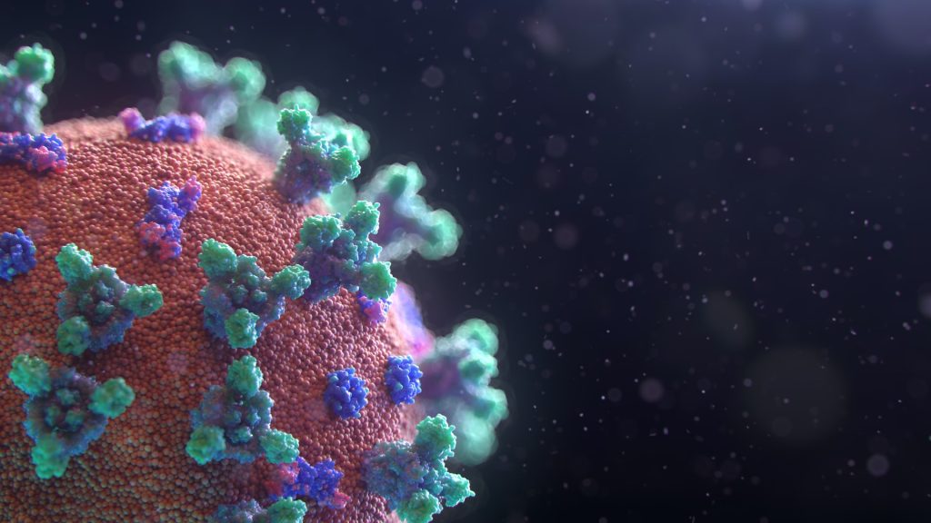
Image by Fusion Medical on Unsplash

The novel coronavirus SARS-CoV-2 has an enzyme that can counteract a cell’s innate defence mechanism against viruses, explaining why it is more infectious than the previous SARS and MERS-causing viruses. This discovery, from Kobe University, may point the way to the development of more effective drugs against this and possibly similar, future diseases.

When a virus attacks, the body’s immune response has two basic layers of defence: the innate and the adaptive immune systems. While the adaptive immune system grows stronger against a specific pathogen as the body is exposed to it multiple times and which forms the basis of vaccinations, the innate immune system is an assortment of molecular mechanisms that work against a broad range of pathogens at a basic level. The Kobe University virologist SHOJI Ikuo says, “The new coronavirus, however, is so infectious that we wondered what clever mechanisms the virus employs to evade the innate immune system so effectively.”

Shoji’s team previously worked on the immune response to hepatitis viruses and investigated the role of a molecular tag called “ISG15” the innate immune system attaches to the virus’s building blocks. Having learned that the novel coronavirus has an enzyme that is especially effective in removing this tag, he decided to use his team’s expertise to elucidate the effect of the ISG15 tag on the coronavirus and the mechanism of the virus’s countermeasures.

In a paper in the Journal of Virology, the Kobe University-led team is now the first to report that the ISG15 tag gets attached to a specific location on the virus’s nucleocapsid protein, the scaffold that packages the pathogen’s genetic material. For the virus to assemble, many copies of the nucleocapsid protein need to attach to each other, but the ISG15 tag prevents this, which is the mechanism behind the tag’s antiviral action. “However, the novel coronavirus also has an enzyme that can remove the tags from its nucleocapsid, recovering its ability to assemble new viruses and thus overcoming the innate immune response,” explains Shoji.

The novel coronavirus shares many traits with the SARS and MERS viruses, which all belong to the same family of viruses – which also have an enzyme that can remove the ISG15 tag. But their versions are less efficient at it than the one in the novel coronavirus, Shoji’s team found. And in fact, it has been reported recently that the previous viruses’ enzymes have a different primary target. “These results suggest that the novel coronavirus is simply better at evading this aspect of the innate immune system’s defence mechanism, which explains why it is so infectious,” says Shoji.

But understanding just why the novel coronavirus is so effective also points the way to developing more effective treatments. The Kobe University researcher explains: “We may be able to develop new antiviral drugs if we can inhibit the function of the viral enzyme that removes the ISG15 tag. Future therapeutic strategies may also include antiviral agents that directly target the nucleocapsid protein, or a combination of these two approaches.”

Source: Kobe University

Men and Women Use Different Biological Systems to Reduce Pain

Photo by Sasun Bughdaryan on Unsplash

In a new study evaluating meditation for chronic lower back pain, researchers at University of California San Diego School of Medicine have discovered that men and women utilise different biological systems to relieve pain. While men relieve pain by releasing endogenous opioids, the body’s natural painkillers, women rely instead on other, non-opioid based pathways. The study was published in PNAS Nexus.

Synthetic opioid drugs, such as morphine and fentanyl, are the most powerful class of painkilling drugs available. Women are known to respond poorly to opioid therapies, which use synthetic opioid molecules to bind to the same receptors as naturally-occurring endogenous opioids. This aspect of opioid drugs helps explain why they are so powerful as painkillers, but also why they carry a significant risk of dependence and addiction.

“Dependence develops because people start taking more opioids when their original dosage stops working,” said Fadel Zeidan, PhD, professor of anaesthesiology and Endowed Professor in Empathy and Compassion Research at UC San Diego Sanford Institute for Empathy and Compassion. “Although speculative, our findings suggest that maybe one reason that females are more likely to become addicted to opioids is that they’re biologically less responsive to them and need to take more to experience any pain relief.”

The study combined data from two clinical trials involving a total of 98 participants, including both healthy individuals and those diagnosed with chronic lower back pain. Participants underwent a meditation training program, then practiced meditation while receiving either placebo or a high-dose of naloxone, a drug that stops both synthetic and endogenous opioids from working. At the same time, they experienced a very painful but harmless heat stimulus to the back of the leg. The researchers measured and compared how much pain relief was experienced from meditation when the opioid system was blocked versus when it was intact.

The study found:

  • Blocking the opioid system with naloxone inhibited meditation-based pain relief in men, suggesting that men rely on endogenous opioids to reduce pain.
  • Naloxone increased meditation-based pain relief in women, suggesting that women rely on non-opioid mechanisms to reduce pain.
  • In both men and women, people with chronic pain experienced more pain relief from meditation than healthy participants.

“These results underscore the need for more sex-specific pain therapies, because many of the treatments we use don’t work nearly as well for women as they do for men,” said Zeidan.

The researchers conclude that by tailoring pain treatment to an individual’s sex, it may be possible to improve patient outcomes and reduce the reliance on and misuse of opioids.

“There are clear disparities in how pain is managed between men and women, but we haven’t seen a clear biological difference in the use of their endogenous systems before now,” said Zeidan. “This study provides the first clear evidence that sex-based differences in pain processing are real and need to be taken more seriously when developing and prescribing treatment for pain.”

Source: University of California – San Diego