Month: September 2024

Unhealthy Commodities – Like Alcohol and Social Media – are Connected with Poor Mental Health

Commercial determinants such as social media, air pollution associated with depression and suicide

Photo by Inzmam Khan

“Unhealthy commodities” such as tobacco, alcohol, ultra-processed foods, social media, and fossil fuels, as well as impacts of fossil fuel consumption such as climate change and air pollution are associated with depression, suicide, and self-harm, according to a study published August 28 by Kate Dun-Campbell from the London School of Hygiene & Tropical Medicine, and colleagues.

Globally, around one out of every eight people currently live with a mental health disorder. These disorders – including depression, suicide, anxiety, and other diseases and disorders – can have many underlying causes. Some of those causes could be related to commercial determinants of health – the ways in which commercial activities and commodities impact health and equity. Commercial determinants of health can be specifically unhealthy, such as alcohol or tobacco consumption, unhealthy food, and the use of fossil fuels. To further understand how these unhealthy commodities might impact mental health, the authors of this study performed an umbrella synthesis of 65 review studies examining connections between six specific commodities – tobacco, alcohol, ultra-processed foods, gambling, social media, and fossil fuels. The author also included studies looking at mental health impacts of fossil fuel use such as climate change and air pollution.

The umbrella review found evidence for links between depression and alcohol, tobacco, gambling, social media, ultra-processed foods and air pollution. Alcohol, tobacco, gambling, social media, climate change and air pollution were associated with suicide, and social media was also associated with self-harm. Climate change and air pollution were also linked to anxiety. The review brought together many different methodologies and measurements, and could not establish the underlying cause of the negative mental health outcomes. But the results indicate that unhealthy commodities should be considered when researchers attempt to understand and improve mental ill health. 

The authors add: “Our review highlights that there is already compelling evidence of the negative impact of unhealthy products on mental health, despite key gaps in understanding the impact of broader commercial practices.”

The study was published in PLOS Global Health.

Provided by PLOS

AI-enabled ‘Digital Stethoscope’ can Diagnose Peripartum Cardiomyopathy Twice as Often

Source: CC0

New research from Mayo Clinic suggests that artificial intelligence (AI) could improve the diagnosis of peripartum cardiomyopathy, a potentially life-threatening and treatable condition that weakens the heart muscle of women during pregnancy or in the months after giving birth. Researchers used an AI-enabled digital stethoscope that captures electrocardiogram (ECG) data and heart sounds to identify twice as many cases of peripartum cardiomyopathy as compared to regular care, according to a news release from the American Heart Association.

Identifying a weak heart pump caused by pregnancy is important because the symptoms, such as shortness of breath when lying down, swelling of hands and feet, weight gain, and rapid heartbeat, can be confused with normal symptoms of pregnancy.

Dr Demilade Adedinsewo, a cardiologist at Mayo Clinic, shared research insights during a late-breaking science presentation at the American Heart Association’s Scientific Sessions 2023.

Women in Nigeria have the highest reported incidence of peripartum cardiomyopathy. The randomised pragmatic clinical trial enrolled 1195 women receiving pregnancy care in Nigeria. Approximately half were evaluated with AI-guided screening using the digital stethoscope, and half received usual obstetric care in addition to a clinical ECG. An echocardiogram was used to confirm when the AI-enabled digital stethoscope predicted peripartum cardiomyopathy. Overall, 4% of the pregnant and postpartum women in the intervention arm of the clinical trial had cardiomyopathy compared to 2% in the control arm, suggesting that half are likely undetected with usual care.

Watch: Dr Adedinsewo explains the red flags for heart failure during pregnancy

Source: Mayo Clinic

White Matter may Aid Recovery from Spinal Cord Injuries

View of the spinal cord. Credit: Scientific Animations CC4.0

Injuries, infection and inflammatory diseases that damage the spinal cord can lead to intractable pain and disability but some degree of recovery may be possible. The question is, how best to stimulate the regrowth and healing of damaged nerves.

At the Vanderbilt University Institute of Imaging Science (VUIIS), scientists are focusing on a previously understudied part of the brain and spinal cord – white matter, which is made up of axons that relay signals. Their discoveries could lead to treatments that restore nerve activity through the targeted delivery of electromagnetic stimuli or drugs.

In a recent paper published in the Proceedings of the National Academy of SciencesAnirban Sengupta, PhD, John Gore, PhD, and their colleagues report the detection of signals from white matter in the spinal cord in response to a stimulus that are as robust as grey matter signals.

“In the spinal cord, the white matter signal is quite large and detectable, unlike in the brain, where it has less amplitude than the grey matter (signal),” said Sengupta, research instructor in Radiology and Radiological Sciences at Vanderbilt University Medical Center.

“This may be due to the larger volume of white matter in the spinal cord compared to the brain,” he added. Alternatively, the signal could represent “an intrinsic demand” in metabolism within the white matter, reflecting its critical role in supporting grey matter.

For several years, Gore, who directs the VUIIS, and his colleagues have used functional magnetic resonance imaging (fMRI) to detect blood oxygenation-level dependent (BOLD) signals, a key marker of nervous system activity, in white matter.

Last year, they reported that when participants undergoing fMRI perform a task, like wiggling their fingers, BOLD signals increase in white matter throughout the brain.

The current study monitored changes in BOLD signals in the white matter of the spinal cord at rest and in response to a vibrotactile stimulus applied to the fingers in an animal model. In response to stimulation, white matter activity was higher in “tracts” of ascending fibres that carry the signal from the spine to the brain.

This result is consistent with white matter’s known neurobiological function, the researchers noted. White matter contains non-neuronal glial cells that do not produce electrical impulses, but which regulate blood flow and neurotransmitters, the signaling molecules that transmit signals between nerve cells.

Much remains to be learned about the function of white matter in the spinal cord. But the findings from this research may help in improved understanding of diseases that affect white matter in the spinal cord, including multiple sclerosis, Sengupta said.

“We will be able to see how activity in the white matter changes in different stages of the disease,” he said. Researchers also may be able to monitor the effectiveness of therapeutic interventions, including neuromodulation, in promoting recovery following spinal cord injury.

Source: Vanderbilt University Medical Center

Inflammation Leaves a Long-lasting Impression on Intestinal Stem Cells

Irritable bowel syndrome. Credit: Scientific Animations CC4.0

Researchers at Baylor College of Medicine, the University of Michigan and collaborating institutions have discovered that inflammation in the gut leaves long-term marks on intestinal stem cells (ISCs) that reduce their ability to heal the intestine, even after inflammation has receded. This is important because it affects ISCs’ response to future challenges. The study appeared in Cell Stem Cell.

“We study graft-vs-host disease (GVHD), a major cause of mortality after bone marrow transplantation, a potentially curative therapy for many blood diseases. One of our goals is to better understand GVHD and identify strategies to control it,” said corresponding author Dr Pavan Reddy, professor and director of the Dan L Duncan Comprehensive Cancer Center at Baylor and previously at the University of Michigan.

“GVHD is an inflammatory reaction in which immune T cells from the bone marrow transplant donor attack the host gut cells, mainly ISCs,” said first author Dr Dongchang Zhao, in Reddy’s lab.

Although many ISCs perish during GVHD, survivors remain. However, it’s not known whether they are fully functional or can return to their full functionality after the resolution of GVHD, which has fundamental implications for host resilience and repair.

“In the current study, we investigated the consequences of inflammation on ISCs in well-defined clinically relevant models of GVHD,” Reddy said.

“Using cellular and animal models, we found that exposure to inflammation drove ISCs to change their metabolism in ways that resulted in the accumulation of succinate, a product of cellular processes, which in turn reprogramed the epigenome,” Zhao said.

The epigenome is a system of chemical marks on the DNA that regulates the genes expressed by the cell. Inflammation-led epigenome reprogramming changed the expression of genes involved in cell reproduction. Overall, reprogrammed ISCs were less able of regenerating, a first step toward healing the intestine.

“We then investigated whether ISCs would be able to recuperate their regenerative ability after inflammation had resolved,” Reddy said. “We found that ISCs had not overcome their initial exposure to inflammation. Despite mitigating GVHD inflammation for 28 days, ISCs retained a reduced regenerative capacity that led to poor recovery and increased mortality from challenges, such as non-lethal radiation exposure, in animal models. More research is on the way to design strategies to help ISCs ‘forget’ their encounter with inflammation and enhance their resilience against immune attacks.”

Source: Baylor College of Medicine

Finerenone Reduces Worsening Heart Failure and Cardiovascular Death in Clinical Trial

Right side heart failure. Credit: Scientific Animations CC4.0

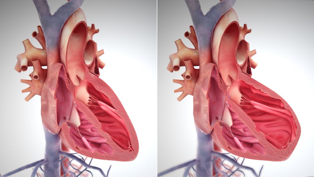
Finerenone reduced the composite of total first and recurrent heart failure (HF) events (hospitalisations for HF or urgent HF visits) and cardiovascular death in patients with HF and mildly reduced or preserved ejection fraction, according to an international clinical trial led by investigators from Brigham and Women’s Hospital.

Heart failure events and cardiovascular death were less common in the finerenone group than in the placebo group. Overall, the rate of serious adverse events was similar across the groups, but rates of hyperkalaemia were higher for the group taking finerenone. Results were presented at the European Society of Cardiology Congress 2024 and published simultaneously in the New England Journal of Medicine.

“We saw benefit regardless of the ejection fraction and even in patients who were on other approved therapies,” said trial principal investigator and corresponding author Scott Solomon, MD, the director of the Clinical Trials Outcomes Center at Mass General Brigham and the Edward D. Frohlich Distinguished Chair at Brigham and Women’s Hospital. “This drug represents a new drug class that may become a pillar of therapy for this disease.”

HF is the progressive decline in the heart’s ability to fill with and pump blood. It affects over 60 million people worldwide. Approximately half of all people living with HF have mildly reduced or preserved left ventricular ejection fraction, a condition with limited treatment options. These findings suggest that the non-steroidal mineralocorticoid receptor antagonist finerenone could represent a new therapeutic option for patients.

The FINEARTS-HF trial, funded by Bayer, assigned 6000 patients to receive either finerenone or placebo in addition to their existing therapies. The trial’s limitations include few Black patients, although the percentage of Black patients was proportional to their regional population. “Our group continues to study novel therapies for heart failure,” Solomon said. “There’s huge residual risk in these patients and so more room for new therapies.“

Source: Brigham and Women’s Hospital

fMRI Discovers Where Love Resides in the Brain

The image represents a statistical average of how different types of love light up different regions of the brain. Photo: Pärttyli Rinne et al 2024, Aalto University.

We use the word ‘love’ in a bewildering range of contexts, from sexual adoration to parental love or the love of nature. Now, more comprehensive imaging of the brain may shed light on why we use the same word for such a diverse collection of human experiences.

“You see your newborn child for the first time. The baby is soft, healthy and hearty – your life’s greatest wonder. You feel love for the little one.”

The above statement was one of many simple scenarios presented to 55 parents, self-described as being in a loving relationship. Researchers from Aalto University utilised functional magnetic resonance imaging (fMRI) to measure brain activity while subjects mulled brief stories related to six different types of love.

“We now provide a more comprehensive picture of the brain activity associated with different types of love than previous research,” says Pärttyli Rinne, the philosopher and researcher who coordinated the study. “The activation pattern of love is generated in social situations in the basal ganglia, the midline of the forehead, the precuneus and the temporoparietal junction at the sides of the back of the head.”

Love for one’s children generated the most intense brain activity, closely followed by romantic love.

“In parental love, there was activation deep in the brain’s reward system in the striatum area while imagining love, and this was not seen for any other kind of love,” says Rinne. Love for romantic partners, friends, strangers, pets and nature were also part of the study, which was published in the journal Cerebral Cortex.

According to the research, brain activity is influenced not only by the closeness of the object of love, but also by whether it is a human being, another species or nature.

Unsurprisingly, compassionate love for strangers was less rewarding and caused less brain activation than love in close relationships. Meanwhile, love of nature activated the reward system and visual areas of the brain, but not the social brain areas.

Pet-owners identifiable by brain activity

The biggest surprise for the researchers was that the brain areas associated with love between people ended up being very similar, with differences lying primarily in the intensity of activation. All types of interpersonal love activated areas of the brain associated with social cognition, in contrast to love for pets or nature – with one exception.

Subjects’ brain responses to a statement like the following, on average, revealed whether or not they shared their life with a furry friend:

“You are home lolling on the couch and your pet cat pads over to you. The cat curls up next to you and purrs sleepily. You love your pet.”

“When looking at love for pets and the brain activity associated with it, brain areas associated with sociality statistically reveal whether or not the person is a pet owner. When it comes to the pet owners, these areas are more activated than with non-pet owners,” says Rinne.

Love activations were controlled for in the study with neutral stories in which very little happened. For example, looking out the bus window or absent-mindedly brushing your teeth. After hearing a professional actor’s rendition of each ‘love story’, participants were asked to imagine each emotion for 10 seconds.

This is not the first effort at finding love for Rinne and his team, which includes researchers Juha Lahnakoski, Heini Saarimäki, Mikke Tavast, Mikko Sams and Linda Henriksson. They have undertaken several studies seeking to deepen our scientific knowledge of human emotions. The group released research mapping subjects’ bodily experiences of love a year ago, with the earlier study also linking the strongest physical experiences of love with close interpersonal relationships.

Not only can understanding the neural mechanisms of love help guide philosophical discussions about the nature of love, consciousness, and human connection, but also, the researchers hope that their work will enhance mental health interventions in conditions like attachment disorders, depression or relationship issues.

Source: Aalto University

Brain Ages at Different Paces According to Social and Physical Environments

An international study employing advanced measurements of brain ageing on a wide range of participants found that people from more disadvantaged countries and backgrounds had older biological ages for their brains compared to chronological ages. The results are published in Nature Medicine.

The pace at which the brain ages can vary significantly among individuals.  This difference between biological and chronological ages may be affected by environmental factors like pollution and social factors like income or health inequalities, especially in older people and those with dementia. Until now, it was unclear how these combined factors could either accelerate or delay brain ageing across diverse geographical populations. 

The study used advanced brain clocks based on deep learning of brain networks, involved a diverse dataset of 5306 participants from 15 countries. By analysing data from functional magnetic resonance imaging (fMRI) and electroencephalography (EEG), the researchers quantified brain age gaps in healthy individuals and those with neurodegenerative conditions such as mild cognitive impairment (MCI), Alzheimer’s disease, and frontotemporal lobe degeneration (FTLD). 

Participants with a diagnosis of dementia, particularly Alzheimer’s disease, exhibited the most critical brain age gaps. The research also highlighted sex differences in brain ageing, with women in Latin American and Caribbean countries showing greater brain age gaps, particularly in those with Alzheimer’s disease. These differences were linked to biological sex and gender disparities in health and social conditions. Variations in signal quality, demographics, or acquisition methods did not explain the results. These findings underscore the role of environmental and social factors in brain health disparities. 

The findings of this study have profound implications for neuroscience and brain health, particularly in understanding the interaction between macro factors (exposome) and the mechanisms that underlie brain ageing across diverse populations in healthy ageing and dementia. The study’s approach, which integrates multiple dimensions of diversity into brain health research, offers a new framework for personalised medicine. This framework could be crucial for identifying individuals at risk of neurodegenerative diseases and developing targeted interventions to mitigate these risks. Moreover, the study’s results highlight the importance of considering the biological embedding of environmental and social factors in public health policies. Policymakers can reduce brain age gaps and promote healthier ageing across populations by addressing issues such as socioeconomic inequality and environmental pollution. 

Source: University of Surrey

Competition Law has again Worked to Fight a Bad Drug Patent, but We Need Other Solutions

By Fatima Hassan and Leena Menghaney

A Competition Commission probe recently resulted in a patent on an important tuberculosis medicine being dropped in South Africa. Twenty years ago, a similar Competition Commission case resulted in a settlement that helped drive down the prices of several antiretrovirals, thereby helping to set the stage for the country’s HIV treatment programme. Fatima Hassan and Leena Menghaney connect the dots between the two landmark cases and map out what has and has not changed over the last two decades.

In the late 1990s and early 2000s, South Africa faced a major uncontrolled AIDS epidemic, worsened by state sponsored AIDS denialism. South Africa was at the epicentre of a global epidemic, with hundreds of thousands of people getting sick and dying, needlessly, because lifesaving antiretroviral medicines were out of reach.

This was in the main because of the Mbeki government’s deadly science denialism denying public sector patients antiretrovirals and the high cost of some of these medicines, which at the turn of the century was available in the private sector but only for the very rich or medically insured. The private sector price for the combination of three antiretrovirals needed by most people living with HIV was exorbitant.

This was because of patent monopolies held at the time by multinational pharmaceutical companies, particularly GlaxoSmithKline (GSK) and Boehringer Ingelheim (BI). In essence, people in South Africa living with HIV had to beg to live – by seeking donations and charity or pressuring their respective medical schemes to provide coverage. Meanwhile, lifesaving antiretrovirals were generally available in the Global North and in some parts of the Global South where governments like those in Thailand and Brazil had taken action to reduce prices.

Hundreds of thousands of people in South Africa died prematurely because they did not get access to these medicines in time.

The landmark Hazel Tau case

Looking for a way to challenge the high prices of key antiretrovirals, activists turned to South Africa’s newly revamped post-apartheid competition law. In September 2002, the Treatment Action Campaign, Hazel Tau, a woman living with HIV and several others lodged a complaint with the country’s Competition Commission. They alleged that the price that GSK and BI were charging for important antiretrovirals was excessive and anti-competitive, undermining not just Competition Law but also the right to health as enshrined in the country’s still fairly new Constitution.

The Competition Commission agreed to investigate. Several months later, they announced that there was a prima facie case of excessive pricing and that they would be referring the matter to the Competition Tribunal (the next phase of a complaint to the Competition authorities). Almost immediately after that announcement, TAC was approached by GSK and BI to “settle” the matter. This meant there would be no public hearings, and the companies would not have to defend their pricing decisions in the dock.

The terms of the settlement, negotiated by the TAC’s legal team, mirrored what TAC had publicly demanded at the beginning of the case. Most importantly, GSK and BI agreed to grant voluntary licenses to several generic manufacturers that would allow them to make and sell the antiretrovirals in question. It was this generic competition that would drive down the prices of antiretrovirals in the years that followed.

Even though the Competition Commission only has jurisdiction in South Africa, the licenses included many other African countries, which meant those countries could also benefit from the generic competition and lower prices. The settlement (including the terms of the voluntary licenses) was agreed to by the Competition Commission, made an order and publicly announced, leading to the conclusion of the complaint.

The case, which came to be known as the Hazel Tau case, would in the years to come be recognised as one of the foundations that made large HIV treatment programmes possible in South Africa and other African countries. Despite this victory, the ongoing effects of AIDS denialism meant that it would in reality be several years before the more affordable generic antiretrovirals would be made widely available in South Africa.

20 years later, the spotlight is on TB drugs

HIV has not been the only health crisis to affect SA. According to the World Health Organization (WHO), Tuberculosis (TB) is one of the leading infectious causes of death globally, and drug-resistant TB (DR-TB) remains a public health crisis. The WHO estimates that around 304 000 people fall ill with TB in South Africa per year, and it claims over 50 000 lives, which means it remains one of the country’s top killers. While TB rates are slowly declining, there is concern that rates of drug-resistant forms of TB (DR-TB) are increasing. DR-TB requires newer, more expensive treatments.

Republished from Spotlight under a Creative Commons licence.

Read the original article

Scientists Develop a Way to Turbocharge Genetic Therapy

Source: Pixabay CC0

Gene therapy, the idea of fixing faulty genes with healthy ones, has held immense promise. But a major hurdle has been finding a safe and efficient way to deliver those genes.

Now, researchers at the University of Hawaiʻi’s John A. Burns School of Medicine (JABSOM) have made a significant breakthrough in gene editing technology that could revolutionise how we treat genetic diseases. Their new method offers a faster, safer, and more efficient way to deliver healthy genes into the body, potentially leading to treatments for hundreds of conditions.

Current methods can fix errors in genes, but they can also cause unintended damage by creating breaks in the DNA. Additionally, they struggle to insert large chunks of genetic material such as whole genes.

The new technique, developed by Dr Jesse Owens along with his team Dr Brian Hew, Dr Ryuei Sato and Sabranth Gupta, from JABSOM’s Institute for Biogenesis Research and Cell and Molecular Biology Department, addresses these limitations. They used laboratory evolution to generate a new super-active integrase capable of inserting therapeutic genes into the genome at record-breaking efficiencies.

“It’s like having a “paste” function for the human genome,” said Dr Owens. “It uses specially engineered ‘integrases’ to carefully insert healthy genes into the exact location needed, without causing breaks in the DNA. This method is much more efficient, with success rates of up to 96% in some cases.”

“This could lead to faster and more affordable treatments for a wide range of diseases, potentially impacting hundreds of conditions with a single faulty gene,” said Dr. Owens.

Faster treatment development and a broader application

The implications of this research extend beyond gene therapy. The ability to efficiently insert large pieces of DNA has applications in other areas of medicine.

When making cell lines to produce therapeutic proteins, the gene encoding the protein is usually randomly inserted into the genome, and it rarely lands in a location in the genome that is good for production. This is like searching for a needle in a haystack. Additionally, finding a cell with the gene inserted correctly and producing the desired protein can take many months.

Instead of searching for a needle in a haystack, Dr Owens’ technique makes a stack of needles. It delivers the gene directly to the desired location, significantly speeding up the development process.

“JABSOM takes pride in nurturing talented researchers like Jesse Owens, whose work has the power to create a global impact,” said Sam Shomaker, dean of the University of Hawaiʻi John A. Burns School of Medicine. “This research, conducted in our lab in the middle of the Pacific, has the potential to significantly improve the way we treat genetic diseases.”  

Dr Owens’ team is exploring how this technique could accelerate the development and manufacture of biologics and advanced therapies such as antibodies. Currently, finding the right cell line for efficient production can be a time-consuming process. However, Dr Owens’ new genome engineering tool can reduce the cell line development timeline and accelerate the manufacture of life-saving therapeutics. 

Source: University of Hawaii at Manoa

Treatment with Dopamine Alleviates Symptoms in Alzheimer’s Disease

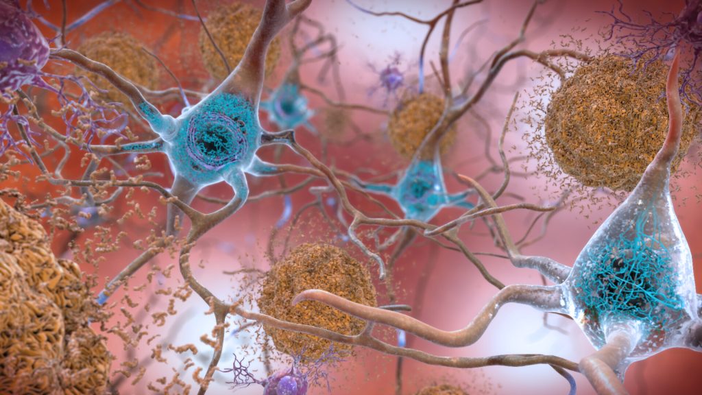
Neurons in the brain of an Alzheimer’s patient, with plaques caused by tau proteins. Credit: NIH

A new way to combat Alzheimer’s disease has been discovered by Takaomi Saido and his team at the RIKEN Center for Brain Science (CBS) in Japan. Using mouse models, the researchers found that treatment with dopamine could alleviate physical symptoms in the brain as well as improve memory. Published in Science Signaling, the study examines dopamine’s role in promoting the production of neprilysin, an enzyme that can break down the harmful plaques in the brain that are the hallmark of Alzheimer’s disease. If demonstrated in human clinical trials, it could lead to a fundamentally new way to treat the disease.

The formation of hardened plaques around neurons is one of the earliest signs of Alzheimer’s disease, often beginning decades before behavioural symptoms such as memory loss are detected. These plaques are formed from pieces of the peptide beta-amyloid that accumulate over time. In the new study, Saido’s team at RIKEN CBS focuses on the enzyme neprilysin because previous experiments showed that genetic manipulation that produces excess neprilysin in the brain (a process called upregulation) resulted in fewer beta-amyloid plaques and improved memory in mice.

Neprilysin by itself cannot be a medication as it cannot enter the brain from the blood stream, so the researchers screened molecules to determine which ones can naturally upregulate neprilysin in the correct parts of the brain. The team’s previous research led them to narrow down the search to hormones produced by the hypothalamus, and they discovered that applying dopamine to brain cells cultured in a dish yielded increased levels of neprilysin and reduced levels of free-floating beta-amyloid.

Now the serious experiments began. Using a DREADD system, they inserted tiny designer receptors into the dopamine producing neurons of the mouse ventral tegmental area. By adding a matching designer drug to the mice’s food, the researchers were able to continuously activate those neurons, and only those neurons, in the mouse brains. As in the dish, activation led to increased neprilysin and decreased levels of free-floating beta-amyloid, but only in the front part of the mouse brain. But could the treatment remove plaques? Yes. The researchers repeated the experiment using a special mouse model of Alzheimer’s disease in which the mice develop beta-amyloid plaques. Eight weeks of chronic treatment resulted in significantly fewer plaques in the prefrontal cortex of these mice.

The DREADD system is an incredible system for precise manipulation of specific neurons. But it is not very useful for human clinical settings. The final experiments tested the effects of L-DOPA treatment. L-DOPA is a dopamine precursor molecule often used to treat Parkinson’s disease because it can enter the brain from the blood, where it is then converted into dopamine. Treating the model mice with L-DOPA led to increased neprilysin and decreased beta-amyloid plaques in both frontal and posterior parts of the brain. Model mice treated with L-DOPA for three months also performed better on memory tests than untreated model mice.

Tests showed that neprilysin levels naturally decreased with age in normal mice, particularly in the frontal part of the brain, perhaps making it a good biomarker for preclinical or at-risk Alzheimer’s disease diagnoses. How dopamine causes neprilysin levels to increase remains unknown, and is the next research topic for Saido’s group.

“We have shown that L-DOPA treatment can help reduce harmful beta-amyloid plaques and improve memory function in a mouse model of Alzheimer’s disease,” explains Watamura Naoto, first author of the study. “But L-DOPA treatment is known to have serious side effects in patients with Parkinson’s disease. Therefore, our next step is to investigate how dopamine regulates neprilysin in the brain, which should yield a new preventive approach that can be initiated at the preclinical stage of Alzheimer’s disease.”

Source: RIKEN