Learning new languages, sending emails, attending a virtual class, or speaking to loved ones halfway around the world are just some of the tasks accomplished by touching a button on a smartphone. Unfortunately, the ease and convenience of modern devices have also come with a painful crick in the neck. The sedentary nature of work and prolonged use of hand-held devices and computers have contributed to a sharp increase in neck pain.
While fatigue in neck muscles has long been suspected of causing pain, the actual mechanical changes in the spine and muscles that precede weakness remain an outstanding question.
Now, using high-precision X-ray imaging to track spine movements during neck exertion tasks, Texas A&M University researchers have discovered that sustained neck exertions cause muscle fatigue that then exaggerate the cervical spine curvature. This leads to neck pain.
“We are talking about subtle movements of the neck in statically held positions, which are hard to capture. They are also highly complex because there are so many individual pieces in the neck, or as we call, motion segments,” said Dr Xudong Zhang, professor in the Department of Industrial and Systems Engineering. “With this study, we have, for the first time, provided unequivocal evidence that fatigue causes mechanical changes that increase the risk.”
Zhang said this understanding can help to make informed decisions about how we work and the design of products (e.g., head-mounted wearables) that can potentially reduce the risk of neck pain.
Neck pain is prevalent
Neck pain is one of the most common musculoskeletal disorders, and globally, around 2500 people out of 100 000 have some form of neck pain. In fact, by 2050, the estimated global number of neck pain cases is projected to increase by 32.5%. An important risk factor for neck pain is bad posture sustained over long periods. Consequently, working long hours on the computer in a stooped position or prolonged use of smart devices are important contributors to neck pain.
Neck posture is maintained dynamically by the bones of the spine pulled into position by the muscles that attach to them. Although the neck is highly flexible, it is also very unstable.
“The muscle drives movements by producing force,” said Zhang. “We hypothesised that when different muscles’ force production abilities diminish, the bone positions change and that can be captured.”
Measuring fatigue
To test their idea, they recruited healthy volunteers in a “sustained-till-exhaustion” neck exertion task. The subjects maintained their necks in the neutral, 40° extended (bent backwards) and 40° bent forward for a certain duration. The investigators used electromyography (EMG) to measure muscle electrical activity. In particular, they objectively measured muscle fatigue through changes in the frequency of the EMG signal. In addition, they used high-precision, dynamic X-ray technology to track small-amplitude cervical spine movements that were of the order of a few degrees.
“We imagined the cervical spine as a cantilever bridge,” said Zhang. “If there is excessive and/or repeated stress on the bridge, it might sag or buckle; similarly, if the muscles get fatigued, the cervical spine may deflect.”
The researchers’ experimental paradigm validated that sustained exertions indeed lead to EMG signals of fatigue. Biomechanically, the muscular fatigue modified the spine’s mechanics, which then increases the propensity for injury.
Further investigations
As a next step, the researchers will develop dynamic biomechanical models, a novel approach that promises to provide a more realistic understanding of the muscular events that precede fatigue. Unlike the model in this study that assumes static neck exertions, the dynamic model will capture subtle but consequential changes in the muscles and bones over time.
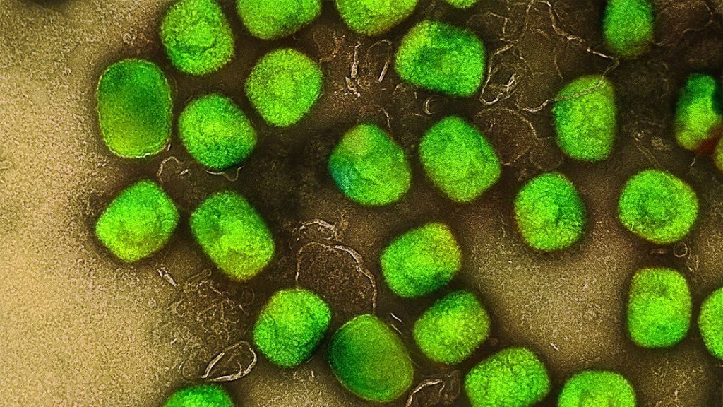
The African Centre for Disease Control and World Health Organization have raised the alarm following a drastic uptick in mpox cases. This surge is being driven by a new strain of the virus. Elri Voigt reports about what we know so far and potential implications for South Africa.
Mpox, a viral illness first identified in Africa in 1970, made headlines in 2022 when it spread across the globe for the first time. Since then, the outbreak has evolved, with multiple strains of the virus circulating in different countries. A new strain, known as clade Ib, first discovered in the Democratic of the Republic of Congo (DRC), is responsible for much of the most recent surge in mpox cases.
These recent developments are complex, and the situation is likely to change. This was the common theme of a special session on the mpox outbreak during the World Health Organization (WHO) Regional Committee for Africa meeting at the end of August. This session took place two weeks after the WHO declared the outbreak to be a Public Health Emergency of International Concern.
“We don’t have one outbreak. We have multiple outbreaks in one,” Dr Jean Kaseya, the Director General of the African Centre for Disease Control (CDC) remarked.
These outbreaks are caused by different clades of the mpox virus. Clades are a classification system based on the genetic similarities between different strains of a virus, explained Professor Tulio de Oliveira, Director of the Centre for Epidemic Response and Innovation (CERI) at Stellenbosch University (SU). “So, what it means is that when we see a genetic change [in a virus] that’s really visible and that may have impacted it, normally we call it a different clade or genotype or variant,” he said.
This is similar to classifying different strains of SARS-CoV-2 as variants, Dr Duduzile Ndwandwe, a molecular biologist working for Cochrane South Africa, an intramural research unit within the South African Medical Research Council, told Spotlight.
She explained that the different mpox clades and sub-clades have mutated so they have genetic differences but still fall under the umbrella of mpox.
“In a nutshell…it’s just talking about the differences in the genome sequence of the virus, how many mutations [it has] or how big the mutations are in that virus’s strain of mpox,” she said.
‘Jump in evolution’
Dr Aida Sivro, senior scientist at the Centre for the AIDS programme of Research in South Africa (CAPRISA), in 2022 told Spotlight that there are two clades of the mpox virus, which were then referred to as the Central African Clade (clade I) and the West African Clade (clade II).
Since then, clade I went through a big jump in evolution and a sub-clade emerged in the DRC, now called clade Ib, De Oliveira told Spotlight. The previous outbreak in 2022 was mostly driven by another sub-clade called clade IIb.
To further complicate matters, there’s a third strain of the virus also circulating – clade Ia.
At the moment, the DRC accounts for about 90% of mpox cases in the African Region, according to Dr Fiona Braka, the Emergency Response Manager for WHO’s AFRO region. She explained that right now the situation is not fully understood because a lack of diagnostics and testing capabilities is limiting understanding of the true burden of disease.
What we do know, she said, is that there are two distinct outbreaks in the DRC. Based on the information currently available, clade Ia is circulating in regions in the country where mpox is considered endemic and affecting mostly children. While clade Ib is spreading mostly among adults in the eastern provinces of South Kivu and North Kivu.
The clade Ib strain has since spread from the DRC to neighbouring countries Burundi, Rwanda, Uganda and Kenya, according to Braka. Sweden and Thailand have also identified one case each.
As of 1 September, the WHO reported that there have been 3 751 confirmed cases of mpox and 32 deaths across 14 countries in African in 2024 alone. But there are many more suspected cases of mpox that have not been tested.
Implications for South Africa
De Oliveira said at this point, South Africa shouldn’t be overly concerned about mpox, but it should be alert. The best way to do this is to make sure the public know what the symptoms are so they can present for diagnosis and treatment if they suspect they have the virus.
In a similar vein, Ndwandwe said the public shouldn’t panic, but we as a country need to remain vigilant. She added that because clade Ib is spreading on the African continent, there is a risk of it spreading to South Africa through cross-border travel, making it a public health concern.
This year, 24 cases of mpox have been reported in South Africa. Three people have died, while 19 have recovered. Two people are still considered to have active disease, with the most recent case identified in early August.
But this doesn’t necessarily mean there aren’t more cases of mpox in the country. “What we do suspect is that we may have milder cases that are actually not reported,” Nevashan Govender, the operation manager of the Emergency Operations Center at the National Institute for Communicable Diseases (NICD) told Spotlight.
He said so far, all the cases in the country have been caused by clade IIb and no cases of clade Ib have been identified.
A polymerase-chain-reaction (PCR) test is the gold standard test used to determine whether someone has mpox. But genome sequencing would need to be done to identify what clade they have.
Lots of unknowns around new strain
At the moment, there are a lot of unknowns around clade Ib.
What is of concern, according to Braka is the severity of disease seen especially in people who are immunocompromised and in pregnant women and children. Ndwandwe added to this and said there is a concern that clade Ib has higher fatality rates than clade IIb.
De Oliveira cautioned against jumping to conclusions about the severity of this new clade without sufficient data. He said we don’t know for sure yet if clade Ib is causing more severe disease than IIb. What we do know from mpox in general, he said, is that when someone is immunocompromised in some way, they tend to develop more severe symptoms.
Govender echoed De Oliveira’s caution that we don’t yet know enough about clade Ib to say definitively if it is for example more transmissible than other clades
“It’s not to say that it isn’t [more transmissible], but there is just not a lot of evidence stating that it is absolutely true…There’s a lot of knowledge and information gaps,” he said.
The NICD in a recent update also stressed that there are a lot of unknowns about this new strain. It added: “South Africa continues to prioritise enhanced surveillance and raising awareness for mpox.”
The state of vaccines and treatment for mpox
Spotlight reported previously that the smallpox vaccine, which hasn’t been routinely administered in South Africa since the 1980s when smallpox was eradicated, is thought to offer some degree of protection against mpox. However, it’s difficult to predict just how much protection the smallpox vaccine would provide, Sirvo told Spotlight for that previous article.
There are currently three vaccines against mpox that have been approved in some countries, a spokesperson from the vaccine alliance Gavi told Spotlight. These are LC16m8, JYNNEOS and ACAM2000.
LC16m8 is a third-generation small pox vaccine manufactured by KM Biologics. According to WHO, from 2022 it had mainly been used in Japan.
The JYNNEOS vaccine is a third-generation smallpox vaccine, manufactured by Bavarian Nordic, Ndwandwe said, and it was used during the outbreak in 2022. She added that this vaccine is considered the preferred option due to its safety profile and targeted protection against mpox.
ACAM2000 is a second-generation vaccine for smallpox and manufactured by Emergent BioSolutions. But it was only approved by the FDA for use in those at high risk for mpox at the end of August this year. It was not widely used during the 2022 outbreak but was available in some places under a compassionate use protocol (a means of providing medicines or vaccines that have not yet been registered).
In 2022, the Centre for Disease Control (CDC) recommended that JYNNEOS be used as the primary vaccine against mpox because it was associated with fewer side effects than ACAM2000.
While these vaccines exist, it doesn’t mean everyone can access them easily. Countries on the African continent have so far relied on vaccine donations facilitated by the WHO, with an initial 10 000 doses expected to arrive in Africa sometime this month.
Vaccine manufacturers KM Biologics and Bavarian Nordic have submitted proposals to the WHO for emergency use listing (EUL), according to WHO Director-General Dr Tedros Adhanom Ghebreyesus. He added this will allow UNICEF and the vaccine alliance GAVI to buy the vaccines to supply to countries that haven’t issued their own national regulatory approval yet.
The treatment options for mpox are also limited. According to this WHO factsheet on mpox, some antivirals have received emergency use authorisation in some countries and are being evaluated in clinical trials. However, so far there is no proven effective antiviral treatment for mpox.
Tecovirimat, which was approved to treat smallpox, is one of these antivirals being evaluated. According to the CDC, studies in animals have shown the antiviral might help treat mpox but it is still considered an investigational drug for mpox. The drug has been used in some cases of severe mpox.
When asked about this, Ndwandwe agreed more research needs to be conducted to fully understand the evidence around using Tecovirimat. “But what we know now is that the fact that it was authorised for compassionate use, there is some benefit to using that treatment, given that there isn’t any other [treatment,” she said.
Mpox vaccine and treatment availability in South Africa
According to De Oliveira, a small batch of vaccines against mpox and an antiviral drug were made available to South Africa through donations during the outbreak earlier this year.
But the country would need more vaccines if cases increase to protect those at risk for severe disease.
At the moment, South Africa does not have access to any mpox vaccines and has asked for a donation of 40 000 vaccine doses, Foster Mohale, spokesperson for the health department told Spotlight. The country has requested the JYNNEOS vaccine, based on the recommendation by the National Advisory Group on Immunisation.
He added that South Africa’s request to its international partners and the WHO is ongoing support with access to tecovirimat should the need increase. He also requested the WHO’s assistance in procuring the 40 000 vaccine doses to vaccinate high-risk groups if mpox cases increase.
When asked if the department will be entirely reliant on donations of mpox vaccines or would seek to procure its own if cases increase, Mohale said it depends. “South Africa has been in communication with the vaccine manufacturer, Bavarian Nordic, and will consider procurement if needed,” he added.
Because there is a shortage of mpox vaccines and treatment and uncertainty about the sustainability of donated supplies, Ndwandwe said: “Our best defence at this point in time is to prevent [the spread of mpox cases] as much as possible and detect the cases as they start, early on.”
Symptoms of mpox
Govender said the NICD is urging people not to panic but to stay informed on the signs and symptoms of mpox using some of the accurate information available from either the National Department of Health or the NICD.
“The first line of defence for any public health emergency and outbreak comes from when people take initiative to protect themselves,” he said.
Mpox, which is spread by close contact, either household or sexual contact, with someone who has the virus, could initially manifest in flu-like symptoms or the characteristic mpox rash. These include a fever, sore throat, muscle aches, headaches and swollen lymph nodes, according to the WHO factsheet on mpox. The rash starts flat and then becomes a blister filled with fluid, which eventually dries and falls off. The rash can occur on someone’s palms or soles of their feet, face, mouth and throat and sometimes the genital areas.
Children, pregnant women and those who are immunocompromised are most at risk for developing severe disease or dying, the factsheet stated. This includes people living with HIV whose viral load is not well controlled.
Mpox is a virus and as with all viral infections it’s the immune system that fights it off, Ndwandwe explained. However, if someone is immunocompromised, so has a weakened immune system, there is a greater chance that the mpox virus will overtake their immune system and cause severe disease.
This is one of the reasons why we would be concerned about the disease in South Africa, Professor Helen Rees, the Co-Chair of the Incident Management Team (IMT) on mpox, previously told eNCA.
“We have many people living with HIV in the country, many of whom are on antiretroviral therapy, their immune system is good. But we have many others, who don’t know what their status is and might be vulnerable to severe mpox,” she said.
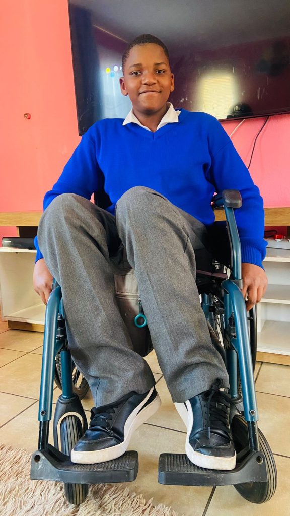
Spinal cord injury survivor is a capable and helpful big brother
Kamogelo Sodi, who was injured in a car crash when he was just six years old, says he learned valuable skills on how to regain his independence at the Netcare Rehabilitation Hospital. The teenager enjoys cooking for himself, taking care of his three younger brothers, and playing basketball when he’s not studying hard to achieve his dream of being a medical practitioner one day.
5 September 2024: At 14 years old, Kamogelo Sodi of Alberton enjoys listening to music, chatting with his friends on social media and working hard at school towards his dream of becoming a neurosurgeon one day. He cooks for himself when he’s hungry and loves looking after his three little brothers. He also likes playing basketball. The difference between him and most other teenagers is that he does all this from his wheelchair.
“Since I’ve been in a wheelchair, I’ve become more confident,” says the vivacious teenager. “I was extremely shy, and I didn’t have a lot of friends, but now I have loads of friends.”
In 2016, when he was just six years old, Kamogelo’s life changed forever. He was in a devastating car crash, which left him with fractures in the lumbar region of his spine, resulting in complete paraplegia.
Once discharged from the hospital, where he had emergency surgery, Kamogelo was sent to the Netcare Rehabilitation Hospital to learn how to cope with, as his mother Reshoketswe Sodi calls it, his new normal. He was to stay there for almost six months.
Mrs Sodi, a radiation therapist, says the enduring care of the doctors, occupational therapists and physiotherapists there helped support Kamogelo and their family on their journey towards accepting and learning to cope with this difficult transition in his life. “It was important for me that he continued his schoolwork while there. When the social worker asked me what I wanted to happen, the first thing I said was that I didn’t want to break the routine of what he had been doing and that I wanted him to continue with school.
“It’s been a struggle, but with the help of the occupational therapists and physiotherapists, it has been an easier journey. We saw real progress when they taught Kamogelo something, and he grasped it, putting all his energy into it by thinking positively about it. It’s been hard, but with the support of the team from Netcare Rehabilitation Hospital, we managed it,” she says.
“After he was discharged, initially, we lived in a flat on the seventh floor. When the lifts weren’t working, like during load shedding, I’d have to carry him upstairs on my back – there was no other way to take him up. I’m so fortunate that I had a lot of support from my family and friends who’ve been pillars of strength for us.”
Kamogelo remembers his first visit to the Netcare Rehabilitation Hospital in Auckland Park. “When I first got to the hospital, I was lost. I didn’t know how to use a wheelchair. I was still so young. But they were so kind and taught me everything I needed to know.
“At first, I struggled to move around. I battled to transfer myself from place to place, but they showed me what to do, and over time, I started getting used to it. I managed to start moving myself around, and I began to enjoy it. From that day forward, I didn’t like people pushing me around. The staff also taught me how to transfer myself from my wheelchair to the car. It was a bit difficult at first, but I learned to push myself up properly so my bottom wouldn’t scrape on the wheelchair.
“It does help you become more independent, but you must be consistent. You don’t need to complain about things, you just need to listen to the people who want to help you learn to be independent.”
Later, in 2022, when he was 12 years old, Kamogelo returned to the Netcare Rehabilitation Hospital after he developed a severe pressure sore.
Dr Anrie Carstens, a doctor at the Netcare Rehabilitation Hospital, said Kamogelo was operated on at Netcare Milpark Hospital under the care of a plastic surgeon who did a flap to close the wound. “When the doctor was happy with his progress, Kamogelo came to us to help him because you get weak after surgery. The wound had healed, but the skin was delicate, so we had a graded seating approach for him to build up his strength and so that the areas of the skin didn’t break down. Another area of focus for Kamogelo was spasticity at the ankles. We worked on relaxing the ankles to get to a ninety-degree angle so he could sit better in his chair with his feet positioned well in the footrest.”
When homesickness inevitably struck, the staff comforted Kamogelo. “I began to miss home, and I cried and said I wanted to go home. They spoke nicely to me and said they first had to help me so I could go back home with no problems so my parents wouldn’t have to worry about me because of the pressure sore.”
Kamogelo said the staff also taught him valuable techniques to help him empty his bladder and bowels and assisted him in his journey to independence. “I was worried it would be painful and was a bit hesitant to try them out. But, doing it daily helped my routine and helped me become independent.”
Charne Cox, a physiotherapist at Netcare Rehabilitation Hospital, describes Kamogelo as bubbly, intelligent and with lovely manners. “He’s so motivated and tried so hard in therapy. He manages to go to school each day, not because of us, but because of his character.”
She says as children grow, their needs change. “The pressure sore developed because his seating in his wheelchair was not adequate because he had grown so much. We collaborated with the wheelchair manufacturer to re-evaluate and reassess the wheelchair seating, and they made him a new wheelchair. He was getting heavier, and his feet weren’t in alignment, so it was trickier for him to safely transfer from the wheelchair to the bed, for instance. It was good to re-educate him on pressure relief and pressure sores. It’s vital that adolescents are taught to take responsibility for themselves.”
Cox also helped Kamogelo work towards getting his feet in a better position.
“Children are so good about learning to use a wheelchair. Kamogelo was so motivated to move and be independent. He absorbed the information we gave him to enable him to go up ramps, turn and even do wheelies because he liked to explore.
“Children want to learn and have fun. They want to be independent. It’s amazing to help give them the tools to be the best new person they can be. Unfortunately, sometimes we can’t fix the injury, but we can give them the best opportunity to be as independent as possible. It’s so satisfying to know that Kamogelo is going to school and playing basketball.”
Kamogelo is determined to pursue a career as a neurosurgeon. “As long as I follow the path that I want to do and enjoy it, I will continue pursuing that path. Academically, I was the top achiever from grade four to grade six at my school.”
When he’s not at school, he loves going around the estate he lives in, getting fresh air, and being a good big brother to his three younger brothers. “They’re a handful, but what can I say – they’re my brothers, and I love them,” he says with a laugh.
Asked who his hero is, Kamogelo is quick to say his mother and father are both his heroes. His mom clearly thinks he’s a hero too. She’s smiling as she speaks about her son. “He’s playful and has a great sense of humour. He’s helpful in the house. Instead of wanting us to help him, thanks to the skills he learned at Netcare Rehabilitation Hospital, Kamogelo always says, ‘Let me give you a hand. Let me help you.’”
As global climate change causes extreme heat waves to become more common around the world, epidemiological studies have shown that heat kills more women than men. Now, a new study by researchers at Penn State has found that older women are physiologically more vulnerable to high heat and humidity than older men, and that women between the ages of 40 and 64 are as vulnerable as men 65 years of age or older. This is the first study to determine this disparity exists due to physiological differences rather than from a preponderance of women at old age due to greater longevity.
Led by Olivia Leach, doctoral candidate in kinesiology at Penn State, and her adviser, W. Larry Kenney, professor of physiology and kinesiology at Penn State, the researchers demonstrated that middle-aged and older women were affected by heat at lower temperature/humidity combinations than middle-aged and older men. The results, published in the American Journal of Physiology-Regulatory, Integrative and Comparative Physiology, were somewhat unexpected, according to Leach, because there are no differences in heat vulnerability based on biological sex in adults younger than 30.
While the researchers did not directly compare middle-aged men to middle-aged women, the physiological responses of middle-aged women were similar to the responses of older men in the study, which demonstrated that middle-aged women are more vulnerable to heat than men of the same age.
“In addition to demonstrating that middle-aged and older women are at greater risk from extreme heat, we also identified what levels of heat and humidity are safe for women as they age,” Leach said. “This information is presented as a temperature/humidity curve based on a person’s age, and it can be useful for setting policies designed to keep people safe during a heat wave.”
The researchers tested the heat thresholds of 72 participants between 40 and 92 years of age in a specialized environmental chamber in Kenney’s laboratory. Before the experiment, participants swallowed a tiny device encased in a capsule that measured their core temperature throughout the experiment.
During the study, participants entered the specialised environmental chamber where they performed light physical activity to simulate the effort of minimal day-to-day tasks – the types of things people would need to do even during a heat wave. The researchers then gradually increased the temperature and/or humidity in the chamber until the participant’s body could no longer adequately cool itself, and their core temperature began to rise.
The study is part of the PSU HEAT, or Human Environmental Age Thresholds, project, led by Kenney. For five years, researchers in the PSU HEAT project have examined the levels of combined heat and humidity that humans can tolerate before their core temperatures begin to rise. When core temperatures rise, people become vulnerable to heat-related illnesses including heat exhaustion, heat stroke and even death.
“We’re not saying that people who experience a certain temperature will necessarily become sick or die,” Kenney said. “We are identifying the limits of livability – the thresholds where people can no longer continue their daily life unimpeded. Once people reach these temperatures, they need to take actions like seeking air conditioning to cool their bodies.”
Previous research by Kenney and others demonstrated that people become increasingly vulnerable to heat as they age, because their ability to efficiently sweat and pump blood to the skin – two primary cooling mechanisms – decreases. Sweat evaporation carries heat away from the body, while extra blood pumped to the skin dissipates heat to the environment and supports sweating.
To date, the PSU HEAT project has conducted more than 600 experiments on nearly 200 participants between ages 18 and 92, but the results of this experiment still yielded surprises, according to Leach.
“Among young adults, there is no difference in heat vulnerability between men and women,” Leach said. “Young people tend to be healthier, so any measurable health metric – from blood pressure to cholesterol – is more homogeneous among young people than it is among older people.”
As with other health measures, older adults have a wide range in their vulnerability to heat, Leach explained.
“We have examined many factors that might explain who faces the most risk in a heat wave,” Leach said. “We found that age and biological sex are the two most important factors that can predict whether a healthy adult would be at risk from high heat and humidity.”
While cardiovascular health and certain medications can affect a person’s sensitivity to heat, biological sex and age appear to be the two primary drivers of heat vulnerability among healthy people, the researchers said.
“Other factors – for example someone’s cardiovascular fitness or their body mass – have little impact on how vulnerable a person is to heat at rest or during light activity,” Leach continued. “Older women really are at greater risk from heat than other people. As governments and other social leaders prepare for extreme heat to become more common, the vulnerability of older women needs to factor into their planning.”
Ball and stick 3D model of testosterone. Source: Wikimedia CC0
A treatment paradox has recently come to light in prostate cancer: Blocking testosterone production halts tumour growth in early disease, while elevating the hormone can delay disease progression in patients whose disease has advanced.
The inability to understand how different levels of the same hormone can drive different effects in prostate tumours has been an impediment to the development of new therapeutics that exploit this biology.
Now, a Duke Cancer Institute-led study appearing in Nature Communications, provides the needed answers to this puzzle.
The researchers found that prostate cancer cells are hardwired with a system that allows them to proliferate when the levels of testosterone are very low. But when hormone levels are elevated to resemble those present in the normal prostate, the cancer cells differentiate.
“For decades, the goal of endocrine therapy in prostate cancer has been to achieve absolute inhibition of androgen receptor function, the protein that senses testosterone levels,” said lead investigator Rachid Safi, PhD, research assistant professor in the Department of Pharmacology and Cancer Biology, at Duke University School of Medicine.
“It’s been a highly effective strategy, leading to substantial improvements in overall survival,” he said. “Unfortunately, most patients with advanced, metastatic disease who are treated with drugs to inhibit androgen signaling will progress to an aggressive form of the disease for which there are limited therapeutic options.”
Using a combination of genetic, biochemical, and chemical approaches, the research team defined the mechanisms that enable prostate cancer cells to recognise and respond differently to varying levels of testosterone, the most common androgenic hormone.
It turned out to be rather simple. When androgen levels are low, the androgen receptor is encouraged to “go solo” in the cell. In doing so, it activates the pathways that cause cancer cells to grow and spread. However, as androgens rise, the androgen receptors are forced to “hang out as a couple,” creating a form of the receptor that halts tumour growth.
“Nature has designed a system where low doses of hormones stimulate cancer cell proliferation and high doses cause differentiation and suppress growth, enabling the same hormone to perform diverse functions,” McDonnell said.
In recent years, clinicians have begun treating patients with late-stage, therapy resistant prostate cancers using a monthly, high-dose injection of testosterone in a technique called bi-polar androgen therapy, or BAT. The inability to understand how this intervention works has hindered its widespread adoption as a mainstream therapeutic approach for prostate cancer patients.
“Our study describes how BAT and like approaches work and could help physicians select patients who are most likely to respond to this intervention,” McDonnell said. “We have already developed new drugs that exploit this new mechanism and are bringing these to the clinic for evaluation as prostate cancer therapeutics.”
Researchers from Japan have discovered a certain oral bacteria associated with rheumatoid arthritis causes inflammation, through macrophages and an inflammatory enzyme, caspase-11. Their results appear in the International Journal of Oral Science.
Periodontal disease, which affects the gums and tissues that surround the teeth, is one of the most prevalent dental conditions worldwide, and besides tooth loss is associated with other health effects. Over the past few decades, clinical studies have revealed that the periodontal pathogen Aggregatibacter actinomycetemcomitans (A. actinomycetemcomitans) is closely related to the onset and worsening of rheumatoid arthritis (RA), a serious autoimmune disease that affects joints. However, what goes down at the molecular level remains largely unexplored and unclear.
In this study, a research team from Tokyo Medical and Dental University (TMDU) in Japan sought to fill this knowledge gap through detailed mechanistic studies in an animal model.
First, the researchers conducted preliminary experiments to confirm whether A. actinomycetemcomitans infection influenced arthritis in mice. To this end, they used the collagen antibody-induced arthritis mouse model, which is a well-established experimental model that mimics several aspects of RA in humans. They found that infection with this specific bacterium led to increased limb swelling, cellular infiltration into the lining of the joints, and higher levels of the inflammatory cytokine interleukin-1β (IL-1β) within the limbs.
Notably, these symptoms of worsening RA could be suppressed by administering a chemical agent called clodronate that depletes macrophages. This demonstrated that macrophages were somehow involved in aggravating RA caused by A. actinomycetemcomitans infection.
Further investigation using macrophages derived from mouse bone marrow revealed that A. actinomycetemcomitans infection increased the production of IL-1β. In turn, this triggered the activation of a multiprotein complex known as the inflammasome, which plays a key role in initiating and modulating the body’s inflammatory response to infections.
The researchers added yet one more piece to this puzzle using caspase-11-deficient mice. In these animals, inflammasome activation due to A. actinomycetemcomitans was suppressed. Most importantly, caspase-11-deficient mice exhibited less deterioration of arthritis symptoms, hinting at the important role that caspase-11 plays in this context. “Our research findings provide new insights into the link between periodontal pathogenic bacteria and the exacerbation of arthritis through inflammasome activation, offering important information on the long-debated relationship between periodontal disease and systemic diseases,” highlights Professor Toshihiko Suzuki, one of the lead authors of the study.
With any luck, these efforts will contribute to the development of novel therapeutic strategies to manage RA. “The findings of this research may pave the way for advances in clinical treatments for RA induced by infection with A. actinomycetemcomitans. Our suggestion to inhibit inflammasome activation could attenuate the expansion of inflammation to joints, resulting in a recovery from arthritis symptoms,” says lead author Dr Tokuju Okano. “Moreover, the outcome of our work could contribute to the development of treatment strategies for not only arthritis but also other systemic diseases, such as Alzheimer’s disease, which is also related to periodontal pathogenic bacteria,” he predicts.
The method can be used to explore treatment effects in people underrepresented in clinical trials
Researchers used real-world clinical data to attempt to emulate a randomised controlled trial testing the effectiveness of two blood thinners, apixaban and warfarin, to prevent stroke in patients with non-valvular atrial fibrillation. The study, led by Emma Maud Powell at the London School of Hygiene and Tropical Medicine, UK, and publishing August 29th in the open-access journal PLOS Medicine, provides a method to explore the effects of treatments in patients who are underrepresented or excluded from clinical trials.
Patients experiencing atrial fibrillation – a potentially dangerous medical condition in which the upper chambers of the heart beat irregularly – will often be prescribed blood thinners such as apixaban or warfarin to prevent a stroke. However, these treatment recommendations are based on results from randomized controlled trials, and it is unknown if they are applicable to populations of patients who were not included in the trial or present only in very low numbers.
In the new study, researchers used routinely collected health data from patients in the United Kingdom to attempt to emulate a previous randomized controlled trial that compared the effectiveness of apixaban and warfarin. They attempted to emulate the patient eligibility, selection and analysis approaches as the previous trial. They found that patients prescribed apixaban had similar outcomes to patients prescribed warfarin, but unlike the previous trial, they did not find that apixaban was superior. The researchers observed the differences in results may have been linked to higher quality of warfarin control, sub-optimal dosing of apixaban, and differences in the ethnicity of patients and use of concomitant medications compared with the clinical trial population.
Overall, the study established that using an existing randomised controlled trial (the reference trial) as a guide for the design of observational analysis of real patient data is an effective and valid way to estimate the treatment effects and risks of blood thinners given to patients with atrial fibrillation. The methods developed in this study can be used to investigate the effects of these medications in patient groups that are excluded from or underrepresented in these clinical trials, such as the elderly, those with multiple conditions and people with a higher risk of bleeding. This method can also help medical researchers to understand whether results from randomized controlled trials are transferable to “real-world” practices, and provides a framework that can be adapted to investigate treatment effects for other conditions.
The authors add, “Our study aimed to emulate a reference trial in oral anticoagulants in patients with atrial fibrillation using routinely collected UK healthcare data. Reference-trial informed design provides a framework for the study of treatment effects in patient groups excluded from or under-represented in trials.”
5 September 2024, International Spinal Cord Injury Day is commemorated on Thursday 5 September, drawing attention to the many ways people can be affected by spinal cord injury, creating awareness of prevention, and highlighting the possibilities for a fulfilling life after injury.
According to the World Health Organization, globally, over 15 million people are living with spinal cord injuries. Most of these cases are due to trauma, including falls, road traffic injuries or violence.
Jessica Morris, an occupational therapist at the Netcare Rehabilitation Hospital in Auckland Park, says one of the most critical aspects of care for those who’ve been impacted by spinal cord injuries is the importance of successful rehabilitation through a holistic, integrated approach from a multidisciplinary team.
“Many people just think it’s just about mobility. It’s so much more than that. Rehabilitation is complex because many different areas of our patients’ lives are affected.” Morris says they are fortunate that their team has so many different practitioners who can contribute to treating spinal cord injury patients, helping them regain a level of independence, which is vital to their self-confidence and sense of empowerment.
Dr Anrie Carstens, a general practitioner with a particular interest in physical medicine and rehabilitation who practises at the Netcare Rehabilitation Hospital, says the message of Spinal Cord Injury Awareness Day has relevance all year round, as people with spinal cord injuries need to be incorporated into society.
“It’s an opportunity to tell people not to be nervous to talk to someone in a wheelchair. They’re just like you or me, and they just have special ways of moving around and managing their pain and different aspects of their bodies. With the help of proper rehabilitation, the person can be better integrated as a functional, contributing member of society.”
Dr Carstens says people should also be aware that if they or their loved ones are ever impacted by a spinal cord injury, professional support is available. “Don’t just go straight home after your hospital stay and try to do everything on your own. Instead, come to a specialised spinal cord injury unit like ours, with therapists, doctors and nursing staff who are well versed in spinal cord injury and know the finer nuances necessary to optimally treat the person and show them how best to cope with their injury.
“In the multidisciplinary approach, every practitioner has a role in getting the person back into the real world, whether it means going back home, back to school, back to work or wherever they were before their injury occurred.”
From doctors and nurses with specialised skills to physiotherapists, occupational therapists, social workers and psychologists, speech therapists, a prosthetist and dieticians, the team provides a broad person focussed rehabilitation service to both adults and children. Their aim is to optimise their patients’ independence level using specialised equipment and teaching specific techniques to help overcome the obstacles a person may face.
Dr Carstens says it’s rewarding work for the staff at the hospital, who build up enduring relationships with those they care for. “One of the highlights is to compare and see what the patient was like when you admitted them and then see on discharge how much they’ve grown, how they’ve gained confidence and become more independent. What’s even better is to see them after they’ve been discharged and observe how well they’ve coped and how they’ve integrated and adjusted to their environment. We build a relationship with our patients because they stay with us for quite a while, and we usually have checkups every year after the person is discharged, often for life. We get to see them grow and thrive outside the healthcare setting, and we need more awareness about how much it is possible for people with spinal cord injuries to achieve.”
By analysing the immune system of female-to-male transgender individuals, Swedish researchers demonstrate the role of sex hormones in regulating the immune system. This newfound knowledge, published in Nature, explains differences between men and women, particularly in terms of immune signalling, and can be used to develop new immunological medications according to researchers.
Sex differences in the immune system are regulated both by genetics and by sex hormones. However, immunological comparisons between men and women can never fully distinguish the significance of genetic versus hormonal variations.
Now, three Swedish research groups led by Karolinska Institutet and Uppsala University has conducted a unique study analysing the regulation and adaptation of the immune system over time in 23 trans men who have undergone gender-affirming testosterone treatment, starting at the age of 18–37 years.
“We have followed individuals who were assigned female sex at birth and later received testosterone treatment in adulthood. Their genetic profile remains unchanged, while their hormone profile shifts entirely from typically female to male hormone levels,” says Petter Brodin, paediatrician and professor of paediatric immunology at the Department of Women’s and Children’s Health, Karolinska Institutet, who led the study together with Nils Landegren, assistant professor at Uppsala University, and Olle Kämpe, Professor at the Department of Medicine, Solna, Karolinska Institutet. “This unique change allows us, for the first time, to identify which parts of a person’s immune system are directly regulated by sex hormones rather than genetic sex differences.”
The researchers can now demonstrate that increased testosterone levels and the accompanying reduction in oestrogen particularly affect the balance between two crucial immune signalling systems: antiviral interferon type 1 (IFN-1) and proinflammatory signals such as tumour necrosis factor alpha (TNFα).
Specifically, they found that testosterone modulates a cross-regulated axis between type-I interferon and tumour necrosis factor. This is mediated by functional attenuation of type-I interferon responses in both plasmacytoid dendritic cells and monocytes. Conversely, testosterone potentiates monocyte responses leading to increased tumour necrosis factor, interleukin-6 and interleukin-15 production and downstream activation of nuclear factor kappa B-regulated genes and potentiation of interferon-γ responses, primarily in natural killer cells.
The immune system changes throughout life
They also have a hypothesis about why the immune system needs to be dynamically regulated by hormones throughout life.
“All individuals must be able to adjust their immune systems over the course of their lives to be optimally regulated for the conditions and challenges we face. During puberty and sexual maturation, new demands arise, and the immune system must be regulated differently to enable pregnancy in women and muscle growth in men,” says Petter Brodin.
By regulating these key functions via sex hormones, this can be achieved, and in women, it is dynamically controlled even during a menstrual cycle,” he adds.
The results of the study open an entirely new field of research, according to Nils Landegren.
“The newfound knowledge will help us better influence people’s immune systems even without using sex hormones. For example, new drugs can be developed to impact these regulatory mechanisms and thus rebalance the immune response, especially for women with the autoimmune rheumatic disease SLE,” he explains.
However, the results also have a more direct implications for transgender individuals.
“This research is also of crucial for transgender individuals undergoing gender-affirming hormone therapy, and I believe that this group deserves significantly more scientific attention and follow-up to ensure their long-term health,” says Petter Brodin.
Researchers from the Center for Healthy Aging, Department of Cellular and Molecular Medicine at the University of Copenhagen have made a breakthrough in lifespan research. They have discovered that a particular protein known as OSER1 has a great influence on longevity.
”We identified this protein that can extend longevity. It is a novel pro-longevity factor, and it is a protein that exists in various animals, such as fruit flies, nematodes, silkworms, and in humans,” says Professor Lene Juel Rasmussen, senior author behind the new study.
Because the protein is present in various animals, the researchers conclude that new results also apply to humans:
”We identified a protein commonly present in different animal models and humans. We screened the proteins and linked the data from the animals to the human cohort also used in the study. This allows us to understand whether it is translatable into humans or not,” says Zhiquan Li, who is a first author behind the new study and adds:
“If the gene only exists in animal models, it can be hard to translate to human health, which is why we, in the beginning, screened the potential longevity proteins that exist in many organisms, including humans. Because at the end of the day we are interested in identifying human longevity genes for possible interventions and drug discoveries.”
Paves the way for new treatment
The researchers discovered OSER1 when they studied a larger group of proteins regulated by the major transcription factor FOXO, known as a longevity regulatory hub.
“We found 10 genes that, when – we manipulated their expression – longevity changed. We decided to focus on one of these genes that affected longevity most, called the OSER1 gene,” says Zhiquan Li.
When a gene is associated with shorter a life span, the risk of premature aging and age-associated diseases increases. Therefore, knowledge of how OSER1 functions in the cells and preclinical animal models is vital to our overall knowledge of human aging and human health in general.
“We are currently focused on uncovering the role of OSER1 in humans, but the lack of existing literature presents a challenge, as very little has been published on this topic to date. This study is the first to demonstrate that OSER1 is a significant regulator of aging and longevity. In the future, we hope to provide insights into the specific age-related diseases and aging processes that OSER1 influences,” says Zhiquan Li.
The researchers also hope that the identification and characterization of OSER1 will provide new drug targets for age-related diseases such as metabolic diseases, cardiovascular and neuro degenerative diseases.
“Thus, the discovery of this new pro-longevity factor allows us to understand longevity in humans better,” says Zhiquan Li.