Month: September 2024

Ignore Antifungal Resistance at Your Peril, Scientists Warn

Candida Auris

Without immediate action, humanity will potentially face further escalation in resistance in fungal disease, a renowned group of scientists from the across the world has warned. The commentary – published in ‘The Lancet’ this week – was coordinated by scientists at The University of Manchester, the Westerdijk Institute and the University of Amsterdam. According to the scientists most fungal pathogens identified by the World Health Organization – accounting for around 3.8 million deaths a year – are either already resistant or rapidly acquiring resistance to antifungal drugs.

The authors argue that the currently narrow focus on bacteria will not fully combat antimicrobial resistance (AMR). September’s United Nations meeting on antimicrobial resistance (AMR) must, they demand, include resistance developed in many fungal pathogens.

Devastating health impacts

Resistance is nowadays the rule rather than the exception for the four currently available antifungal classes, making it difficult – if not impossible – to treat many invasive fungal infections. Fungicide resistant infections include Aspergillus, Candida, Nakaseomyces glabratus, and Trichophyton indotineae, all of which can have devastating health impacts on older or immunocompromised people.

Dr Norman van Rhijn from The University of Manchester coordinated the comment with Professor Ferry Hagen from the University of Amsterdam and the Westerdijk Institute in the Netherlands.

Dr van Rhijn said: “Most people agree that resistant bacterial infections constitute a significant part of the AMR problem. However many drug resistance problems over the past decades have also been the result of invasive fungal diseases largely underrecognized by scientists, governments, clinicians and pharmaceutical companies. The threat of fungal pathogens and antifungal resistance, even though it is a growing global issue, is being left out of the debate.”

Unlike bacteria, the close similarities between fungal and human cells which, say the experts, means it is hard to find treatments that selectively inhibit fungi with minimal toxicity to patients.

Back to square one

Professor Ferry Hagen added: “Despite the huge difficulties in developing them, several promising new agents including entirely new classes of molecules, have entered clinical trials in recent years. But even before they reach the market after years of development, fungicides with similar modes of action are developed by the agrochemical industry resulting in cross-resistance. That sets us back to square one again. It is true many essential crops are affected by fungi, so antifungal protection is required for food security. But the question is, at what price?”

The scientists recommend:

  • Worldwide agreement on restricting the use of certain classes of antifungal molecules for specific applications.
  • Collaboration on solutions and regulations that ensure food security and universal health for animals, plants, and humans.
  • Adding priority to AMR to fungal infections at the UN’s meeting in September.

Source: Universiteit van Amsterdam

Can a Cancer Diagnosis Cause Mental Health and Cardiovascular Problems in Family Members?

Study’s findings reveal increased risks, indicating the need for interventions to reduce stress for patients and their relatives.

Photo by Alex Green on Pexels

New research suggests that a family member’s cancer diagnosis may increase first-degree relatives’ and spouses’ risks of developing psychological and cardiovascular illnesses. The findings are published by Wiley online in CANCER, a peer-reviewed journal of the American Cancer Society.

Having a family member diagnosed with cancer can be a stressful and traumatic experience for the entire family. Because stress influences not only mental health but also cardiovascular health, investigators explored whether a cancer diagnosis contributes to negative psychological and cardiovascular outcomes in family members.

Using data from the Utah Population Database, the researchers identified 77 938 first-degree relatives and spouses of 49 284 individuals diagnosed with genitourinary cancer between 1990 and 2015, and they compared them with 81 022 relatives and spouses of 246 775 individuals not diagnosed with cancer.

The team found that 7.1% of relatives and spouses were diagnosed with a psychological illness within 5 years of a family member’s cancer diagnosis, and 7.6% were diagnosed with a cardiovascular illness. Compared with controls, they had 10%, 5%, and 4% higher risks of developing a psychological condition at 1, 3, and 5 years after a family member’s cancer diagnosis. They also had 28%, 16%, and 14% higher risks of developing cardiovascular disease at 1, 3, and 5 years.

Parents of children with cancer experienced the highest risks of developing negative health outcomes – a nearly 4-times increased risk at 1-year compared with other relatives. Also, a diagnosis of kidney or bladder cancer appeared to be the most stressful among genitourinary cancer types, while testis cancer was the least.

“A diagnosis of cancer is a life-changing event for patients and their families. With our group’s unique access to the Utah Population Database, we were able to create multi-generational networks highlighting the impact of a cancer diagnosis on families,” said lead author Mouneeb Choudry, MD, of the Mayo Clinic, in Phoenix, Arizona. “As health care professionals, we should take a multidisciplinary approach to addressing the stress of a cancer diagnosis by helping mitigate financial toxicity, treatment burden, and emotional impact on both the patient and their family.”

Source: Wiley

A Cuffless Smartphone App that Can Measure Blood Pressure

Photo by Ivan Samkov on Pexels

Researchers at the University of Pittsburgh are pioneering a new approach to blood pressure monitoring, using the devices we carry with us every day. Ramakrishna Mukkamala, professor of bioengineering at Pitt’s Swanson School of Engineering, is passionate about developing accessible blood pressure (BP) detection tools. Instead of designing a new medical device to monitor BP, Mukkamala decided to take advantage of the sensors readily available in smartphones and figure out how to detect blood pressure with them. 

“The most significant thing you can do to reduce your risk of cardiovascular disease is to lower high blood pressure through lifestyle changes, but in underserved populations, many people don’t have access to blood pressure cuffs, regular doctor’s appointments, or even know it’s a problem,” Mukkamala said. “But they do have smartphones.”

Mukkamala’s team harnessed tools already built into most smartphones, like motion-sensing accelerometers, front cameras, and touch sensors to build an Android smartphone application that can measure an individual’s pulse pressure. The user performs a hand-raising motion while holding the smartphone to make a measurement. The results of the project, published in Scientific Reports, demonstrate a promising new technology that could uniquely help reduce the burden of systolic hypertension globally, particularly in underserved populations. 

Designing blood pressure technology for a touchscreen 

Turning a smartphone into a monitoring device is no easy task, as Vishaal Dhamotharan, graduate student in the Cardiovascular Health Tech Laboratory, found out through multiple iterations of app development. Because smartphones don’t have force sensing tools, a crucial element of the project was figuring out how to replicate the effects of a traditional blood pressure exam using only a cell phone, which the team solved by using a familiar force – gravity.

“Because of gravity, there’s a hydrostatic pressure change in your thumb when you raise your hands up above your heart, and using the phone’s accelerometer, you’re able to convert that into the relative change in pressure.” Dhamotharan said. 

By pairing this hand-raising motion with guided thumb maneuvers on the smartphone, the team was able to calculate each participant’s pulse pressure, the difference between systolic and diastolic numbers. For example, an individual with a BP measurement of 120/80 has a pulse pressure of 40. For Sanjeev Shroff, collaborator and bioengineering department chair, this publication is a promising advancement for blood pressure measurement devices. 

“Development of a cuffless blood pressure measurement device that does not require any external calibration is the holy grail – such a device currently does not exist,” Shroff said. “The research work reported in this publication is an important step in the right direction, and is also encouraging for additional work aimed at obtaining systolic, diastolic, and mean pressures.”

Although pulse pressure isn’t typically used in cardiovascular disease monitoring, the study revealed its significance as a metric for detecting hypertension, according to Céderick Landry, assistant professor at the University of Sherbrooke and former postdoctoral researcher in the lab. 

“Guidelines typically require doctors to measure both systolic and diastolic blood pressure, and pulse pressure is just the difference between the two.” Landry said. “We showed that if you only have access to pulse pressure, it’s still very correlated with hypertension, so part of our challenge now is changing the mentality on how to best measure things.”

Hypertension management within reach

This app could bring blood pressure monitoring software to any smartphone owner, enabling consistent self-monitoring and easy sharing of results with healthcare providers. This innovation is especially promising for managing hypertension, which can often be lowered through lifestyle changes such as reducing salt intake, quitting smoking and exercising regularly. 

“This app would be really useful in low-income settings where people may not even have existing access to blood pressure tools.” Dhamotharan said. “Being able to measure blood pressure more frequently would allow an individual to track any significant changes in blood pressure, monitor for hypertension, and be able to manage their conditions with that knowledge.” 

“The research is here – we just need some help making the technology better.” Landry said. “This is the first method of its kind, and even better, it’s something that we can start implementing right now.”

Source: University of Pittsburgh

Anaesthesia Experiment Hints at Consciousness Arising from Quantum Effects

Photo by Bruce Christianson on Unsplash

For decades, one of the most fundamental and vexing questions in neuroscience has been: what is the physical basis of consciousness in the brain? Most researchers favour classical models, based on classical physics, while a minority have argued that consciousness must be quantum in nature, and that its brain basis is a collective quantum vibration of ‘microtubule’ proteins inside neurons.

New research from Wellesley College published in eNeuro has yielded important experimental results relevant to this debate, by examining how anaesthesia affects the brain of rat models. Volatile anaesthetics are currently believed to cause unconsciousness by acting on one or more molecular targets including neural ion channels, receptors, mitochondria, synaptic proteins, and cytoskeletal proteins.

Anaesthetic gases including isoflurane bind to cytoskeletal microtubules (MTs) and dampen their quantum optical effects, potentially contributing to causing unconsciousness. This idea is supported by the observation that taxane chemotherapy, consisting of MT-stabilising drugs, reduces anaesthesia effectiveness during surgery in human cancer patients.

Lead researcher professor Mike Wiest and his research team found that when they gave rats the brain-penetrant MT–stabilising drug epothilone B (epoB), it took the rats significantly longer (69s) to fall unconscious under 4% isoflurane, as measured by loss of righting reflex (LORR).

The effect could not be accounted for by tolerance from repeated exposure to isoflurane.

Their results suggest that binding of the anesthetic gas isoflurane to MTs causes unconsciousness and loss of purposeful behaviour in rats (and presumably humans and other animals). This supports the idea that consciousness is a quantum state tied to MTs.

“Since we don’t know of another (ie, classical) way that anesthetic binding to microtubules would generally reduce brain activity and cause unconsciousness,” Wiest says, “this finding supports the quantum model of consciousness.”

It’s hard to overstate the significance of the classical/quantum debate about consciousness, says Wiest, an associate professor of neuroscience at Wellesley. “When it becomes accepted that the mind is a quantum phenomenon, we will have entered a new era in our understanding of what we are,” he says. The new approach “would lead to improved understanding of how anaesthesia works, and it would shape our thinking about a wide variety of related questions, such as whether coma patients or non-human animals are conscious, how mysterious drugs like lithium modulate conscious experience to stabilize mood, how diseases like Alzheimer’s or schizophrenia affect perception and memory, and so on.”

More broadly, a quantum understanding of consciousness “gives us a world picture in which we can be connected to the universe in a more natural and holistic way,” Wiest says. Wiest plans to pursue future research in this field, and hopes to explain and explore the quantum consciousness theory in a book for a general audience.

Source: Wellesley College

Gene Therapy for Inherited Blindness Results in 100-fold Vision Improvement

Photo by Victor Freitas on Pexels

People with a rare genetic mutation that causes significant vision loss early in childhood experienced a 100-fold improvement in vision after receiving a corrective gene therapy. Some patients even experienced a 10 000-fold improvement in their vision after receiving the highest dose of the therapy, according to researchers from the Perelman School of Medicine at the University of Pennsylvania who co-led the clinical trial published in The Lancet.

“That 10 000-fold improvement is the same as a patient being able to see their surroundings on a moonlit night outdoors as opposed to requiring bright indoor lighting before treatment,” said the study’s lead author, Artur Cideciyan, PhD, a research professor of Ophthalmology and co-director of the Center for Hereditary Retinal Degenerations. “One patient reported for the first time being able to navigate at midnight outdoors only with the light of a bonfire.”

A total of 15 people participated in the Phase 1/2 trial, including three paediatric patients. Each patient had Leber congenital amaurosis as the result of mutations in the GUCY2D gene, which is essential to producing proteins critical for vision. This specific condition, which affects less than 100 000 people worldwide and is abbreviated as LCA1, causes significant amount of vision loss as early as infancy.

All subjects had severe vision loss with their best measure of vision being equal or worse than 20/80 – meaning if a typically-sighted person could see an object clearly at 80 feet (24m), these patients would have to move up to at least 20 feet (6m) to see it. Glasses provide limited benefit to these patients because they correct abnormalities in the optical focusing ability of the eye, and are unable to address medical causes of vision loss, such as genetic retinal diseases like LCA1.

The trial tested different dosage levels of the gene therapy, ATSN-101, which was adapted from the AAV5 microorganism and was surgically injected under the retina. For the first part of the study, cohorts of three adults each received either a low, mid, ore high dose. Evaluations were held between each level of dosage to ensure that they were safe before upping the dosage for the next cohort. A second phase of the study involved only administering the high dosage levels to both an adult cohort of three and a paediatric cohort of three, again after safety reviews of the previous cohorts.

Improvements were noticed quickly, often within the first month, after the therapy was applied and lasted for at least 12 months. Observations of participating patients are also ongoing. Three of six high-dosage patients who were tested to navigate a mobility course in varying levels of light achieved the maximum-possible score. Other tests used eye charts or measured the dimmest flashes of light patients perceived in a dark environment.

Of the nine patients who received the maximum dosage, two had the 10 000-fold improvement in vision.

“Even though we previously predicted a large vision improvement potential in LCA1, we did not know how receptive patients’ photoreceptors would be to treatment after decades of blindness,” said Cideciyan. “It is very satisfying to see a successful multi-center trial that shows gene therapy can be dramatically efficacious.”

Primarily, the study sought to determine the safety of the gene therapy and its varying dosage levels. Researchers did find some patients had side effects, but the overwhelming majority were related to the surgical procedure itself. The most common side effect was conjunctival haemorrhage, the breakage of small blood vessels underneath the clear surface of the eye, which healed. Two patients had eye inflammation that was reversed with a course of steroids. No serious side effects were related to the study drug.

This work comes on the heels of another successful ophthalmological trial at Penn restoring sight in patients with a different form of LCA. Earlier in 2024, CRISPR-Cas9 gene editing was used to improve the sight of many patients with a form of LCA tied to mutations in the CEP290 gene. Co-led by one of the new paper’s co-authors, Tomas S. Aleman, MD, professor in ophthalmology and co-director with Cideciyan of the Center for Hereditary Retinal Degenerations, the study used similar tests and was the first time children were involved in any gene editing work.

“The treatment success in our most recent clinical trials together with our earlier experience brings hope for a viable treatment for about 20 percent of infantile blindness caused by inherited retinal degenerations,” Aleman said. “The focus now is on perfecting the treatments and treating earlier manifestations of these conditions once safety is confirmed. We hope similar approaches will lead to equally positive outcomes in other forms of congenital retinal blindness.”

Moving forward, approval of this experimental medicine for clinical use requires a randomised controlled trial.

Source: University of Pennsylvania School of Medicine

Higher Odds of Miscarriage, Needing Fertility Treatment for Women with Asthma

Credit: Pixabay CC0

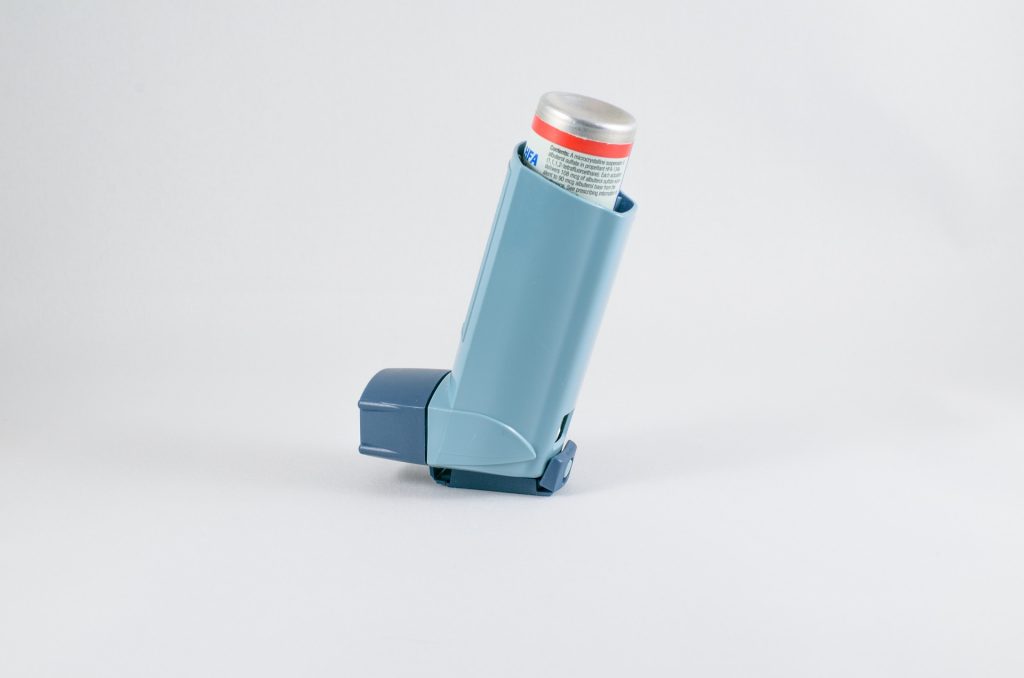
Women who are being treated for asthma are more likely to miscarry and need fertility treatment to get pregnant, according to a large study presented at the European Respiratory Society (ERS) Congress in Vienna, Austria. The study also suggests that most women with asthma are able to have babies.

The study was presented by Dr Anne Vejen Hansen from the department of respiratory medicine at Copenhagen University Hospital, Denmark.

She said: “Asthma is common in women of reproductive age. Previous studies have shown that it takes women with asthma longer to get pregnant than those without asthma when undergoing fertility treatment, and that asthmatic women who succeed in getting pregnant have more often had fertility treatment than non-asthmatic women. But most existing studies are on women who have actually got pregnant, so we wanted to examine fertility outcomes on a national scale, to also include those that might not become pregnant at all.”

The team analysed reproductive outcomes for all Danish women born from 1976 to 1999, following them from 1994 to 2017. In total, 769,880 women were included and followed; anyone who took anti-asthma medication on a regular basis was classified as asthmatic.

They found that women with asthma experienced a higher degree of foetal loss compared to women without asthma (17.0% vs. 15.7%) and more use of fertility treatment (5.6% vs. 5.0%). However, the proportion who subsequently gave birth was 77% in women with and without asthma, suggesting that asthma does not seem to affect the number of live births.

Dr Vejen Hansen said: “We found that women fulfilling the definition of asthma had a higher rate of foetal loss and an increased use of fertility treatment. The more severe the asthma and the more flare ups the women experienced, the more likely they were to need fertility treatment. Why this is, is not clear. It might be related to systemic inflammation throughout the body, including women’s reproductive organs.

“But the numbers also show that these same women who redeem asthma medication still have as many live births in the end as women who don’t. This suggests that most women with asthma probably do manage to become pregnant and have babies in the end.

“We also plan to investigate the possible effect of male asthma on fertility, and, therefore, have another similar registry-based study in the pipeline.”

Professor Lena Uller is Chair of the ERS group on Airway Pharmacology and Treatment and Head of the Respiratory Immunopharmacology research group at Lund University, Sweden, and was not involved in the research. She said: “It’s reassuring that women seem to have the same live birth rate regardless of their asthma. However, the results also indicate that women with asthma should take into consideration potential reproductive challenges in their family planning. If women with asthma are worried about their fertility, they should speak to their doctor.

“The results of this study also underscore the importance of managing asthma in reproductive-aged women. The fact that the more severe the asthma, the more the problems with fertility, suggests that uncontrolled asthma is the problem and we should be helping women to get their asthma under control.”

Source: European Respiratory Society

SIU Takes Aim at Ballooning Dodgy Medical Litigation that is Costing the Government Billions

Photo by Scott Graham on Unsplash

By Sandiso Phaliso for GroundUp

Payments of medical related legal claims (medico-legal) against the Department of Health ballooned to R2.7-billion in 2023. In 2013, it was R265-million. This is according to the Special Investigating Unit (SIU) when it briefed the Standing Committee on Public Accounts (SCOPA) last week.

SIU head advocate Andy Mothibi told SCOPA it found evidence of collusion between attorneys, touts, nurses and doctors, in both public and private healthcare. Some law firms also withdrew claims when the SIU started investigating them. This had stopped about R3-billion in fraudulent claims, he said.

Claims under investigation included those targeting families with children born with cerebral palsy, false claims of medical malpractice in state hospitals, and collusion between state healthcare workers and rogue lawyers to unlawfully secure private medical records to initiate claims against the government.

They uncovered cases of agents of rogue law firms impersonating officials of the South African Social Security Agency to secure powers of attorney on behalf of victims by claiming to be securing them social grants. He said they found two attorneys pursuing identical claims for the same individual in two different courts, and for vastly different amounts, in one case for R7.5-million and R25-million for the same patient and same condition

Mothibi said the health sector experienced an explosion of medical practice litigation cases in 2015, directed against health institutions and individual medical practitioners in both public and private practice.

Mothibi said in one case a claimant demanded R70-million for a supposedly botched circumcision at a Limpopo hospital when no circumcision had been performed.

Read the SIU presentation to Parliament

In 2017, the SIU started targeting provinces with the highest share of claims. At that stage, the Eastern Cape’s contingent liability for medico-legal claims was R15.9-billion; in Gauteng, it was R21.2-billion.

In the Eastern Cape, most medico-legal claims emanated from one Johannesburg-based law firm, Nonxuba Attorneys Incorporated. In five years, from 2012 to 2017, the firm submitted 44 claims totaling R497-million against the provincial health department. Nine claims for children born with cerebral palsy were identical each demanding R15-million.

“This was suspicious and indicated a lot of cut-and-paste on the part of this legal firm,” said Mothibi.

The company has, according to Mothibi’s presentation, been charged.

We have been unable to get hold of Nonxuba Attorneys and Business Day has previously reported that the company’s owner, Zuko Nonxuba, has been suspended from legal practice.

Also, in the Mthatha High Court claims increased from 46 to 529 between 2010 and 2016. There was collusion, said Mothibi, between some officials in the Office of the State Attorney, whereby out-of-court settlements for hefty sums were entered into without the mandate or even the knowledge of the department.

MP Veronica Mente-Nkuna (EFF) wanted to know the names of the legal firms implicated besides Nonxuba Attorneys and what the legal bodies have done about their operating licences.

She asked why the Department of Health had not conducted its investigations before the claims were paid. Who was responsible for the loss of money through these fraudulent claims, she asked.

Republished from GroundUp under a Creative Commons Attribution-NoDerivatives 4.0 International License.

Read the original article

Carriers of Sickle Cell are at Increased Risk for Certain Conditions

Sickle cell disease. Credit: National Institutes of Health

Individuals that have sickle cell trait, who did not which increases the risk of blood clots, a risk that is the same among diverse human populations that may not traditionally be associated with sickle cell disease. The study provides estimated clinical risks for people with sickle cell trait, which can inform clinical practice guidelines.

The study, published in Blood Advances, was led by researchers at National Human Genome Research Institute (NHGRI), part of NIH, The Johns Hopkins University School of Medicine, Baltimore, and the company 23andMe, South San Francisco, California. The researchers examined the largest and most diverse set of people with sickle cell trait to date, which includes data from over 19 000 people of various ancestral backgrounds with sickle cell trait. 

While people with sickle cell trait typically do not have any associated health complications, they are carriers for sickle cell disease. In rare cases, sickle cell trait has been found to be a risk factor for health complications such as muscle breakdown, presence of blood in the urine and kidney disease.

Previous research investigating the relationship between sickle cell trait and blood clots have only included individuals of African genetic ancestry and self-identified Black participants because of the incorrect assumption that the genetic carrier state only affects those who identify as Black or African American. While sickle cell trait in the United States is most prevalent in individuals who self-identify as Black or African American, individuals from all ancestral backgrounds may have sickle cell trait. Sickle cell trait is often found in individuals living in or from West and Central Africa, Mediterranean Europe, India and the Middle East.

“Because sickle cell trait is often associated with people who identify as Black or African American, it is not widely studied in other populations, a bias that has led to unintended harm for those with sickle cell trait,” says Vence Bonham Jr, J.D., who co-led the study and serves as acting deputy director and associate investigator at NHGRI. “In particular, the racialisation of sickle cell trait has resulted in biased estimations of health risks. The results of our study will help clinicians properly contextualise the risk of blood clots amongst people with sickle cell trait without unintended bias.”  

Individuals in this study are part of the 23andMe research program and have volunteered to participate in the research online and provided informed consent, which includes allowing their de-identified data to be analysed and subsequently shared with research collaborators. Using data from this research cohort, which consists of over four million participants, researchers calculated the risk of blood clots in the veins, also known as venous thromboembolism. Through statistical analyses, participants were grouped based on their genetic similarities into genetic ancestry groups. The study found that people with sickle cell trait have a 1.45-fold higher risk of venous thromboembolism than those without sickle cell trait, a risk that is similar across all studied genetic ancestry groups.

To help clinicians estimate the risk of blood clots in people with sickle cell trait in comparison to other genetic carrier states, the researchers analysed risk in people who are carriers for Factor V Leiden, a well-known inherited blood-clotting disorder. The study found that carriers for Factor V Leiden, which is more prevalent in people of European genetic ancestries, had an even higher risk of venous thromboembolism than people with sickle cell trait.

The researchers found that people with sickle cell trait have a higher risk of pulmonary embolism than those without sickle cell trait.

While previous studies have demonstrated that in individuals with sickle cell trait, the risk of blood clots occurring in the lungs is higher than the risk of clots occurring only in the legs, this study supports the link more definitively with a larger sample size.

“This study, therefore, provides important insights about patterns of venous blood clots and suggests a unique mechanism of blood clotting in people with sickle cell trait,” said Rakhi Naik, M.D., clinical director for the Division of Hematology at Johns Hopkins University, Baltimore, who co-led the study. “Knowing the risks of blood clots in people with sickle cell trait is important for situations such as surgeries or hospitalizations, which add to the risk of developing serious blood clots.”

Source: NIH/National Human Genome Research Institute

Research on the Hepatitis C Virus Reveals its Mysteries

Hepatitis C virus. Credit: Scientific Animations CC4.0

Around 58 million people suffer from chronic inflammation caused by the hepatitis C virus, and 300 000 people die from the disease every year. So far, no treatment has successfully managed to reduce the global prevalence of hepatitis C, prompting scientists to start looking for a vaccine. But limited knowledge of the protein complex that enables the virus to infect the cells has made this difficult.

A new study by a cross-disciplinary research team at the University of Copenhagen is about to change that. It is out now in the journal Nature.

“We are the first ever to identify the protein complex at the surface of the hepatitis C virus that enables it to bind to our cells,” says Associate Professor Jannick Prentø.

“This knowledge of the structure of the protein complex will enable us to design vaccine candidates that can prevent the virus from infecting the cells,” says Postdoc Elias Augestad.

The protein complex helps the virus bind to the cells. In the corona virus, it is a so-called spike protein with the well-known spikes. In the hepatitis C virus, the structure is different, but the function of the protein complex is the same.

Paves the way for vaccine development

The study can be considered a blueprint for HCV vaccine development. Scientists hope to be able to use the new knowledge to develop a vaccine which will make the immune system produce antibodies that bind effectively to the surface of the hepatitis C virus and thus render it harmless.

“Expressing and cleaning up the protein complex is extremely difficult, which is why it has not been done before. The structure of these proteins on the surface of the hepatitis C virus makes them extremely vulnerable. Researchers did not know what they were dealing with, and therefore, whenever someone tried to reproduce these protein structures in the lab they would fall apart before they could get a chance to study them,” says Associate Professor Jannick Prentø.

“But we managed to describe their structure, and this has enabled us to reproduce these protein complexes outside the cell and study them closely,” says Associate Professor Pontus Gourdon.

Source: University of Copenhagen

24hrs Oxygen Therapy for Hypoxaemia no Better than 15hrs

Photo by Samuel Ramos on Unsplash

There were no differences in quality of life, symptoms, hospital admissions or mortality between a group of patients with pulmonary disease and low oxygen levels in the blood that received oxygen therapy at home for 24 hours a day, and a group that received the same therapy for 15 hours a day, according to a study in the New England Journal of Medicine.

“This has considerable significance for patients with chronic obstructive pulmonary disease and other pulmonary diseases who are undergoing long-term oxygen therapy at home,” says study leader Magnus Ekström of Lund University.

Every year, there are about a million patients in the US alone with severe sub-optimal oxygen levels who begin oxygen therapy at home. The patient must wear a nasal oxygen mask 24 hours a day, which is often felt to be restrictive and burdensome. In addition, the gas is cold and dry, which may cause problems such as dryness, ulcers and inflammation of the airways. Using the equipment may also be perceived as stigmatising by patients.

The aim of the therapy is to prolong life, but its application differs and the scientific evidence for the therapy’s efficacy has been inadequate. 

In a randomised, controlled large multicentre study, the researchers have now examined the effect of different durations of oxygen therapy on important health outcomes. A total of 241 patients with chronic and severely low oxygen levels in the blood from 20 different clinics in Sweden were randomised to receive oxygen therapy for either 24 hours a day or 15 hours a day. The patients were followed for one year, and all completed the study. There was a high level of compliance with the prescribed daily therapy duration.  

“There were no differences between the groups regarding hospital admission or the risk of dying. And again, when we compare self-reported quality of life and physical activity, symptoms and fatigue between the groups, we see no differences or indication of advantages if oxygen is used for more than 15 hours a day,” says Magnus Ekström, researcher in respiratory and palliative medicine at Lund University.

 The results differ from previous studies conducted in the 1970s, which suggested that therapy for 24 hours a day could increase the survival rate. 

“The older studies were small and only included patients with chronic obstructive pulmonary disease, not least because it is difficult to recruit such seriously ill individuals for a randomised study. Also, the patients included in the older studies differ from those who start oxygen therapy at home nowadays. The strength of this study is  that we have been able to include twice as many patients, and that it represents the reality nowadays in which many of those receiving oxygen therapy at home are older, also have cardiovascular diseases and are women,” says Josefin Sundh, adjunct senior lecturer at Örebro University and pulmonologist at the University Hospital in Örebro, who was co-leader of the study.

“The group treated with oxygen 15 hours a day received this during the night when, in general, oxygenation is poorer. The results show that it seems to be safe for this type of patient to be without oxygen for quite a large part of the day. This is important as it may reduce the side effects of the therapy and mean that the patients can adapt it more to their everyday life,” says Magnus Ekström.

The researchers based the study on data from Swedevox, the Swedish National Registry for Respiratory Failure. 

The researchers are now moving on to examine whether therapy using a high flow rate of warmed-up, more humidified oxygen at night can improve prognoses and patient well-being.

Source: Lund University