Atrial fibrillation, a rapid, irregular heartbeat that can lead to stroke or sudden death, is three times more common than previously thought, affecting nearly 5% of the population, according to new estimates from UC San Francisco.
A-Fib, as the condition is commonly known, has been on the rise for at least the past decade, driven by the aging of the population, along with increasing rates of hypertension, diabetes and obesity. Earlier projections had estimated that 3.3 million U.S. adults had atrial fibrillation, but these have not been updated in more than two decades.
“Atrial fibrillation doubles the risk of mortality, is one of the most common causes of stroke, increases risks of heart failure, myocardial infarction, chronic kidney disease and dementia, and results in lower quality of life,” said first author Jean Jacques Noubiap, MD, PhD, a postdoctoral scholar at UCSF with a specialty in global cardiovascular health.
“Fortunately, atrial fibrillation is preventable, and early detection and appropriate treatment can substantially reduce its adverse outcomes,” he said.
Rising numbers reflect need for better prevention and treatment
UCSF investigators reviewed the medical records of nearly 30 million adult patients who received some form of acute or procedural care in California from 2005 to 2019. About 2 million of these people had been diagnosed with A-Fib, and the numbers grew over time, rising from 4.49% of the patients treated between 2005 and 2009 to 6.82% of the patients treated between 2015 and 2019.
The data were standardised to reflect the entire country, and researchers estimated the current national prevalence to be at least 10.55 million. They also found that during the study timeframe, A-Fib patients skewed younger, were less likely to be female and more likely to have hypertension and diabetes.
A-Fib has a broad spectrum of complications from shortness of breath and light-headedness to blood clots, stroke and even heart failure. Studies have shown that people with A-Fib are up to five times more likely to have a stroke. The authors said that by outlining the scope of the problem, these new estimates can help guide health care planning, resource allocation and public health interventions.
“Physicians recognise that atrial fibrillation is often encountered in essentially every field of practice,” said senior and corresponding author Gregory M. Marcus, MD, MAS, a cardiologist and electrophysiologist at UCSF Health. “These data provide objective evidence to demonstrate that prior projections severely underestimated how common it truly is.”
Digital technologies may reveal it is even more common than the current analysis indicates.
“With the growing use of consumer wearables designed to detect atrial fibrillation combined with safer and more effective means to treat it, this current prevalence of atrial fibrillation in health care settings may soon be dwarfed by future health care utilisation that will occur due to the disease,” Marcus said.
In a review of 27 different studies, a Johns Hopkins Children’s Center team concludes that some video games created as mental health interventions can be helpful – if modest – tools in improving the mental well-being of children and teens with depression and attention-deficit/hyperactivity disorder (ADHD). They did not significantly help with anxiety, however.
A report on the review of studies from peer-reviewed journals between 2011 and March 20, 2024, was published in JAMA Pediatrics.
“We found literature that suggests that even doubling the number of paediatric mental health providers still wouldn’t meet the need,” says Barry Bryant, MD, a resident in the Department of Psychiatry and Behavioral Sciences in the Johns Hopkins University School of Medicine and first author of the new study.
In a bid to determine if so-called “gamified digital mental health interventions,” or video games designed to treat mental health conditions, benefited those with anxiety, depression and ADHD, the research team analysed their use in randomised clinical trials for children and adolescents.
Bryant and child and adolescent psychologist Joseph McGuire, PhD, identified 27 such trials from the US and around the world. The studies overall included 2911 participants with about half being boys and half being girls, ages six to 17 years old.
The digital mental health interventions varied in content, but were all created with the intent of treating ADHD, depression and anxiety. For example, for ADHD, some of the games involved racing or splitting attention, which required the user to pay attention to more than one activity to be successful in gameplay. For depression and anxiety, some of the interventions taught psychotherapy-oriented concepts in a game format. All games were conducted on technology platforms, such as computers, tablets, video game consoles and smartphones. The video games are available to users in a variety of ways. Some are available online, while others required access through specific research teams involved in the studies.
The research team’s analysis found that video games designed for patients with ADHD and depression provided a modest reduction (both with an effect size of .28) in symptoms related to ADHD and depression, such as improved ability to sustain attention and decreased sadness, based on participant and family feedback from the studies. (An effect size of .28 is consistent with a smaller effect size, where as in-person interventions often produce moderate [.50] to large [.80] effects.) By contrast, video games designed for anxiety did not show meaningful benefits (effect size of .07) for reducing anxiety symptoms for participants, based on participant and family feedback.
Researchers also examined factors that led to improved benefit from digital mental health interventions. Specific factors related to video game delivery (i.e., interventions on computers and those with preset time limits) and participants (i.e., studies that involved more boys) were found to positively influence therapeutic effects. Researchers say these findings suggest potential ways to improve upon the current modest symptom benefit.
“While the benefits are still modest, our research shows that we have some novel tools to help improve children’s mental health – particularly for ADHD and depression – that can be relatively accessible to families,” says Joseph McGuire, Ph.D., an author of the study and an associate professor of psychiatry and behavioural sciences in the school of medicine. “So if you are a paediatrician and you’re having trouble getting your paediatric patient into individual mental health care, there could be some gamified mental health interventions that could be nice first steps for children while waiting to start individual therapy.”
The team cautioned that their review did not indicate why certain video game interventions performed better than others. They also note that some of the trials included in the study used reported outcome measures, and the studies did not uniformly examine the same factors which could have influenced the effects of the treatment. Some of the video games included in the studies are not easily accessible to play.
The researchers also noted that while video game addiction and the amount of screen time can be concerns, those children who played the games studied in a structured, time-limited format tended to do best.
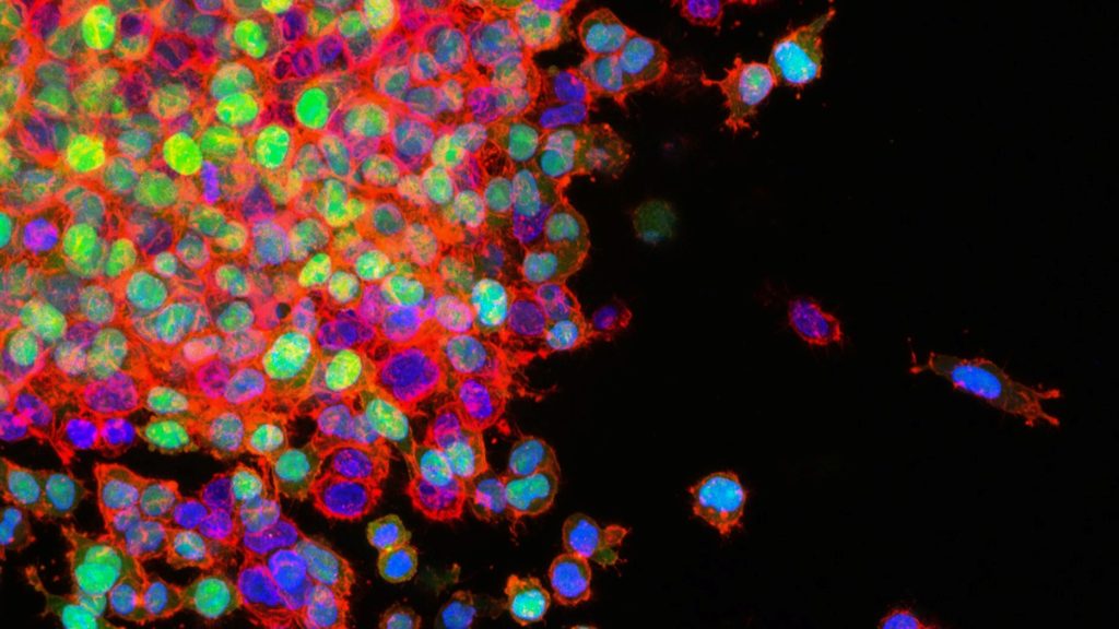
Lung cancer metastasis. Credit: National Cancer Institute
In research published in Nature Communications, scientists have tested a combination of treatments in mice with lung cancer and shown that these allow immunotherapies to target non-responsive tumours.
The study findings, from Francis Crick Institute, in collaboration with Revolution Medicines, show that targeting tumours in different ways simultaneously might increase response to treatments.
The scientists tested a combination of tool compounds in mice with lung cancer. These compounds were used to represent:
Targeted drugs which block a cancer-causing protein called KRAS G12C. These have been approved for use in lung cancer, but often fail to benefit patients in the long term because the tumours develop resistance to these medicines over time.
Immunotherapy drugs. These are designed to stimulate the immune system to fight the tumour, but only 20% of people with lung cancer respond, as tumours often block immune cells from entering.
The researchers combined a newly identified KRAS G12C inhibitor, with a compound that blocks a protein called SHP2, which inhibits cancer cells and can also activate tumor immunity.
These two inhibitors were combined with an immune checkpoint inhibitor, which blocks proteins that help the cancer cells hide from the immune system.
Obstructive sleep apnoea may be a risk factor for developing abdominal aortic aneurysms, according to researchers from the University of Missouri School of Medicine and NextGen Precision Health.
Abdominal aortic aneurysms occur when the aorta swells and potentially ruptures, causing life-threatening internal bleeding. Obstructive sleep apnoea is characterised by episodes of a complete or partial airway collapse with an associated drop in oxygen saturation or arousal from sleep. It can increase the risk of developing cardiovascular problems. Citing studies that indicate a higher prevalence of abdominal aortic aneurysms in patients with obstructive sleep apnoea, MU researchers examined the link between the two using mouse models.
The research team found that intermittent hypoxia caused by obstructive sleep apnoea increased the susceptibility of mice to develop abdominal aortic aneurysms.
“Chronic intermittent hypoxia by itself is not enough to cause abdominal aortic aneurysms, but for a patient with obstructive sleep apnoea who also has additional metabolic problems like obesity, our findings suggest it may help degrade aortic structures and promote aneurysm development,” said Luis Martinez-Lemus, study author and a professor of medical pharmacology and physiology.
Intermittent hypoxia happens during obstructive sleep apnoea when throat muscles relax and block the flow of air into the lungs. According to the research, the loss of oxygen triggers certain enzymes called MMPs. The increased enzyme activity can degrade the extracellular matrix, which acts like a cell scaffolding network, weakening the aorta.
“Patients with abdominal aortic aneurysms usually don’t notice any symptoms, except for some back and belly pain, until the aneurysm bursts. Once that happens, it’s crucial to get the patient to surgery quickly so doctors can repair the aorta,” said Neekun Sharma, the lead author of the study. “Learning how these aneurysms develop can help us find ways to monitor or slow down their progression, especially for patients who have obstructive sleep apnoea.”
Craig Comrie, chairperson of the Health Funders Association
Wednesday, 25 September 2024, The passage of the National Health Insurance (NHI) Bill into an Act of law has set the stage for one of the most significant overhauls of South Africa’s healthcare system. As the government embarks on this ambitious plan, the stakes have never been higher. The NHI Act is more than a mere piece of legislation; it stands as a test of constitutional rights and the nation’s commitment to fostering a more inclusive and equitable society.
The Health Funders Association (HFA) welcomes the meeting between President Cyril Ramaphosa and Business Unity South Africa (BUSA) leadership on Tuesday, 17 September, to discuss the NHI as a positive step.
As a member of BUSA, we find it encouraging that the Minister and Deputy Minister of Health, along with other senior officials, were part of this constructive and forward-looking discussion. We can only hope that these recent discussions will mark the start of a series of engagements with key stakeholders, as the South African government must engage in open dialogue with all stakeholders, including private healthcare providers, medical schemes, and the general public. HFA will provide industry input to BUSA’s presentation to the President to help propose workable solutions to set South Africa’s healthcare train on the right track with inclusive mechanisms that will benefit all.
Constitutional rights and freedom of choice
The critical questions about the Act’s constitutional validity, economic feasibility, and potential impact on both public and private healthcare sectors, specifically the role of medical schemes, remain unanswered.
Recently, during a Q&A session, the President reaffirmed his belief that the Act is constitutionally sound but he declined to share specifics on how this conclusion was reached. In response, the Health Funders Association (HFA) will test various aspects of the NHI’s constitutionality, which is crucial for establishing a stable healthcare framework that delivers quality health services for all South Africans.
This is a critical juncture for politicians, policymakers and every South African who relies on the nation’s healthcare services – private and public sectors alike. Against this background, the HFA is seeking to strike a balance between much-needed healthcare reforms and the hard-hitting risks of the NHI Act that demand amendments.
Government has been adamant about the NHI’s transformative potential to address inequalities in healthcare access, contending that the NHI is aligned with Section 27(1) of the Constitution, which guarantees the right to access healthcare services, including reproductive healthcare.
On the other hand, this part of the Act cannot be read without the consideration of Section 36 which indicates that existing rights should not be compromised if there are alternative ways to achieve, in this case, universal health coverage. This is a key part of responding to the President’s invitation for alternative proposals that stand a more realistic chance of achieving a partnership with the private sector for improving healthcare for consumers.
Any alternative proposals provided by the private sector will come with the need to amend the current. The Act, if left unchallenged in its current form, can be likened to letting the healthcare train run on a single track of public sector inefficiency. By adding the private sector as a parallel track – both heading in the same direction towards universal healthcare coverage, we can stabilise and accelerate the journey.
The medical scheme sector effectively provides funding to the majority of services in the private sector with less than 10% of health consumers paying for services out of pocket in the private sector. This population group serves a significant portion of the taxpaying population, yet its role in the new system remains ambiguous. Will private healthcare consumers be forced to rely solely on centrally procured services, or will there be room for a coexistence that stands a better chance of laying the track towards a successful healthcare future that can benefit all South Africans?
Transparency and collaboration are essential in addressing the questions surrounding the NHI’s financial viability, human resource capacity, and broader economic impact. The future of South Africa’s healthcare system hinges on finding a balanced approach that ensures quality, accessibility, and economic sustainability for all citizens.
The government proposes funding the NHI through general taxes and mandatory payroll contributions. However, this plan has been met with scepticism as it raises considerable questions about the economic burden on taxpayers, particularly given the country’s high unemployment rate of 33.5% and sluggish economic growth rates of around 0.4% in the second quarter of this year. Critics argue that increasing tax rates to fund the NHI could backfire, reducing overall tax revenue, as highlighted by the Laffer Curve, which suggests that higher taxes can lead to lower revenue if they exceed an optimal rate.
Where will the healthcare professionals come from?
The public healthcare sector faces its own set of challenges. Reports indicate that the quality of care in public health facilities is often subpar, with systemic inefficiencies and resource constraints leading to poor health outcomes. The sector is grappling with high vacancy rates for healthcare professionals, exacerbating the strain on an already overburdened system. As it is, South Africa is already facing a severe shortage of medical personnel, with the vacancy rate for doctors ranging from 22.4% in the Free State to 5.5% in the Western Cape, while the national average vacancy rate for nurses stands at 14.7%.
The current system is struggling to fill thousands of vacancies for doctors, nurses and allied healthcare professionals, with over 2 000 unfunded vacant posts for medical doctors across nine provinces requiring an estimated R2.4 billion to fill them. Many provinces report alarming doctor vacancy rates: 18.5% in Mpumalanga, 17.6% in Limpopo, and 11.3% in KwaZulu-Natal, among others. The shortage is not just a numbers game; it affects the quality of healthcare that can be delivered. These figures raise serious concerns about the system’s capacity to deliver reliable and accessible healthcare services hampered by corruption and lack of proper leadership and management.
We must consider the strain this places on those simply trying to cope with the current healthcare demand from a growing population. The NHI aims to provide universal health coverage, but where will the necessary healthcare professionals come from?
The way forward
The NHI is not the only solution to South Africa’s healthcare challenges and is certainly not a panacea in its current form. The government must take a more collaborative approach, engaging with all stakeholders, including the private healthcare sector, medical schemes and the general public. Open dialogue is crucial to finding sustainable solutions that work for everyone. Critical decisions must be made, and these should not be taken behind closed doors. The nation’s healthcare needs are too important to be dictated by a select few without broader consultation.
The government has a constitutional obligation to ensure every South African has access to quality healthcare services. However, this must not come at the expense of freedom of choice, job losses and economic stability.
I urge President Cyril Ramaphosa to protect the constitutional rights of all South Africans and engage in meaningful dialogue with all stakeholders. The future of South Africa’s healthcare system depends on our collective ability to find innovative and inclusive solutions.
Joshua Lupton, MD, has no memory of his own cardiac arrest in 2016. He only knows that first responders resuscitated his heart with a shock from a defibrillator, ultimately leading to his complete recovery and putting him among fewer than one in 10 people nationwide who survive cardiac arrest outside of a hospital.
He attributes his survival to the rapid defibrillation he received from first responders – but not everybody is so fortunate.
Now, as lead author on an observational study published in JAMA Network Open, he and co-authors from Oregon Health & Science University say the study suggests the position in which responders initially place the two defibrillator pads on the body may make a significant difference in returning spontaneous blood circulation after shock from a defibrillator.
Researchers used data from the Portland Cardiac Arrest Epidemiologic Registry, which comprehensively recorded the placement position of defibrillation pads from July 1, 2019, through June 30, 2023. For purposes of the study, researchers reviewed 255 cases treated by Tualatin Valley Fire & Rescue, where the two pads were placed either at the front and side or front and back.
They found placing the pads in front and back had 2.64-fold greater odds of returning spontaneous blood circulation, compared with placing the pads on the person’s front and side.
The current common knowledge among health care professionals is that pad placement – whether front and side, or front and back – is equally beneficial in cardiac arrest. The researchers cautioned that their new study is only observational and not a definitive clinical trial. Yet, given the crucial importance of reviving the heartbeat as quickly as possible, the results do suggest a benefit from placing the pads on the front and back rather than the front and side.
“The key is, you want energy that goes from one pad to the other through the heart,” said senior author Mohamud Daya, MD, professor of emergency medicine in the OHSU School of Medicine.
Placing the pads in the front and back may effectively “sandwich” the heart, raising the possibility that the electrical current will be delivered more comprehensively to the organ.
However, that’s not readily possible in many cases. For example, the patient may be overweight or positioned in such a way that they can’t be easily moved.
“It can be hard to roll people,” said Daya, who also serves as medical director for Tualatin Valley Fire & Rescue. “Emergency medical responders can often do it, but the lay public may not be able to move a person. It’s also important to deliver the electrical current as quickly as possible.”
In that respect, pad placement is only one factor among many in successfully treating cardiac arrest.
Lupton survived his cardiac arrest and went on to complete medical school at the very hospital where he spent several days recovering in the intensive care unit – Johns Hopkins University in Baltimore. The episode led him to alter the focus of his research so that he could examine ways to optimise early care for cardiac arrest patients.
The results of the new study surprised him.
“I didn’t expect to see such a big difference,” he said. “The fact that we did may light a fire in the medical community to fund some additional research to learn more.”
Although new cancer diagnoses largely returned to pre-pandemic levels by 2021, the recovery does not account for the potential missed diagnoses due to delays in screening and other medical care in early 2020. Credit: National Cancer Institute
Cancer incidence trends in 2021 largely returned to what they were before the COVID pandemic, according to a study by researchers at the National Institutes of Health (NIH). However, there was little evidence of a rebound in incidence that would account for the decline in diagnoses in 2020, when screening and other medical care was disrupted, according to findings published in the Journal of the National Cancer Institute. One exception was breast cancer, where the researchers did see an uptick in diagnoses of advanced-stage disease in 2021.
A previous study showed that new cancer diagnoses fell abruptly in early 2020, as did the volume of pathology reports, suggesting that many cancers were not being diagnosed in a timely manner. To determine whether these missed diagnoses were caught in 2021, possibly as more advanced cancers, researchers from NIH’s National Cancer Institute (NCI) compared observed cancer incidence rates for 2021 with those expected from pre-pandemic trends using data from NCI’s Surveillance, Epidemiology, and End Results Program.
A full recovery in cancer incidence should appear as an increase over pre-pandemic levels (also known as a rebound) to account for the missed diagnoses. The researchers looked at cancer overall, as well as five major cancer types that vary in how they are typically detected: through screening (female breast and prostate cancer), due to symptoms (lung and bronchus and pancreatic cancer), or incidentally during other medical procedures (thyroid cancer).
Cancer incidence rates overall and for most specific cancers approached pre-pandemic levels, with no significant rebound to account for the 2020 decline. However, in addition to an uptick in new diagnoses of advanced breast cancer in 2021, the data also provided some evidence of an increase in diagnoses of advanced pancreatic cancer. Also, new diagnoses of thyroid cancers in 2021 were still below pre-pandemic levels.
The researchers concluded that 2021 was a transition year that was still affected by new variants and new waves of COVID-19 cases, which continued to impact medical care. They said the findings highlight the need for ongoing monitoring to understand the long-term impacts of the pandemic on cancer diagnoses and outcomes.
Parkinson’s disease, a debilitating nervous system disorder, is treated with medications that sometimes cause impaired decision-making and poor impulse control. Now, researchers from Fujita Health University in Japan have identified a structure in the brain called the external globus pallidus which may be responsible for this side effect, paving the way for new treatments.
Parkinson’s disease (PD), also known simply as Parkinson’s, is a disorder of the nervous system that affects millions of people worldwide. The nerve cell damage associated with Parkinson’s can cause tremors, slowed movements, problems with balance, and many other symptoms which worsen gradually over time. Although there is no cure, there are medications available that can treat PD symptoms. Some of these medications, however, have previously unexplained side effects – including impaired decision-making that leads to potentially harmful behaviours such as pathological gambling, binge eating and compulsive shopping.
Now, in a study published in the International Journal of Molecular Sciences, researchers at Fujita Health University in Japan, led by Assistant Professor Hisayoshi Kubota from the Division of Behavioral Neuropharmacology, International Center for Brain Science (ICBS), Fujita Health University, have investigated the mechanism by which a drug called pramipexole or PPX impairs the decision-making process in mice with Parkinson’s disease. The research was co-authored by Professor Taku Nagai from the Division of Behavioral Neuropharmacology, International Center for Brain Science (ICBS), and Professor Hirohisa Watanabe from the Department of Neurology, School of Medicine, both at Fujita Health University.
To take a closer look at the findings of this study, we first need to understand how PPX works to alleviate PD symptoms. PD mainly results from a loss of nerve cells or neurons that produce a compound called dopamine. Some neurons are dependent on dopamine for their regular functioning – they have structures called ‘dopamine receptors’ which can be thought of as locks which can then be activated using dopamine as the ‘key’. Drugs like PPX can imitate the function of dopamine and bind to these receptors instead, especially in patients with PD who lack dopamine-producing neurons.
To study the effects of PPX on PD, the researchers injected the brains of mice with a toxin called 6-hydroxydopamine (or 6-OHDA). 6-OHDA damages neurons in a very similar manner to that observed in the brains of patients with PD. The mice were treated with PPX and then subjected to a touchscreen-based ‘gambling task’ to test their decision-making skills. Interestingly, these mice picked the high-risk/high-reward option much more often – they opted for a disadvantageous outcome where they received a large reward (of strawberry milkshake), which also comes with an increased risk of a large punishment by exposure to flashing lights.
But which part of the brain is responsible for this behaviour? Investigating the brains of mice treated with PPX revealed that a region deep inside the brain called the external globus pallidus (GPe) was hyperactivated, or showed a much higher level of neuron activity. The researchers then chemically inhibited the neurons in the GPe, which actually reduced disadvantageous risk-taking activity in the mice. This proved that hyperactivation of the GPe was indeed responsible for poor decision-making in the mice treated with PPX.
This study has huge implications for treating patients with Parkinson’s disease. “Our findings could lead to the development of new medications or interventions that specifically target the external globus pallidus,” explains Dr. Kubota. “This would help to prevent or reduce decision-making impairments in Parkinson’s disease patients.“
Besides helping medical professionals develop better treatments for Parkinson’s disease, these findings can also help improve awareness among affected patients, their families, as well as the general public. Dr. Kubota, explains that “Investigating how Parkinson’s disease medications affect decision-making will help the public to better understand the complexity of the disease and its treatment.” He also says “This will benefit patients, their families and carers, and motivate them to consider early care and preventive strategies.”
These findings shed new light on the complex processes in the brain that aid our everyday decision-making skills, and promise to improve quality of life for patients affected by Parkinson’s disease. Maybe we can take away some important lessons from this study as well, and think twice before we indulge in poor decision-making in our daily lives!
In a remarkable achievement, CareFirst, the innovative healthcare web app developed by First Care Solutions, has secured 1st place in the Health Section at the 2024 Stuff Awards. Held annually by Stuff Magazine, these prestigious awards recognise excellence in digital innovation and technological advancement.
CareFirst has quickly emerged as a game-changer in South Africa’s healthcare landscape, offering users:
Instant medical consultations with qualified doctors, available 24/7 – 365
AI-powered Vital Scanning: Advanced technology to monitor key health metrics including blood pressure, respiratory rate, and heart rate.
Comprehensive Medical Services: Convenient access to doctors’ consultations, prescriptions, sick notes, and referrals.
‘’I’m really proud of what our team has created. CareFirst is a platform that can really transform the way people access healthcare,’’ says Dr Steve Holt, Chief Executive Officer – at First Care Solutions.
CareFirst represents a shift towards more accessible, efficient and personalised care, promising a future where quality care is available to everyone, regardless of their location or time constraint and this award is a testament to that.
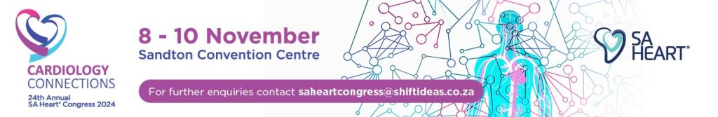
The SA Heart Annual Congress will take place from 8–10 November at the Sandton Convention Centre, Johannesburg. The three-day Congress, themed ‘Cardiology Connections,’ will promote collaboration and dialogue among local and international Cardiology professionals. The congress offers a unique platform for experts, practitioners, and researchers worldwide to share insights on the latest advancements and challenges in cardiovascular medicine.
The dynamic programme includes keynote speeches, panel discussions, workshops, and networking sessions. The agenda covers a comprehensive range of cardiology topics, designed to provide practical knowledge and inspire innovation in the field. Attendees will gain critical insights into the latest developments that have the potential to enhance patient care.
“We are excited to welcome a distinguished international and local faculty,” says Dr Ahmed Vachiat, SA Heart Congress Convenor. “At the core of SA Heart is the mission to advance cardiovascular care through education, research, and advocacy. By connecting healthcare professionals from across sectors, this Congress will drive forward our vision of improving cardiovascular care for all in South Africa. We are also grateful for the invaluable support of our local experts, whose contributions consistently uphold international standards of excellence.”
A significant focus this year is strengthening connections among various special interest groups, including the Society of Cardiovascular Interventions (SASCI), Cardiovascular Imaging Society of South Africa (CISSA), Cardiovascular Arrhythmia Society of South Africa (CASSA), Heart Failure Association of South Africa (HEFFSA), Intervention Society of Cardiovascular Allied Professionals (ISCAP), South African Society of Cardiovascular Research (SASCAR), and the Paediatric Society of Cardiology (PCSSA).
Joint sessions and interdisciplinary programmes will enable these groups to work together to enhance healthcare delivery for all patients in need of cardiac intervention and treatment. Workshops and scientific sessions will feature innovative learning approaches aimed at facilitating knowledge exchange and professional growth.
A cardiovascular team from the Mayo Clinic – Prof Vuyi Nkomo (Imaging Cardiologist), Prof Sorin Pislaru (Chair, Structural Heart Disease), and Dr Juan Crestanello (Chair, Cardiothoracic Surgery) – will conduct an echocardiography workshop and contribute to various specialist workshops on Friday morning, November 8th.
Dr Thomas Alexander, a respected interventional cardiologist based in India, will share insights on establishing STEMI networks in South Africa. Prof Stylianos Pyxaras from Germany and Dr Andrew Ludwiniec from the UK will discuss chronic total occlusions and complex coronary interventions. Prof Azfar Zaman and Prof Roy Gardner also from the UK and leaders in their field, as well as Prof Thierry Lefevre from France, will join esteemed local experts in addressing important cardiovascular topics.
A new addition to this year’s programme is the Imbizo on Rheumatology and Cardiac diseases. Over 40 Abstracts have been submitted and research sessions guided by SASCAR will be keeping delegates up to date with the latest in the field of Cardiology.
In addition, an excellent parallel paediatric programme will feature global leaders, Prof Krishna Kumar, from India and Prof McDaniel from the USA, with a pre-congress workshop and highly interactive sessions that will incorporate insights from local experts.
“This year, a Heartbeat Stage will feature insightful talks, engaging presentations, and a special networking address,” says Dr Vachiat. “We are honoured to have Dr Imtiaz Sooliman from Gift of the Givers, who will share his thoughts on ‘Connecting Hearts and Social Responsibility’.”