A new study led by an investigator from Brigham and Women’s Hospital, evaluated a cuffless monitor that uses optical sensors to record blood pressure continually and efficiently, without disruption to the patient. The study, published in Frontiers in Medicine, highlights promising advancements in hypertension diagnosis, risk assessment and management that may be enabled by use of cuffless devices.
“The successful management of hypertension depends on patients being able to take blood pressure measurements easily and reliably outside of the traditional doctor’s office setting,” said corresponding author Naomi Fisher, MD, of the Division of Endocrinology, Diabetes and Hypertension at Brigham and Women’s Hospital. “Cuffless devices have the potential to revolutionise hypertension management. They provide many more readings than traditional devices, during both the day and night, which can help confirm the diagnosis of hypertension and guide medication titration.”
Medical guidelines increasingly recommend the incorporation of at-home blood pressure monitoring into hypertension diagnosis and management. This is because isolated blood pressure readings taken at a clinician’s office may be inaccurate: for some, blood pressure tends to rise in medical settings (“white coat hypertension”) while others have normal blood pressure during examination despite hypertensive readings at home (“masked hypertension”).
Time-in-target-range (TTR) describes how often a patient’s blood pressure is in the normal range, and it is emerging as a promising metric of cardiovascular risk. But TTR requires more frequent blood pressure readings that can feasibly be obtained by patients with traditional blood pressure cuffs, which can be inconvenient, burdensome and sometimes uncomfortable for patients.
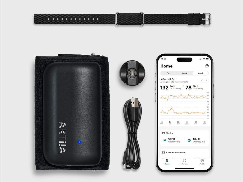
Fisher, who designed and led the study, collaborated with co-authors from Aktiia SA, a Swiss biotechnology company, to analyse over 2.2 million blood pressure readings from 5189 subjects in Europe and the U.K. who wore a cuffless wrist monitor manufactured by Aktiia. On average, the Aktiia device collected 29 readings per day, a substantial increase from the number of blood pressure readings patients typically take with home devices (guidelines recommend four per day, which is more than most patients measure). Over a 15-day period, the researchers obtained an average of 434 readings from each patient.
By calculating TTR over a 15-day period, the researchers were able to risk stratify participants by percentage of readings in target range and compare these classifications to those generated via traditional measurement patterns, using either 24-hour or week-long daytime monitoring schedules. They found that the traditional methods misclassified 26 and 45 percent of subjects, respectively, compared to the reference TTR. They determined that continual monitoring for seven days is required to obtain 90 percent or greater accuracy in hypertension risk classification, a frequency of measurement that may only be possible with cuffless monitors.
Though the cuffless device studied here has not been approved by the US Food and Drug Administration, it has been validated in multiple studies and is available for over-the-counter purchase in Europe and the UK. Work to evaluate and set standards for such devices in the U.S. is ongoing.
“For the first time, by using a cuffless device, we can collect continual out-of-office blood pressure readings and use these data to calculate a new metric, time-in-target-range, which shows great promise as a predictor of risk,” Fisher said. “The use of cuffless devices could create a shift in the paradigm of blood pressure monitoring and hypertension management.”
Dr Ali Bashashati observes an endometrial cancer sample on a microscope slide. Credit: University of British Columbia
A discovery by researchers at the University of British Columbia promises to improve care for patients with endometrial cancer, the most common gynaecologic malignancy. Using artificial intelligence (AI) to spot patterns across thousands of cancer cell images, the researchers have pinpointed a distinct subset of more stubborn endometrial cancer that would otherwise go unrecognised by traditional pathology and molecular diagnostics.
The findings, published in Nature Communications, will help doctors identify patients with high-risk disease who could benefit from more comprehensive treatment.
“Endometrial cancer is a diverse disease, with some patients much more likely to see their cancer return than others,” said Dr Jessica McAlpine, professor at UBC. “It’s so important that patients with high-risk disease are identified so we can intervene and hopefully prevent recurrence. This AI-based approach will help ensure no patient misses an opportunity for potentially lifesaving interventions.”
AI-powered precision medicine
The discovery builds on work by Dr McAlpine and colleagues in the Gynaecologic Cancer Initiative, who in 2013 helped show that endometrial cancer can be classified into four subtypes based on the molecular characteristics of cancerous cells, with each posing a different level of risk to patients.
Dr McAlpine and team then went on to develop an innovative molecular diagnostic tool, called ProMiSE, that can accurately discern between the subtypes. The tool is now used across parts of Canada and internationally to guide treatment decisions.
Yet, challenges remain. The most prevalent molecular subtype, encompassing approximately 50% of all cases, is largely a catch-all category for endometrial cancers lacking discernible molecular features.
“There are patients in this very large category who have extremely good outcomes, and others whose cancer outcomes are highly unfavourable. But until now, we have lacked the tools to identify those at-risk so that we can offer them appropriate treatment,” said Dr McAlpine.
Dr McAlpine turned to long-time collaborator and machine learning expert Dr.Ali Bashashati, an assistant professor of biomedical engineering and pathology and laboratory medicine at UBC, to try and further segment the category using advanced AI methods.
Dr Bashashati and his team developed a deep learning AI model that analyses images of tissue samples collected from patients. The AI was trained to differentiate between different subtypes, and after analysing over 2300 cancer tissue images, pinpointed the new subgroup that exhibited markedly inferior survival rates.
“The power of AI is that it can objectively look at large sets of images and identify patterns that elude human pathologists,” said Dr Bashashati. “It’s finding the needle in the haystack. It tells us this group of cancers with these characteristics are the worst offenders and represent a higher risk for patients.”
Bringing the discovery to patients
The team is now exploring how the AI tool could be integrated into clinical practice alongside traditional molecular and pathology diagnostics.
“The two work hand-in-hand, with AI providing an additional layer on top of the testing we’re already doing,” said Dr McAlpine.
One benefit of the AI-based approach is that it’s cost-efficient and easy to deploy across geographies. The AI analyses images that are routinely gathered by pathologists and healthcare providers, even at smaller hospital sites in rural and remote communities, and shared when seeking second opinions on a diagnosis.
The combined use of molecular and AI-based analysis could allow many patients to remain in their home communities for less intensive surgery, while ensuring those who need treatment at a larger cancer centre can do so.
“What is really compelling to us is the opportunity for greater equity and access,” said Dr Bashashati. “The AI doesn’t care if you’re in a large urban centre or rural community, it would just be available, so our hope is that this could really transform how we diagnose and treat endometrial cancer for patients everywhere.”
By Ben Selier, Vice President: Secure Power, Anglophone Africa at Schneider Electric
The adage, knowledge is king couldn’t be more applicable when it comes to the collection and utilisation of data. And at the heart of this knowledge and resultant information lies the datacentre. Businesses and users count on datacentres, and more so in critical services such as healthcare.
Many hospitals today rely heavily on electronic health records (EHR), and this information resides and is backed up in on-premises datacentres or in the cloud. Datacentres are therefore a major contributor to effective and modernised healthcare.
There are several considerations when designing datacentres for healthcare. For one, hospitals operate within stringent legislation when it comes to the protection of patient information. The National Health Act (No. 61 of 2003), for example, stipulates that information must not be given to others unless the patient consents or the healthcare practitioner can justify the disclosure.
Datacentres form part of critical systems
To add an extra layer of complexity, in South Africa, datacentres should feature built-in continuous uptime and energy backup due to the country’s unstable power supply. Hospitals must therefore be designed to be autonomous from the grid, especially when they provide emergency and critical care.
Typically, datacentres are classified in tiers, with the Uptime Institute citing that a Tier-4 datacentre provides 99.995% availability, annual downtime of 0.4 hours, full redundancy, and power outage protection of 96 hours.
In healthcare and when one considers human lives, downtime is simply not an option. And whilst certain healthcare systems and its resultant availability are comparable to a typical Tier-3 or Tier-4 scenario, critical systems in hospitals carry a higher design consideration and must run 24/7 with immediate availability.
In healthcare, the critical infrastructure of a hospital enjoys priority. What this means is the datacentre is there to protect the IT system which in turn ensures the smooth running of these critical systems and equipment. There is therefore a delicate balance between the critical systems and infrastructure, and the datacentre, one can’t exist without the other.
Design considerations
To realise the above, hospitals must feature a strong mix of alternative energy resources such as backup generators, uninterrupted power supply (UPS) and renewables such as rooftop solar.
Additionally, like most organisations, storage volume and type and cloud systems will also vary from hospital to hospital. To this end, datacentre design for hospitals is anything but cookie cutter; teams need to work closely with the hospital whilst meeting industry standards for healthcare.
When designing healthcare facilities system infrastructure, the following should also be considered:
Software like Building Management Systems (BMS) are not just about building efficiency but also offer benefits such as monitoring and adjusting indoor conditions like temperature control, humidity, and air quality.
The BMS contributes to health and safety and critical operations in hospitals whilst also enabling patient comfort.
Maintenance – both building and systems maintenance transcend operational necessity and become a matter of life or death.
As mentioned, generators are essential when delivering continuous power which means enough fuel must be stored to run it. Here, hospitals must store fuel safely and in compliance with stringent regulations. In South Africa, proactively managing the refuelling timelines is also critical. The response times of refuelling these (fuel) bunkers can be severely hindered by issues such as traffic congestion as a result of outages and lights now working.
Selecting the right equipment for hospitals is therefore a delicate balance between technological advancement and safety. For instance, while lithium batteries offer many benefits, when used in hospitals, it is paramount that it is also stored in dry, cool and safe location.
Here, implementing an extinguishing system is a must to alleviate any potential damage from fire or explosions. That said, lithium batteries are generally considered safe to use but it’s important to be cognisant of its potential safety hazards.
Ultimately, hospitals carry the added weight of human lives which means the design of critical systems require meticulously planning and executed.
Scientists identify a positive molecular feedback loop which could explain stroke-induced memory loss.
Ischaemic and haemorrhagic stroke. Credit: Scientific Animations CC4.0
In learning, neurons communicate with each other, and the connections between them getting stronger with repetition. This is known as long-term potentiation or LTP.
Another type of LTP occurs when the brain is deprived of oxygen temporarily – anoxia-induced long-term potentiation or aLTP. aLTP blocks the former process, thereby impairing learning and memory. Therefore, some scientists think that aLTP might be involved in memory problems seen in conditions like stroke.
Researchers at the Okinawa Institute of Science and Technology (OIST) and their collaborators have studied the aLTP process in detail. They found that maintaining aLTP requires the amino acid glutamate, which triggers nitric oxide (NO) production in both neurons and brain blood vessels. This process forms a positive glutamate-NO-glutamate feedback loop. Their study, published in iScience, indicates that the continuous presence of aLTP could potentially hinder the brain’s memory strengthening processes and explain the memory loss observed in certain patients after experiencing a stroke.
The brain’s response to low oxygen
When there is a lack of oxygen in the brain, the neurotransmitter glutamate is released from neurons in large amounts. This increased glutamate causes the production of NO. NO produced in neurons and brain blood vessels boosts glutamate release from neurons during aLTP. This glutamate-NO-glutamate loop continues even after the brain gets enough oxygen.
“We wanted to know how oxygen depletion affects the brain and how these changes occur,” stated Dr Han-Ying Wang, a researcher in the former Cellular and Molecular Synaptic Function Unit at OIST and lead author of the study,. “It’s been known that nitric oxide is involved in releasing glutamate in the brain when there is a shortage of oxygen, but the mechanism was unclear.”
During a stroke, when the brain is deprived of oxygen, amnesia – the loss of recent memories – can be one of the symptoms. Investigating the effects of oxygen deficiency on the brain is important because of the potential medicinal benefits. “If we can work out what’s going wrong in those neurons when they have no oxygen, it may point in the direction of how to treat stroke patients,” Dr Patrick Stoney, a scientist in OIST’s Sensory and Behavioral Neuroscience Unit, explained.
Brain tissues from mice were placed in a saline solution, mimicking the natural environment in the living brain. Normally, this solution is oxygenated to meet the high oxygen demands of brain tissue. However, replacing the oxygen with nitrogen allowed the researchers to deprive the cells of oxygen for precise lengths of time.
The tissues were then examined under a microscope and electrodes were placed on them to record electrical activity of the individual cells. The cells were stimulated in a way that mimics how they would be stimulated in living mice.
Stopping memory and learning activity
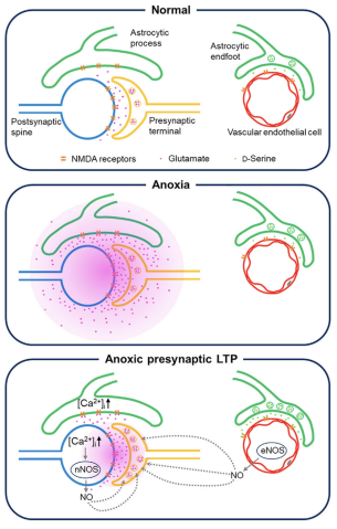
The aLTP process is activated when the brain is temporarily deprived of oxygen and glutamate levels increase. If aLTP is maintained for an extended period, this hijacks the normal functioning of the memory strengthening process (LTP), resulting in memory loss. Blocking nitric oxide (NO) synthesis or the molecular pathways that boost glutamate release eventually stops aLTP. Credit: Wang et al., 2024
The scientists found that maintaining aLTP requires NO production in both neurons and in blood vessels in the brain. Collaborating scientists from OIST’s Optical Neuroimaging Unit showed that in addition to neurons and blood vessels, aLTP requires the activity of astrocytes, another type of brain cell. Astrocytes connect and support communication between neurons and blood vessels.
“Long-term maintenance of aLTP requires continuous synthesis of nitric oxide. NO synthesis is self-sustaining, supported by the NO-glutamate loop, but blocking molecular steps for NO-synthesis or those that trigger glutamate release eventually disrupt the loop and stop aLTP,” Prof. Tomoyuki Takahashi, leader of the former Cellular and Molecular Synaptic Function Unit at OIST, explained.
Notably, the cellular processes that support aLTP are shared by those involved in memory strengthening and learning (LTP). When aLTP is present, it hijacks molecular activities required for LTP and removing aLTP can rescue these memory enhancing mechanisms. This suggests that long-lasting aLTP may obstruct memory formation, possibly explaining why some patients have memory loss after a short stroke.
Prof Takahashi emphasised that the formation of a positive feedback loop formed between glutamate and NO when the brain is temporarily deprived of oxygen is an important finding. It explains long-lasting aLTP and may offer a solution for memory loss caused by a lack of oxygen.
Life Healthcare through its wholly owned subsidiary Life Molecular Imaging Limited (LMI), has entered into a contract with Lantheus Holdings Inc. (“Lantheus”), to sub-license one of LMI’s early-stage novel radiotherapeutic and radio diagnostic products (RM2).
“As part of Life Healthcare’s strategy to monetise LMI’s product development portfolio, we are delighted to have found a partner for our RM2 product”, said Pete Wharton-Hood, Life Healthcare, CEO. “Through this agreement, LMI has secured a partnership for the development of this early-stage diagnostic and therapeutic product through to commercialisation. This exciting opportunity unlocks some of the value in LMI’”, continued Wharton-Hood.
Lantheus will make an upfront payment of $35 million for the sub-licensing rights to RM2, as per the agreement. In addition, several payments will potentially be paid to LMI on the achievement of development and regulatory milestones as well as royalty payments when the product is sold commercially.
The sub-licensing agreement secures Lantheus’ rights to develop the product and complete the early development in collaboration with LMI. “LMI is uniquely positioned to assist in this area, says Wharton -Hood and we are pleased by this development as it showcases and harnesses the specialised, dedicated and focused talent within LMI”. “With Lantheus’ experience in developing and providing access to radiotheranostics in cancer, we are confident in our decision to hand them the reins for this promising theranostic pair and are honored to work with them toward improving the future of people with prostate and breast cancer,” said Ludger Dinkelborg, CEO, Life Molecular Imaging.
Lantheus Holdings, Inc. is listed on NASDAQ in the United States of America and is the leading radiopharmaceutical-focused company committed to delivering life-changing science to enable clinicians to Find, Fight and Follow disease to deliver better patient outcomes. Lantheus has been providing radiopharmaceutical solutions for more than 65 years and has identified value and commercial opportunity in continuing the development of RM2.
LMI is a wholly owned subsidiary in Life Healthcare and is registered in the United Kingdom. The company has a product Neuraceq® which has been approved in many countries and is used to detect amyloid plaque in the brain through a PET-CT Scan and has multiple products in early clinical development. LMI also provides clinical research services for pharmaceutical companies.
Life Healthcare has retained R1bn to provide for funding requirements of LMI as part of the Alliance Medical Group disposal which was concluded earlier this year “This transaction will reduce the quantum required and Life Healthcare will consider distributing a portion of the surplus to shareholders as part of the full year dividend,” stated Wharton-Hood.
About RM2
RM2 is a 9 amino acid peptide that binds to Gastrin Releasing Peptide receptor (GRPr); and can be used to treat multiple malignant tumors like prostate, breast, lung, glioma, and ovarian tumors.
About Life Molecular Imaging
LMI is a wholly owned subsidiary in Life Healthcare and is registered in the United Kingdom. The company has one globally approved product Neuraceq ® that is used to detect amyloid plaque in the brain through a PET-CT scan and has multiple products in early clinical development as well as providing clinical research services for pharmaceutical companies.
New evidence shows higher oxygen concentrations may help prevent deaths of preterm babies
Photo by Hush Naidoo on Unsplash
Giving very premature babies high concentrations of oxygen soon after birth may reduce the risk of death by 50%, compared to lower levels of oxygen says new research led by University of Sydney researchers.
Premature babies sometimes need assisted breathing because their lungs haven’t finished developing, so doctors may give them supplemental oxygen via a breathing mask or breathing tube.
The study, published in JAMA Pediatrics, examined clinical trial data and outcomes of over one thousand premature babies who were given different oxygen concentrations. This included low concentrations of oxygen (~30%), intermediate (~50–65%) or high (~90%).
The study found for babies born prematurely, at less than 32 weeks starting resuscitation with high concentrations of oxygen (90% or greater), could increase chances of survival compared to low levels (21–30%).
When a doctor provides oxygen to babies that need help breathing, there is a device that regulates how oxygen is mixed together to reach the desired concentration. The researchers believe higher initial levels of oxygen may jump-start independent breathing, but more research is required to explore the underlying cause for this effect.
The researchers emphasise that additional large studies will be important to confirm this finding, and that even when starting with high oxygen, it needs to be adjusted to lower levels quickly to avoid hyperoxia (oxygen poisoning).
How the oxygen is delivered during the first 10 minutes of the infant’s life is critical. Doctors may give the baby high levels of oxygen at the start but then monitor vital signs and continually adjust the oxygen to avoid over or under exposure.
If confirmed in future studies, the findings challenge current international recommendations that suggest giving preterm babies the same amount of oxygen as babies born at term, 21%–30% oxygen (room air), rather than extra oxygen.
This study also demonstrates that there may not be a one-size-fits-all approach, and babies born prematurely may have different needs than babies born at term.
“Ensuring very premature infants get the right treatment from the beginning sets them up to lead healthy lives. There is no better time to intervene than immediately after birth,” said lead author Dr James Sotiropoulos from the University of Sydney’s NHMRC Clinical Trials Centre.
“The goal is to find the right balance – how do we give enough oxygen to prevent death and disability, but not damage vital organs.”
“Whilst promising and potentially practice-changing, these findings will need to be confirmed in future larger studies.”
Historically, oxygen with a 100% concentration was used to resuscitate all newborn infants. But due to studies that found high concentrations of oxygen over time can lead to hyperoxia and subsequent organ damage, in 2010 it prompted changes in international treatment recommendations for the use of blended oxygen (starting with low oxygen) for preterm infants.
Hyperoxia still a danger
However, researchers say the change was mainly based on evidence for full-term infants, who have fully developed lungs and who are often not as sick as premature infants. To date, there is little conclusive evidence to guide best practice for premature infants. The researchers emphasise the findings should not minimise the dangers of hyperoxia.
“The debate around exactly how much oxygen is best for extremely premature babies is still ongoing but, ultimately, everyone has the same shared goal of determining the best treatment for newborns,” said Dr Anna Lene Seidler from the NHMRC Clinical Trials Centre.
“Our findings, together with all the other research that is currently happening, may help the most vulnerable preterm infants have the best chance of survival.”
“We are very lucky to work with a highly collaborative international group on this question, some of whom have been studying it for decades. The group’s diverse expertise and experience is a major strength of this work,” said Dr Sotiropoulos.
Nutritionists generally advise everyone to eat more dietary fibre, but a new study suggests that its effects on health can vary, suggesting that recommendations should be tailored to each individual’s gut microbiome. The study, published in Gut Microbes, focused on resistant starch, a category of dietary fibre found in such foods as bread, cereals, green bananas, whole-grain pasta, brown rice and potatoes.
The researchers identified the gut microbe species that change in response to two different types of resistant starch. They found evidence that each individual may have a unique response to eating a resistant starch, with some people benefiting and others experiencing little or no effect. The reason for the variation appears tied to the level of diversity and composition of a person’s gut microbiome.
“Precision nutrition definitely has a use in determining what dietary fibre we should tell people to eat,” said Angela Poole, assistant professor of molecular nutrition and senior author of the study.
“This is critical because we’ve had public messaging advising people to eat more dietary fibre for decades,” Poole said. “At the same time, less than 10% of people eat the recommended intake. Since there are many different types of dietary fibre and carbohydrates, a better strategy would be to collect data on each person and tell them which dietary fibre they can eat to get the most bang for their buck.”
Resistant starch comes in five types, and resists degradation by human digestive enzymes until it reaches the gut. There, it acts as a substrate for certain gut microbes to produce short chain fatty acids, which are important in signaling pathways that regulate glucose and lipid metabolism. Multiple microbe species may work together to create the fatty acids.
In the study, Poole and colleagues tested three dietary treatments on 59 participants over seven weeks.
The team had three different types of crackers manufactured. Two crackers had the same ingredients, except one contained resistant starch type 2, which occurs naturally, and the other contained resistant starch type 4, which is human-made. A third control cracker was digestible by human enzymes, similar to white bread, and the researchers expected none of the bacteria to act on the control.
Subjects were then divided into two groups. The first group ate the resistant starch type 2 cracker first, followed by the control, and then resistant starch type 4. Each cracker type was eaten for 10 days, with five days of no cracker consumption between treatments. The second group reversed the order, also with the control in the middle.
They then sequenced the microbiomes of each participant before and after each treatment. For resistant starch type 2, more than 30 bacteria changed in abundance, including Ruminococcus bromii, which is considered a keystone resistant starch degrader in the human gut. For type 4, more than 20 bacteria changed. And for the control, nothing changed.
“For the resistant starch crackers, we could detect that 20 or 30 of them were changing, but how much they changed and whether they changed at all, for each of those bacteria, depended on the person,” Poole said.
Similarly, each resistant starch type changed different short chain fatty acids, with variable levels of fatty acid increases and decreases based on the individual. For resistant starch type 2, the researchers identified a subset of 13 bacteria that predicted change in amounts of propionate, a type of short chain fatty acid. Also for resistant starch 2, by knowing the diversity of an individual’s gut microbiome, the researchers could roughly predict if two types of short chain fatty acids (acetate and butyrate) were going to increase.
The most surprising result was that the control digestible cracker led to the greatest gains of short chain fatty acids. More work is needed to understand why, but Poole suspects that the order of cracker consumption was key to the result. Since many microbes are involved in making short chain fatty acids, she hypothesises that eating a resistant starch first primed the gut to produce the fatty acids when that person ate the digestible starch.
“That’s one of the major takeaways, maybe I can get away with eating a French baguette some of the time, and it may be better than just eating whole grain all the time,” Poole said. “But I have to test that, and it probably varies between people.”
Dr Tim Forgan at the surgeon’s console of the da Vinci robotic system. (Photo: Biénne Huisman/Spotlight)
By Biénne Huisman
Within South Africa’s beleaguered public health sector – unsettled by budget cuts, understaffing, and divisive NHI legislation – cutting edge surgical robots that have been used to perform more than 600 surgeries at two Cape Town public hospitals are beacons of excellence that offer a glimmer of hope. Spotlight’s Biénne Huisman visited Dr Tim Forgan at Tygerberg Hospital to learn more.
Cutting edge robotic surgery might not immediately come to mind when one thinks of public hospitals, but in a first for public healthcare in South Africa, such systems are being used at two hospitals in the Western Cape.
The da Vinci Xi systems enable surgeons to control operations from a console – steering three arms with steel “hands” equipped with tiny surgical instruments; plus a fourth arm bearing a video camera (the laparoscope). The system translates a surgeon’s hand movements in real time, with enhanced precision, range and visuals, compared to manual surgery.
“It really is next level, it feels like you’re inside the patient,” says colorectal specialist Dr Tim Forgan, Tygerberg Hospital’s da Vinci robotics coordinator. “With this technology we can operate so much finer. You can see ten times better with this robot than with the naked eye; you can see tiny, tiny nerves you wouldn’t normally see. And you can manoeuvre surgical instruments so much better. Because of that, people have way better function after the procedure.”
He explains that the technology allows major surgery to be completed through small incisions – instead of larger cuts made by a doctor’s hand – leading to less bleeding and a faster recovery time.
Over 600 surgeries in two years
Lorraine Gys from Phillipstown in the Northern Cape can attest. On 22 February 2022, the 65-year-old pensioner became the first patient to undergo da Vinci robotic surgery in South Africa’s public sector. Forgan was behind the console, at Tygerberg Hospital.
Gys tells Spotlight: “The next day the sisters offered to wash me, I said to them ‘no, I’m not helpless.’ My recovery was very quick. I was up and about in no time, while the other patients had to be assisted. I was discharged on day four, and back at home I could even continue doing my own chores.”
Two years later, Gys is cancer free. The mother of three, who now lives in Eerste River, recalls how she made news headlines: “Before the operation, Dr Forgan explained everything to me. They asked my permission, saying that media will be there and the [provincial health] minister.”
Indeed, on the day Forgan operated on Gys, removing a cancerous rectal tumour, he was joined in theatre by several onlookers including former Western Cape MEC of Health and Wellness Nomafrench Mbombo.
“Yes it was a circus,” says Forgan, laughing. “A whole bunch of people watching me operate, quite bloody nerve-wracking. Fortunately I’m experienced at having lots of students around watching; plus performing surgery is just so immersive, everything else fades out.”
On that day, also in the operating room was colorectal surgeon Dr Roger Gerjy, keeping an eye. “He’s a very well-known robotic surgeon; a Swedish surgeon who works in Dubai,” says Forgan. “And if there was a problem, Roger would’ve taken over. He was also there to impart tips and tricks: move the instrument like this, shape it like a hockey stick; because with the robot it’s like having your whole arm inside [the body]. He’d give me advice on what to do with my extra floating arm – where to place it and how to manipulate it – because remember you’re controlling three arms at a time.”
Since 2022, the da Vinci robots installed at Cape Town’s two tertiary hospitals: Groote Schuur and Tygerberg, have enabled over 600 minimally invasive surgeries – including colorectal operations, prostatectomies, cystectomies (bladder removal surgery), and gynaecological procedures to treat endometriosis.
Groote Schuur Hospital has the other da Vinci Xi system run by Western Cape public healthcare
A spokesperson for the Western Cape Ministry of Health and Wellness, under former MEC Mbombo, Luke Albert explains: “We can see the immense impact it has for patients and the health system. For example, a traditional open cystectomy patient would require three days of ICU stay, as well as two weeks of hospital stay to recuperate. During this time, on average, 42% of patients require blood transfusions and almost 20% need total parenteral nutrition (when a patient is fed intravenously). A patient undergoing robotic surgery for a cystectomy requires no ICU stay and goes straight to a general ward for no more than six days on average, with no blood transfusions needed.”
Where the money came from
Asked how the department was able to afford R40 million per system for these machines in the context of severe budget cuts, Albert says: “The purchase was applicable to 2021/22 and not the current financial year; with all provincial health departments currently managing the effects of budget cuts.”
Asked the same question, Forgan explains the investment derived from surplus budget discovered within the throes of the COVID-19 pandemic: “There was a surplus because certain services just couldn’t be done. I mean, for us, we couldn’t do elective surgery. And how state funding works; if you don’t spend your [provincial] budget within the financial year, it goes back to central government.”
What it looks like
On a Friday afternoon at Tygerberg Hospital, Forgan is guiding Spotlight along corridors and up grey linoleum stairs, to the theatre where the da Vinci system is used. Dressed in black surgical scrubs bearing his name and a cap; on his feet Forgan is wearing bright pink crocs. In passing, he waves hello to fellow healthcare staff.
Inside the small blindingly white room, Forgan points out the three core components of the da Vinci system. There is a console with two control levers similar to refined joysticks – he demonstrates how to delicately hold them between forefingers and thumbs – a patient-side cart with four interactive metal arms (they are disposable; each arm can be used on twelve patients), and another trolley with a television screen. All connected by blue fibre optic cables.
As we speak, nurses arrive in the theatre, preparing it for upcoming gynaecology procedures scheduled for Monday. Forgan greets them, then continues to expand on his passion for colorectal surgery.
“With colorectal surgery, there’s a high rate of complications, but I really enjoy it, I really enjoy my job. When you have a successful outcome, saving a person from their cancer and prolonging their life through your intervention, that is the reward. Colorectal cancer is a very unpleasant disease, and operating like this can make one hell of a difference in a patient’s life.”
Colorectal cancer on the increase
Forgan adds that colorectal cancer is on the increase: “There aren’t many colorectal surgeons in South Africa, with a dire need for people to operate in this subspecialty. I mean, there are so few of us, we’re all on a WhatsApp group.”
Colorectal or colon cancer is the second most common cancer in South African men (following prostate cancer), and the third most common cancer in women (following breast and cervical cancer), according to the Cancer Association of South Africa.
Originally from Johannesburg, Forgan attended medical school at the University of the Witwatersrand. He qualified as a general surgeon at Stellenbosch University, sub-specialising in colorectal surgery at the University of Cape Town, before studying minimally invasive colorectal surgery at the Academic Medical Centre in Amsterdam.
He is also president of the South African Colorectal Society and runs a part-time private practise with his Tygerberg colleague, Dr Imraan Mia, at Cape Town’s Christiaan Barnard Hospital, where he has 32 all five-star Google reviews.
‘Early adopter’
Forgan considers himself an early adopter. But learning to use the da Vinci system did not happen overnight.
“We trained for ages,” he says. “On the surgical console there’s a simulator, so you spend hours and days and days doing procedures, over and over and over again. You have to get over 95% for each one of the procedures, before you can move on to the next skill.
“Then it’s how to use the machine, how to put it together, what to do if there’s an emergency; what if there’s a power failure and the machine stops working? How to safely remove it from the person. Then we went to the University of Lyon [in France] for two days of hands-on robotics training. And then a proctor – an international expert – comes to your theatre and does the procedures with you. So that was Dr Roger Gerjy, and that’s when we did Lorraine…”
First introduced by American biotechnology company Intuitive Surgical in 1999, the da Vinci Xi systems have sparked some liability lawsuits. An article from the Tampa Bay Times in February cites a lawsuit filed at the United States District Court in West Palm Beach, with a man claiming that a stray electrical arc from a surgical robot burned his wife’s small intestine during a colon cancer procedure, causing her death. The article quotes Intuitive Surgical’s 2023 financial report, which notes 8 606 da Vinci systems in use worldwide, having performed 2 286 000 procedures in 2023. The financial report mentions an undisclosed number of pending lawsuits, which the company disputes.
Nevertheless, Forgan remains an advocate.
Exiting via Tygerberg’s maze of corridors, he continues to reflect on his job. After our meeting, he is set to deliver a talk at the Cape Town International Convention Centre. His manner is earnest. Shrugging, he describes himself as a “glorified plumber”.
Research by West Virginia University has demonstrated that American Heart Association and American Stroke Association guidelines are effective at speeding up hospitals’ response times for stroke treatment and can be mastered even by members of ‘ad hoc‘ medical teams that assemble rapidly on the fly.
When a stroke patient arrives at an emergency room, specialists from across hospital departments – emergency medical services, neurologists, pharmacists, physicians, nurses, radiologists and technicians – rush to coordinate a team response. AHA and ASA guidelines put specific limits on how much time can optimally elapse between the onset of ischaemic stroke, in which blood flow to the brain is blocked, and subsequent events like arrival at the hospital and delivery of an infusion.
But experts have questioned whether the communication of those best practices helps medical teams that assemble temporarily and whose members don’t typically collaborate. In a Journal of Operations Management article, WVU associate professor Bernardo Quiroga and coauthors answer that question using data about more than 8000 patients who received stroke care at a large hospital between 2009 and 2017.
“‘Time is brain’ for stroke victims,” Quiroga explained. “Blocked blood flow to the brain kills almost two million neurons a minute, so your life or ability to walk or talk hinges on how quickly multiple professionals coordinate to restore blood flow. If you’re lucky, you’re treated within the first hour of symptom onset. Better yet, you receive a shot of Tissue Plasminogen Activator, which dissolves clots. TPA works better the earlier it’s given and usually isn’t effective after 4.5 hours.”
In 2010, the AHA and ASA launched Target: Stroke, a program that identifies stroke care best practices and standardises each step in the process. Participating hospitals reduced median treatment times from 79 minutes in 2009 to 51 minutes in 2017, but it wasn’t clear if that improvement was driven by adherence to best practices or by clinicians learning through repetition as they handled more stroke cases.
To figure that out, the researchers investigated whether repeated ‘learning by doing’ decreased the hospital’s stroke care time. Then, they evaluated whether deliberate, ‘induced’ learning and implementation of AHA/ASA best practices decreased the time further.
Learning through repetition worked. The more strokes the hospital treated, the faster it responded. For each doubling of cumulative stroke alerts, ‘door-to-needle time’ – the time to get patients from the hospital door to a TPA infusion – decreased by 10.2%.
Best practices also worked. Specifically, the researchers examined two best practices: the Helsinki Model protocol, which directs that EMS staff keep stroke patients on the stretcher for transport to the CT room rather than transferring them to ER beds; and the Rapid Administration of TPA protocol, which requires the pharmacist to be in the CT room with TPA before completion of the CT scan. Those protocols significantly reduced the hospital’s door-to-needle time beyond improvements from repetition-based learning.
According to Quiroga’s coauthor and former PhD student Brandon Lee, that matters because it demonstrates the efficacy of best practices and shows ad hoc teams learning guidelines and implementing them long-term.
However, Lee emphasised the importance of the presence of the hospital’s stroke advisory committee, which set targets, evaluated stroke teams’ performances and gave feedback.
Without similar “countermeasures to organisational forgetting,” Quiroga acknowledged that best practices aren’t always sustainable, especially on ad hoc teams.
“In the case of the best practice indicated by the Helsinki Model, compliance is difficult because the hospital needs to coordinate with multiple independent EMS systems. Some EMS providers may be reluctant to commit resources to extended time in the CT room, and EMS staff turnover may lead to forgetting,” Quiroga said.
Lee added, “Overall, because ad hoc teams are fluid, information sharing is harder. And when a group of people don’t know each other well, group learning slows. But although ad hoc teams learn more slowly, we determined they still learn.”
The research also assessed whether neurologists’ abilities to meet time goals were affected by their recent experiences treating prior stroke patients.
“As team leaders, neurologists can have an outsized influence on performance,” Quiroga said. “Because other members of the ad hoc team aren’t familiar with each other, they lean on their leader.”
But data showed stroke teams improving response times regardless of how many stroke cases the neurologist had treated individually or what the neurologist’s recent success rate was. Quiroga said that’s good news.
“The implication is that learning and sustaining best practices ensures an even quality of care for patients, regardless of individual neurologists’ experience levels.”
The current method for assessing medication-related liver injury does not accurately reflect some medications’ toxicity to the liver, according to a new study led by University of Pennsylvania researchers. Hepatotoxicity classification has historically been determined by counting individual reported cases of acute liver injury (ALI). Instead, the researchers used real-world health care data to measure rates of ALI within a population and uncovered that some medications’ levels of danger to the liver are being misclassified. Their paper was published in JAMA Internal Medicine.
“From a clinical standpoint, knowing the rate of severe ALI after starting a medication in real-world data will help determine which patients should be monitored more closely with liver-related laboratory tests during treatment,” said senior author Vincent Lo Re, MD, MSCE, an associate professor of Medicine and Epidemiology. “Incidence rates of severe ALI can be a valuable tool for determining a medication’s toxicity to the liver and when patients should be monitored, since incidence rates provide a truer, real-world look at this toxicity. Case reports did not accurately reflect observed rates of ALI because they do not consider the number of persons exposed to a medication, and cases of drug-induced liver injury are often underreported.”
Within the study, 17 different medications had rates that exceeded five severe ALI events per 10 000 person-years. The team determined that 11 of these medications were in lower categories of hepatoxicity by case counts that were likely not reflective of their true risk, since their incidence rates revealed higher levels of toxicity. One of the medications that fell into this group was metronidazole, an antimicrobial that can be used to treat infections in the reproductive or gastrointestinal systems, as well as some dermatological conditions.
Incidence rates, the number of new cases of a disease within a time period divided by the number of people at risk for the disease, are a key measure for examining health in a population because they give a more complete picture than simple counting. For instance, a medication with 60 reports of liver injury would be considered the most hepatotoxic through the traditional method, using the raw number of reported liver injury cases. However, if that medication had 60 observed severe ALI events and was used by five million people, the incidence rate would be very low and likely point to the medication not being dangerous to the liver. However, if 60 severe ALI events were observed within a population of 1,000 patients, it would reflect a higher, potentially more important, rate of injury.
To determine incidence rates, Lo Re and his team, including lead author Jessie Torgersen, MD, MHS, MSCE, an assistant professor of Medicine, examined electronic medical record data on almost 8 million people provided by the United States Veterans Health Administration that had been compiled from 2000 through 2021. Each person did not have pre-existing liver or biliary disease when they began taking any of the 194 medications that were studied. Each of those medications were analysed due to suspicion that they could cause harm to the liver, since each had more than four published reports of liver toxicity associated with their use.
On the other side of the hepatotoxicity coin, the researchers found eight medications that were classified as the most hepatotoxic based on the number of published case reports, but should actually be in the least liver-toxic group, with incidence rates of less than one severe ALI event per 10 000 person-years. For example, rates of severe ALI for statin medications, often used for high cholesterol, were in the group that had fewer than one event per 10 000 person-years.
“The systematic approach that we developed enables successful measurement of the rates of liver toxicity after starting a medication,” Lo Re said. “It wasn’t surprising that the case report counts did not accurately reflect observed rates of severe acute liver injury given the inherent limitations with case reports.”
With these findings, the researchers hope that there might soon be mechanisms established within electronic medical records to alert clinicians to closely monitor the liver-related laboratory tests of patients who start a medication with a high observed rate of severe ALI.
“Importantly, our approach offers a method to allow regulatory agencies and the pharmaceutical industry to systematically investigate reports of drug-induced ALI in large populations,” Lo Re said.