Hosted by Dr Hlombe Makuluma, Medicolegal Advisor at EthiQal, this webinar will be co-presented by two admitted attorneys, Mashooma Parker and Jessica Viljoen, who are both legal advisors within the claims team at EthiQal. The 90-minute session will cover compliance for record-keeping requirements as well as dealing with requests for patient records from patients and third parties.
Participants will gain valuable insights to ethically enhance their practice’s visibility and reach, fostering responsible and compliant advertising practices.
Mashooma Parker is a skilled Legal Advisor within the Claims & Legal team at EthiQal, specialising in medical malpractice. With a strong background in the legal field and a passion for assisting healthcare practitioners, Mashooma brings a wealth of expertise to navigate the complexities that arise with patients and third parties. Hosting the first topic, She will cover the requirements for healthcare practitioners to ensure quality record-keeping compliance with Booklet 9 of the HPCSA’s Ethical Guidelines.
Jessica Viljoen is an admitted attorney and legal advisor specialising in professional indemnity insurance for healthcare practitioners, and medical malpractice law. With her extensive experience within the medico-legal space, including her years of litigation experience, Jessica leverages her industry knowledge to provide legal advice and assistance to all specialties of medical practitioners throughout South Africa. She will present the second part of the talk, which will deal with Patient and Third-party requests for patient records and how to ensure compliance with the Promotion of Access to Information Act 2 of 2000.
The speakers will offer some useful tips from a medico-legal risk management perspective for health practitioners to be cognisant of, as well as to work through some practical examples to illustrate the importance of the topic.
At least one hour’s attendance on the Zoom Platform is required to earn CPD points, and for those unable to watch it live, a recording will be made available.
In a novel study, a team of dermatologists evaluated the effect of ultraviolet (UV) exposure on appetite and weight regulation. They found that UV exposure raises norepinephrine levels, decreases leptin levels, and induces the browning of subcutaneous fat, thereby increasing energy expenditure. These results potentially pave the way for new approaches to prevent and treat obesity and metabolic disorders. Their findings appear in the Journal of Investigative Dermatology, published by Elsevier.
Co-first authors Qing-Ling Quan, MD, PhD, and Eun Ju Kim, PhD, Department of Dermatology, Seoul National University Hospital, explained, “Recent evidence has suggested that UV exposure limits body weight gain in mouse models of obesity. Subcutaneous fat is a critical organ in regulating energy homeostasis. Alongside previous studies on the effects of UV exposure on obesity and metabolic disorders, our team was inspired by our prior discovery that, although UV rays do not directly reach subcutaneous fat when exposed to the skin, they can regulate the metabolism of subcutaneous fat. This led us to hypothesise that skin exposure to UV rays could play a significant role in systemic energy homeostasis, prompting this research.”
Investigators discovered that when exposed to UV radiation consistently, mice fed a normal diet and those on a high-fat diet exhibited increased appetite due to a decrease in leptin, a key hormone in appetite regulation. But there was no weight increase – they found that UV radiation inhibits weight gain by enhancing secretion of the neurotransmitter norepinephrine, which not only decreases leptin but also increases energy expenditure through the “browning” of subcutaneous fat.
The increased energy intake, driven by heightened appetite, is converted to heat and burned before it can accumulate in subcutaneous fat, thus preventing weight gain.
This research provides new insights into the impact of UV exposure on appetite and weight regulation, opening possibilities for novel approaches in the prevention and treatment of obesity and metabolic disorders. Specifically, uncovering the mechanism by which UV radiation prevents weight gain could offer new approaches to dietary regulation and weight loss, providing innovative insights into health and obesity management that could positively impact human health.
Lead investigator Jin Ho Chung, MD, PhD, Department of Dermatology, Seoul National University Hospital, Seoul National University College of Medicine, explained, “This study elucidates the mechanism by which UV exposure can increase appetite while inhibiting weight gain. These findings contribute significantly to understanding the effects of UV radiation on energy metabolism and homeostasis and open new avenues for exploring prevention and treatment strategies for obesity and metabolic disorders. Notably, the fact that UV radiation lowers leptin levels and increases norepinephrine, thereby promoting the browning of subcutaneous fat and increasing energy expenditure, provides a groundbreaking clue for the development of obesity treatment strategies. This research demonstrates that UV exposure not only affects the skin but also plays a deep role in our body’s energy metabolism and homeostasis processes. However, further research is needed on the long-term effects and safety of UV exposure, and there should be significant interest in developing new therapeutic approaches that utilise the efficacy of UV radiation.”
However, as co-corresponding author Dong Hun Lee, MD, PhD, Institute of Human-Environment Interface Biology, Seoul National University, noted, “Because UV exposure can accelerate skin aging and promote skin cancer, it is advisable to minimise UV exposure and protect the skin with sunscreen. Thus, our research team plans to conduct follow-up studies to develop new strategies that could mimic the effects of UV radiation for obesity and metabolic regulation.”
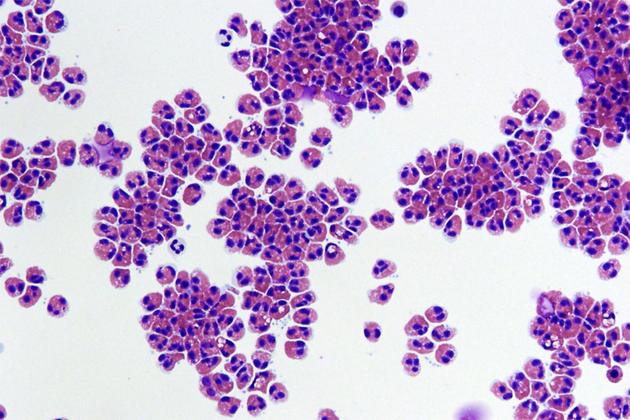
Human eosinophils, a type of white blood cell, are shown isolated from blood. In patients with eosinophilic oesophagitis, these cells store and release packages of inflammatory proteins (red) that can damage the throat and pesophagus.
Credit: Julie Caldwell, Cincinnati Children’s Hospital Medical Center
Eosinophils, specialised immune cells, are identified by their distinctive granules that stain red when treated with an acidic reagent, eosin, which gave them their name. Eosinophils are typically rare in blood and tissues, accounting for about 3% of white blood cells. Their biological roles are poorly understood, but recent studies suggest that eosinophils are involved in regulating fat metabolism, repairing certain tissues, and helping fight different infections and cancers. Now, a new study has found that the cytokine Interleukin-5 (IL-5) acts differently on the production of eosinophils than previously believed.
In common diseases such as allergic asthma and rhinosinusitis, eosinophils are abnormally numerous in the blood and tissues, a condition known as eosinophilia. Eosinophilia is a clinical sign that aids in diagnosing these “eosinophil-associated” diseases and guides their treatment. It is known that eosinophilia is driven by increased production of eosinophils by the bone marrow. Since the 1990s, it has also been known that IL-5 is essential for eosinophilia. This led to the development and market introduction of precision therapies targeting IL-5 with monoclonal antibodies to treat severe forms of eosinophilic diseases. However, the effects of IL-5-blocking treatments on eosinophils remain poorly described.
The Laboratory of Cellular and Molecular Immunology (LCMI) of the University of Liege, under the direction of Fabrice Bureau and Christophe Desmet, aimed to better understand the origin of eosinophils and eosinophilia, and the effects of treatments targeting eosinophils. As Christophe Desmet explains, “these questions previously suffered from a too rudimentary definition of the eosinophil development pathway in our bone marrow.” Two doctoral students from the laboratory, Joseph Jorssen and Glenn Van Hulst, combined their talents in bioinformatics and flow cytometry with the help of the Genomics and Flow Cytometry platforms of the GIGA Institute to finely characterise, using different approaches to analyse the surface protein and messenger RNA composition of eosinophils at various stages of their development. Although the mouse remains a reference model, collaboration with the Hematology Department of Liege University Hospital and GIGA also allowed for a very detailed and updated mapping of eosinophil development in human bone marrow, and to observe its conservation through evolution.
This detailed characterisation work, published in the journal Immunity, provides the community with simple-to-use methods and freely accessible bioinformatics data that will greatly facilitate future studies of eosinophils. Using these resources, the same study showed that IL-5 does not act as previously believed by researchers and clinicians. Most thought that IL-5 promoted the maturation of cells destined to become eosinophils and that IL-5-targeting treatments blocked this maturation. “Our study actually supports the opposite hypothesis,” explains Christophe Desmet: IL-5 slows down the maturation of developing eosinophils, allowing them to multiply longer. By stimulating this “transit amplification,” IL-5 promotes eosinophilia, and by inhibiting this process, IL-5-targeting treatments reduce it.
The researchers also showed that interferon response factor-8, considered an essential promoter of leukocyte production, was not intrinsically required for eosinophil production.
This study thus provides resources, methods, and perspectives to understand the origin of eosinophils, the effects of current precision therapies, and the regulation of eosinophil development and numbers in normal and disease conditions.
A new study led by UC Davis researchers finds widespread differences in brain development between autistic boys and girls ages 2-13. The study, published recently in Molecular Psychiatry, found sex-specific changes in the thickness of the brain’s cortex, or outer layer.
The findings are notable because so few studies have addressed cortical development in autistic girls, who are diagnosed with autism less often than males. Nearly four males are diagnosed with autism for every one female.
“It is clear that this sex bias is due, in part, to underdiagnosis of autism in females,” said Christine Wu Nordahl, a professor in the Department of Psychiatry and Behavioral Sciences and the UC Davis MIND Institute and a senior author on the paper.
“But this study suggests that differences in diagnosis are not the full story – biological differences also exist.”
The cortex is made up of distinct layers comprised of millions of neurons. Until about age 2, the cortex rapidly thickens as new neurons are created. After this peak, the outer cortical layer thins. Previous studies have found that this thinning process is different in autistic children than non-autistic children, but whether autistic boys and girls share the same differences had not been examined.
“It’s important to learn more about how sex differences in brain development may interact with autistic development and lead to different developmental outcomes in boys and girls,” explained Derek Andrews, lead author on the study and an assistant project scientist in the Department of Psychiatry and Behavioral Sciences and at the MIND Institute.
A changing cortex in childhood
The research team studied the brain scans of 290 autistic children – 202 males and 88 females, and 139 non-autistic, typically developing individuals – 79 males and 60 females.
All participants were in the MIND Institute’s Autism Phenome Project (APP), one of the largest longitudinal autism studies in the world.
The project includes the Girls with Autism Imaging of Neurodevelopment (GAIN) study, launched to increase the number of females represented in research.
The researchers took MRI scans at up to four time periods between the ages of 2 and 13.
They found that at age 3, autistic girls had a thicker cortex than non-autistic girls of the same age, comprising about 9% of the total cortical surface. Differences in autistic males when compared to non-autistic males of the same age were much less widespread.
In addition, when compared to males, autistic females had faster rates of cortical thinning into middle childhood. The cortical differences were present across multiple neural networks.
“We found differences in the brain associated with autism across nearly all networks in the brain,” Andrews said.
He noted that it was a surprise at first that the differences were greatest at younger ages. Because autistic girls had a more rapid rate of cortical thinning, by middle childhood, the differences between autistic males and females were much less pronounced.
“We typically think of sex differences as being larger after puberty. However, brain development around the ages of 2-4 is highly dynamic, so small changes in timing of development between the sexes could result in large differences that then converge later,” Andrews explained.
The importance of long-term studies of both sexes
These findings make it clear that longitudinal studies that include both sexes are necessary, Nordahl said.
“If we had only looked at boys at age 3, we may have concluded that there were no differences. If we had both boys and girls, but only investigated differences at 11 years of age, we may have concluded that there were very few sex differences in the cortex. We needed to follow both boys and girls across development to see the full picture,” she explained.
This was why Nordahl, who now directs the APP, launched the GAIN study in 2014. “The APP had a wonderfully large sample of about 150 autistic boys, but only about 30 autistic girls. This was too few autistic girls to really examine how they might be similar or different to boys, so we worked to increase the representation of autistic females in our research,” she said.
GAIN is unique, and Andrews said he hopes other researchers will follow suit in including more autistic girls in autism research. “Autistic females represent about 20% of the autistic population. Any successful effort to understand autism will need to include autistic females.”
Late last year, geneticist Marlena Fejzo and colleagues made the discovery that morning sickness’s most serious presentation, hyperemesis gravidarum (HG), is caused by the hormone GDF15, not human chorionic gonadotropin as previously thought. In a peer-reviewed opinion article published in the journal Trends in Molecular Medicine, Fejzo dispels common morning sickness myths and discusses potential treatments, including sensitising people to GDF15 prior to pregnancy, similar to the way we treat allergies.
“HG can be life threatening and is associated with adverse outcomes that need to be taken seriously,” says Fejzo of the Keck School of Medicine of the University of Southern California. “Now that we know that GDF15 is the most likely cause of HG, we are on the cusp of having treatments that target this hormonal pathway and end the suffering.”
Myth 1: Severe morning sickness is harmless and normal
Pregnant people with HG are essentially starving, Fejzo says, and an increasing number of studies have demonstrated that this has serious short- and long-term clinical implications for both the parent and child. HG is a top predictor of postnatal depression, and 26% of pregnant people with HG report suicidal ideation while 18% meet the full criteria for post-traumatic stress disorder.
For the child, HG is associated with preterm birth, low birth weight, and later in life, autism spectrum disorder, ADHD, depression, social problems, in addition to an increased risk of childhood cancer and respiratory and cardiovascular disease. Still, pregnant people with the condition are often dismissed by their clinicians and families.
“It really is like a teratogen in pregnancy, a factor which interferes with normal foetal development, but it’s still not taken seriously by a lot of medical professionals,” Fejzo says. “A lot of people are brushed off and told, ‘oh that’s normal, it’s okay, just don’t take your pre-natal vitamins; you don’t need them.'”
At its most extreme, individuals with HG can develop Wernicke encephalopathy, a life-threatening swelling of the brain due to thymine (vitamin B1) deficiency. Since individuals with HG can have trouble even swallowing vitamins, the American College of Obstetricians and Gynecologists currently recommends that they replace broad spectrum prenatal vitamins with folic acid, but Fejzo warns that this is likely insufficient, and that thiamine supplementation is also warranted for individuals with HG.
“I believe all women who have hyperemesis should be given vitamin B1 to avoid this serious brain swelling that can lead to permanent brain damage and often leads to foetal death,” Fejzo says.
Myth 2: Morning sickness is caused by human chorionic gonadotropic hormone (hCG) or is psychosomatic
Though it was long thought that morning sickness is caused by hCG, the recent breakthrough has shown that HG’s main cause is actually the hormone GDF15, which is part of a normal stress response. Usually, GDF15 is expressed only in very small amounts, but during early pregnancy it spikes by a huge amount, then wanes, and finally rises again during the third trimester.
A recent Nature study co-authored by Fejzo showed that individuals who suffer from HG can have genetic variants that causes them to have lower levels of circulating GDF15 prior to pregnancy, which makes them extra sensitive when they become pregnant and are suddenly exposed to high levels. This finding has clinical implications for preventing and treating HG, since preliminary research suggests that it might be safe to manipulate GDF15 during or even prior to pregnancy.
“GDF15 may be safe to manipulate in pregnancy or even prior to pregnancy,” says Fejzo. “If we can increase levels of GDF15 before someone becomes pregnant, that might desensitise them, similar to how we try to desensitize people to allergens who have severe allergies,” says Fejzo. “And during pregnancy, we may be able to minimise or get rid of symptoms by blocking GDF15 or its receptors in the brain stem.”
Myth 3: Only humans experience morning sickness
Nausea and appetite loss during gestation is not a uniquely human trait – these symptoms have been observed throughout the animal kingdom, from monkeys, dogs, and cats, to chickens, vipers, and octopuses.
“I always think it’s interesting that the recommendation for cats is that if they’re unable to eat for a day, you should contact your veterinarian, but we don’t have that recommendation out there for women with hyperemesis,” says Fejzo. “If you call your doctor’s office and say you haven’t eaten for a day, they’ll say, ‘that’s normal’ and won’t do anything. There’s more proactive care for cats than humans.”
In addition to preventing ingestion of harmful foods, Fejzo speculates that pregnancy-induced nausea likely evolved to prevent dangerous foraging trips.
“This condition likely evolved because it was probably beneficial to avoid going out searching for food during pregnancy,” says Fejzo. “That may still be true for animals, but people don’t need this anymore, so let’s end the suffering once and for all if we can.”
Now, Fejzo is working toward developing and testing the proposed GDF15-based treatments. She also plans to investigate other genes and variants of GDF15 that might contribute to HG.
The study shows that hyperandrogenism, a key characteristic of polycystic ovary syndrome (PCOS), affects immune cell populations in reproductive, metabolic, and immunological tissues in a PCOS-like mouse model. These findings are of great importance as it is known that immune dysfunction is an essential part of reproductive complications and metabolic disease, which are very common among women with PCOS.
However, as the study shows that hyperandrogenism affects different tissues in unique ways, any possible treatments would have to be carefully tailored to target specific tissue dysfunctions. Since concurrent treatment with an androgen receptor antagonist prevented many changes in the mouse model we used in our study, combination therapies that include both anti-androgens and other drugs that target specific altered immune pathways could be explored.
Alteration of immune cells in adipose tissue
One of the most fascinating findings was the clear alterations of immune cells in adipose tissue, despite an unaltered fat mass of the androgen exposed mice. It is well known that immune cells in adipose tissue contribute to insulin resistance in overweight and obese individuals, but here we have an insulin resistant mouse model that mimics normal weight women with PCOS. The impact of androgens on immune cells in adipose tissue is therefore very interesting considering the high prevalence of insulin resistance and type-2 diabetes among normal weight women with PCOS. Another rather surprising finding was the drastic decrease of eosinophils in the uterus, as these don’t express the androgen receptor. This shows that androgens play a broader and more complex role in modulating the immune environment than only through direct androgen receptor activation on immune cells.
The next step will be to dissect the underlying mechanisms of the immune alterations, and to assess if these do contribute to the reproductive complications and metabolic comorbidities of PCOS. This will include further characterisation of immune cell changes to understand how their function could be affected and link these to reproductive and metabolic mechanisms. We also want to understand how the effect of androgens on eosinophils and mature NK cells is mediated, since neither of them expresses the androgen receptor.
A rare autoimmune disease has been newly described as a COVID-related syndrome, following an investigation by the University of California San Diego School of Medicine and Leeds University.
It started when Pradipta Ghosh, MD, a professor in the Departments of Medicine and Cellular and Molecular Medicine at UC San Diego School of Medicine, received an email from Dennis McGonagle, PhD, professor of investigative rheumatology at the University of Leeds in the UK. This was the beginning of an international collaboration, one that uncovered a previously overlooked COVID-related syndrome and resulted in a paper in eBioMedicine, a journal published by The Lancet.
McGonagle asked if she was interested in collaborating on a COVID-related mystery. “He told me they were seeing mild COVID cases,” Ghosh said. “They had vaccinated around 90 percent of the Yorkshire population, but now they were seeing this very rare autoimmune disease called MDA5 – autoantibody associated dermatomyositis (DM) in patients who may or may not have contracted COVID, or even remember if they were exposed to it.”
McGonagle told of patients with severe lung scarring, some of whom presented rheumatologic symptoms – rashes, arthritis, muscle pain – that often accompany interstitial lung disease. He was curious to know if there was a connection between MDA5-positive dermatomyositis and COVID.
“DM is more common in individuals of Asian descent, particularly Japanese and Chinese,” Ghosh said. “However, Dr McGonagle was noting this explosive trend of cases in Caucasians.”
“But that’s the least of the problem,” Ghosh said. “Because he said, ‘Oh, and by the way, some of these patients are progressing rapidly to death.'”
Ghosh is the founding director of the Institute for Network Medicine at UC San Diego School of Medicine, home to the Center for Precision Computational Systems Network (PreCSN – the computational pillar within the Institute for Network Medicine). PreCSN’s signature asset is BoNE – the Boolean Network Explorer, a powerful computational framework for extracting actionable insights from any form of big-data.
“BoNE is designed to ignore factors that differentiate patients in a group while selectively identifying what is common (shared) across everybody in the group,” Ghosh explained. Previous applications of BoNE allowed Ghosh and her team to identify other COVID-related lung and heart-afflicting syndromes in adults and children, respectively.
As a rheumatologist, McGonagle specialises in inflammatory and autoimmune conditions. Ghosh said that McGonagle’s roster of patients, all within the UK’s National Health System (NHS), helped to facilitate the investigation.
“The NHS has a centralised health care database with comprehensive medical records for a large population, making it easier to access and analyse health data for research purposes,” Ghosh explained.
Ghosh and McGonagle put together a team to probe what they found was indeed an entirely new syndrome.
The study began with McGonagle lab’s detection of autoantibodies to MDA5 – an RNA-sensing enzyme whose functions include detecting COVID and other RNA viruses. A total of 25 patients from the group of 60 developed lung scarring, also known as interstitial lung disease. Ghosh noted that the lung scarring was bad enough to cause eight people in the group to die due to progressive fibrosis. She said that there are established clinical profiles of MDA5 autoimmune diseases.
“But this was different,” Ghosh said. “It was different in behaviour and rate of progression – and in the number of deaths.”
Ghosh and the UC San Diego team explored McGonagle’s data with BoNE. They found that the patients who showed the highest level of MDA5 response also showed high levels of interleukin-15.
“Interleukin-15 is a cytokine that can cause two major immune cell types,” she explained. “These can push cells to the brink of exhaustion and create an immunologic phenotype that is very, very often seen as a hallmark of progressive interstitial lung disease, or fibrosis of the lung.”
BoNE allowed the team to establish the cause of the Yorkshire syndrome – and pinpoint a specific single nucleotide polymorphism that is protective. By right of discovery, the group was able to give the condition a name: MDA5-autoimmunity and Interstitial Pneumonitis Contemporaneous with COVID. It’s MIP-C for short, “Pronounced ‘mipsy,'” Ghosh said, adding that the name was coined to make a connection with MIS-C, a separate COVID-related condition of children.
Ghosh said that it’s extremely unlikely that MIP-C is confined to the United Kingdom. Reports of MIP-C symptoms are coming from all over the world. She said she hopes the team’s identification of interleukin-15 as a causative link will jump start research into treatment.
Using population-wide registry data, researchers investigated whether mental disorders can be transmitted within social networks formed by school classes.
The study is the largest and most comprehensive so far on the spread of mental disorders in social networks, with more than 700 000 ninth-grade pupils from 860 Finnish schools participating. The adolescents were followed from the end of ninth grade for a median of 11 years.
The researchers, from the University of Helsinki, the Finnish Institute for Health and Welfare, the University of Jyväskylä and the University of Manchester, demonstrated that the number of classmates diagnosed with a mental disorder was associated with a higher risk of receiving a mental disorder diagnosis later in life.
“The observed link was the strongest during the first year of follow-up in the study. This was not explained by a number of factors related to parents, school and residential area. The link was most pronounced in the case of mood, anxiety and eating disorders,” says Associate Professor Christian Hakulinen of the University of Helsinki.
Schools well-suited to social network research
According to Hakulinen, prior studies have yielded similar results: for example, American researchers have observed indications of depressive symptoms potentially being transmitted from one individual to another in social networks.
In prior research, however, social networks have typically been chosen independently by the research subjects, which may result in bias in the data. Hakulinen points out that school classes are social networks well suited to research, as people are usually not able to choose their classmates.
“Defining the social networks and following adolescents were made possible by extensive Finnish registers. The findings significantly deepen our understanding of how mental health problems develop and affect other people in our social networks,” he says.
Hakulinen nevertheless notes that the connection observed in the study is not necessarily causal. Furthermore, the study did not investigate how mental disorders can potentially be transmitted between individuals.
“It may be possible, for instance, that the threshold for seeking help for mental health issues is lowered when there are one or more people in your social network who have already sought help for their problems. In fact, this kind of normalisation of diagnosis and treatment can be considered beneficial contagion of mental disorders,” Hakulinen says.
The study involved a total of 713 809 Finnish citizens born between 1985 and 1997. The adolescents were investigated from the end of comprehensive school until they received their first mental disorder diagnosis, relocated from the country or died. At the latest, the follow-up was discontinued at the end of 2019, resulting in a median follow-up period of 11.4 years.
More preventive measures?
Mental disorders are a significant global challenge, adversely affecting individuals, society and the economy. According to Hakulinen, anxiety and mood symptoms in particular have in recent years increased among young people.
Previous studies have shown that, in roughly half of all cases, the onset of mental disorders in adulthood occurs when people are under 18. In fact, Hakulinen emphasises the importance of preventive measures and early intervention.
“When taking preventive measures, it’s worthwhile considering that mental disorders can spread from one adolescent to another,” Hakulinen says.
A new study by Dana-Farber Cancer Institute investigators, which tracked nearly 200 young women treated for breast cancer, found that the majority of those who tried to conceive during a median of 11 years after treatment were able to become pregnant and give birth to a child.
The findings, to be presented at the 2024 Annual Meeting of the American Society of Clinical Oncology (ASCO), are particularly noteworthy because they answer several questions left open by previous studies of pregnancy and live-birth rates among breast cancer survivors, the study authors say.
“Earlier studies were limited because they included select subgroups of patients, followed patients for a relatively short period of time, and didn’t ask participants, during the study period, if they had attempted pregnancy,” says the study’s senior author, Ann Partridge, MD, MPH, the founder and director of the Program for Young Adults with Breast Cancer at Dana-Farber. “This study was designed to address those gaps by tracking pregnancy and live birth rates among a group of breast cancer survivors and patients who indicated they’d attempted to conceive following their cancer diagnosis.”
The patients in the study were participants in the Young Women’s Breast Cancer Study, which is tracking the health of a group of women diagnosed with breast cancer at or under age 40. Of 1213 eligible participants, 197 reported an attempt of pregnancy over a median follow-up period of 11 years. Within this latter group, the median age at the time of diagnosis was 32 years, and most were diagnosed with hormone receptor-positive breast cancer. Participants were periodically surveyed about whether they had tried to become pregnant and whether they had conceived and given birth.
Over the course of the study, 73% of women attempting to conceive achieved a pregnancy and 65% had a live birth, researchers found. Those who opted for fertility preservation by egg/embryo freezing before cancer treatment tended to have a higher live birth rate, while older participants tended to have lower pregnancy and live birth rates
Participants in the study had breast cancers ranging from stage 0, which are non-invasive and confined to the inside of the milk duct, to stage III, in which the cancer has spread to the lymph nodes. There was no statistically significant association with stage of the disease at diagnosis and achieving a pregnancy or live birth.
“For many young women with breast cancer, the ability to have children following treatment is a major concern,” says the study’s first author, Kimia Sorouri, MD, MPH, of Dana-Farber. “The findings of our study can be helpful when counselling patients about fertility issues. The finding that egg/embryo freezing before treatment was associated with a higher live birth rate underscores the need for accessibility to fertility preservation services for this population.”
Between 1990 and 2019, more than 150 000 deaths around the globe were associated with heatwaves each year, according to a new study published May 14 in PLOS Medicineby Yuming Guo of Monash University, Australia, and colleagues.
Heatwaves, periods of extremely high ambient temperature that last for a few days, can impose overwhelming thermal stress on the human body.
Studies have previously quantified the effect of individual heatwaves on excess deaths in local areas, but have not compared these statistics around the globe over such a prolonged period.
In the new study, researchers used data from the Multi-Country Multi-City (MCC) Collaborative Research Network that included daily deaths and temperatures from 750 locations across 43 countries.
With the MCC data, the researchers estimated excess heatwave deaths around the world spanning 1990 to 2019 and mapped the variance in these deaths across continents.
During the warm seasons from 1990 to 2019, heatwave-related excess deaths accounted for 153 078 deaths per year, a total of 236 deaths per 10 million residents or 1% of global deaths.
While Asia had the highest number of estimated deaths, Europe had the highest population-adjusted rate, at 655 deaths per 10 million residents.
A substantial burden of estimated deaths was seen in southern and eastern Europe as well as the area between Northern Africa, the Arabian Peninsula and Southern Asia.
At the national level, Greece, Malta, and Italy had the highest excess death ratios.
Overall, the largest estimated rates of heatwaves deaths were seen in areas with dry climates and lower-middle incomes.
Understanding the regional disparity of heatwave-related mortality is key to planning local adaptation and risk management towards climate change.
“Heatwaves are associated with substantial mortality burden that varies spatiotemporally over the globe in the past 30 years,” the authors say.
“These findings indicate the potential benefit of government actions to enhance health sector adaptation and resilience, accounting for inequalities across communities.”
The authors add, “In the context of climate change, it is crucial to address the unequal impacts of heatwaves on human health. This necessitates a comprehensive approach that not only tackles immediate health risks during heatwaves but also implements long-term strategies to minimize vulnerability and inequality. The strategies include: climate change mitigation policy, heat action plans (e.g., heat early warning system), urban planning and green structure, social support program, healthcare and public health services, education awareness, and community engagement and participation.”