Day: May 20, 2024

Singing Repairs the Language Network of the Brain after Stroke

Photo by Sergio Capuzzimati on Unsplash

Cerebrovascular accidents, or strokes, are the most common cause of aphasia, a speech disorder of cerebral origin. People with aphasia have a reduced ability to understand or produce speech or written language. An estimated 40% of people who have had a stroke have aphasia. As many as half of them experience aphasia symptoms even a year after the original attack.

Researchers at the University of Helsinki previously found that sung music helps in the language recovery of patients affected by strokes. Now, the researchers have uncovered the reason for the rehabilitative effect of singing. The recently completed study was published in the eNeuro journal.

According to the findings, singing, as it were, repairs the structural language network of the brain. The language network processes language and speech in the brain, which has been damaged.

“For the first time, our findings demonstrate that the rehabilitation of patients with aphasia through singing is based on neuroplasticity changes, that is, the plasticity of the brain,” says University Researcher Aleksi Sihvonen from the University of Helsinki.

Singing improves language network pathways

The language network encompasses the cortical regions of the brain involved in the processing of language and speech, as well as the white matter tracts that convey information between the different end points of the cortex.

According to the study results, singing increased the volume of grey matter in the language regions of the left frontal lobe and improved tract connectivity especially in the language network of the left hemisphere, but also in the right hemisphere.

“These positive changes were associated with patients’ improved speech production,” Sihvonen says.

A total of 54 aphasia patients participated in the study, of whom 28 underwent MRI scans at the beginning and end of the study. The researchers investigated the rehabilitative effect of singing with the help of choir singing, music therapy and singing exercises at home.

Singing is a cost-effective treatment

Aphasia has a wide-ranging effect on the functional capacity and quality of life of affected individuals and easily leads to social isolation.

According to Sihvonen, singing can be seen as a cost-effective addition to conventional forms of rehabilitation, or as rehabilitation for mild speech disorders in cases where access to other types of rehabilitation is limited.

“Patients can also sing with their family members, and singing can be organised in healthcare units as a group-based, cost-efficient rehabilitation,” Sihvonen says.

Source: University of Helsinki

Metformin Use Linked to Lower Risk of Developing Blood Cancers

Depiction of multiple myeloma. Credit: Scientific Animations

People who use metformin are less likely to develop a myeloproliferative neoplasm (MPN) over time, indicating that the treatment may help prevent the development of certain types of cancers, according to a study published in Blood Advances.

Metformin is a therapy used to treat high blood sugar in people with type 2 diabetes that increases the effect of insulin, reduces how much glucose is released from the liver and helps the body absorb glucose. A meta-analysis of previous studies connected the therapy with a reduction in the risk of gastrointestinal, breast, and urologic cancers, while a retrospective study of US veterans found that metformin users have a reduced risk for solid and haematological cancers.

Metformin’s anti-inflammatory properties in focus

“Our team was interested in understanding what other effects we see with commonly prescribed treatments like metformin,” said Anne Stidsholt Roug, MD, PhD, chief physician at Aarhus University Hospital and clinical associate professor at Aalborg University Hospital in Denmark. “The anti-inflammatory effect of metformin interested us, as MPNs are very inflammatory diseases. This is the first study to investigate the association between metformin use and risk of MPN.”

MPNs are a group of diseases that affect how bone marrow produces blood cells, resulting in an overproduction of red blood cells, white blood cells, or platelets that can lead to bleeding problems, a greater risk of stroke or heart attack, and organ damage.

Surprisingly strong association

The researchers compared metformin use among patients diagnosed with MPNs and a matched population from the Danish general population between 2010 and 2018. Of the 3816 MPN cases identified from the sample, a total of 268 (7.0%) individuals with MPN had taken metformin as compared to 8.2% (1573 out of 19 080) of the control group of people who had taken metformin but were not diagnosed with MPN. Just 1.1% of MPN cases had taken metformin for more than five years, as compared to 2.0% of controls. The protective effect of metformin was seen in all subtypes of MPN when adjusting for potential confounders.

“We were surprised by the magnitude of the association we saw in the data,” said Daniel Tuyet Kristensen, MD, PhD student, at Aalborg University Hospital and lead author of the study. “We saw the strongest effect in people who had taken metformin for more than five years as compared to those who had taken the treatment for less than a year.” Dr Kristensen added that this makes clinical sense, as MPNs are diseases that develop over a long period of time, like other types of cancer.

The researchers noted that while the protective effect of long-term metformin use was seen in all subtypes of MPN, the study was limited by its registry-based retrospective design. Further, they could not account for risk-modifying lifestyle factors, such as smoking, obesity, and dietary habits.

Dr Roug noted that while the study team were unable to assess exactly why metformin seems to protect against the development of MPN, they hope additional research will be conducted to better understand why this may be. Moving forward, the researchers aim to identify any similar trends with myelodysplastic syndromes and acute myeloid leukaemia in population-level data for future study.

Source: American Society of Hematology

Increase in Global Life Expectancy of Nearly 5 Years by 2050

Photo by Tima Miroshnichenko on Pexels

The latest findings from the Global Burden of Disease Study (GBD) 2021, published in The Lancetforecast that global life expectancy will increase by 4.9 years in males and 4.2 years in females between 2022 and 2050.

The largest increases are expected in countries where life expectancy is lower, such as in sub-Saharan Africa, contributing to a convergence of increased life expectancy across geographies. The trend is largely driven by public health measures that have prevented and improved survival rates from cardiovascular diseases, COVID, and a range of communicable, maternal, neonatal, and nutritional diseases (CMNNs).

This study indicates that the ongoing shift in disease burden to non-communicable diseases (NCDs) like cardiovascular diseases and cancer, and exposure to NCD-associated risk factors, such as obesity and smoking, will have the greatest impact on disease burden of the next generation.

Longer lives, but more years of poor health

As the disease burden continues to shift from CMNNs to NCDs and from years of life lost (YLLs) to years lived with disability (YLDs), more people are expected to live longer, but with more years spent in poor health. Global life expectancy is forecasted to increase from 73.6 years of age in 2022 to 78.1 years of age in 2050 (a 4.5-year increase). Global healthy life expectancy (HALE) – the average number of years a person can expect to live in good health – will increase from 64.8 years in 2022 to 67.4 years in 2050 (a 2.6-year increase).

To come to these conclusions, the study forecasts cause-specific mortality; YLLs; YLDs; disability-adjusted life years (DALYs, or lost years of healthy life due to poor health and early death); life expectancy; and HALE from 2022 through 2050 for 204 countries and territories.

“In addition to an increase in life expectancy overall, we have found that the disparity in life expectancy across geographies will lessen,” said Dr Chris Murray, Chair of Health Metrics Sciences at the University of Washington and Director of the Institute for Health Metrics and Evaluation (IHME). “This is an indicator that while health inequalities between the highest- and lowest-income regions will remain, the gaps are shrinking, with the biggest increases anticipated in sub-Saharan Africa.”

Dr Murray added that the biggest opportunity to speed up reductions in the global disease burden is through policy interventions aimed to prevent and mitigate behavioural and metabolic risk factors.

These findings build upon the results of the GBD 2021 risk factors study, also released today in The Lancet. This accompanying study found that the total number of years lost due to poor health and early death (measured in DALYs) attributable to metabolic risk factors has increased by 50% since 2000. Read more on the risk factors report at https://bit.ly/GBDRisks2021.

Alternative scenarios for 2050

The study also puts forth various alternative scenarios to compare the potential health outcomes if different public health interventions could eliminate exposure to several key risk factor groups by 2050.

“We forecast large differences in global DALY burden between different alternative scenarios to see what is the most impactful on our overall life expectancy data and DALY forecasts,” said Dr Stein Emil Vollset, first author of the study who leads the GBD Collaborating Unit at the Norwegian Institute of Public Health. “Globally, the forecasted effects are strongest for the ‘Improved Behavioural and Metabolic Risks’ scenario, with a 13.3% reduction in disease burden (number of DALYs) in 2050 compared with the ‘Reference’ (most likely) scenario.”

The authors also ran two more scenarios: one focused on safer environments and another on improved childhood nutrition and vaccination.

“Though the largest effects in global DALY burden were seen from the ‘Improved Behavioural and Metabolic Risk’ scenario, we also forecasted reductions in disease burden from the ‘Safer Environment’ and ‘Improved Childhood Nutrition and Vaccination’ scenarios beyond our reference forecast, said Amanda E. Smith, Assistant Director of Forecasting at IHME. “This demonstrates the need for continued progress and resources in these areas and the potential to accelerate progress through 2050.”

“There is immense opportunity ahead for us to influence the future of global health by getting ahead of these rising metabolic and dietary risk factors, particularly those related to behavioural and lifestyle factors like high blood sugar, high body mass index, and high blood pressure,” continued Dr Murray.

Source: Institute for Health Metrics and Evaluation

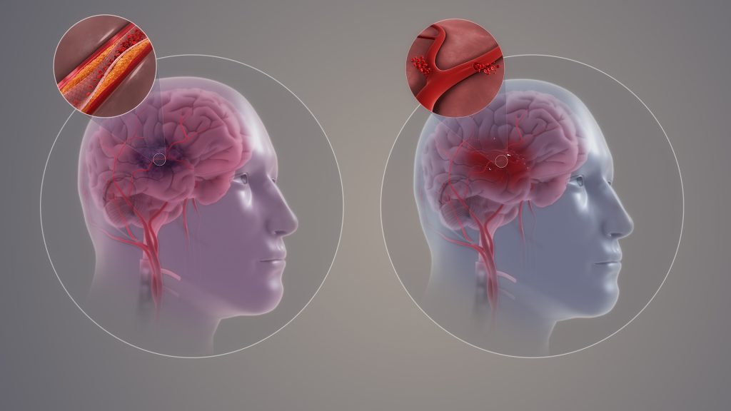
New Blood Test for Ischaemic Stroke is a ‘Game-changer’

Ischaemic and haemorrhagic stroke. Credit: Scientific Animations CC4.0

A new study led by investigators from Brigham and Women’s Hospital has developed a new test by combining blood-based biomarkers with a clinical score to identify patients experiencing large vessel occlusion (LVO) stroke with high accuracy. Their results are published in the journal Stroke: Vascular and Interventional Neurology.

“We have developed a game-changing, accessible tool that could help ensure that more people suffering from stroke are in the right place at the right time to receive critical, life-restoring care,” said senior author Joshua Bernstock, MD, PhD, MPH, a clinical fellow in the Department of Neurosurgery at Brigham and Women’s Hospital.

Most strokes are ischaemic, in which blood flow to the brain is obstructed. LVO strokes are an aggressive type of ischaemic stroke that occurs when an obstruction occurs in a major artery in the brain, causing brain cells to rapidly die off from lack of oxygen. Major medical emergencies, LVO strokes require the swift treatment with mechanical thrombectomy, a surgical procedure that retrieves the blockage.

“Mechanical thrombectomy has allowed people that otherwise would have died or become significantly disabled be completely restored, as if their stroke never happened,” said Bernstock. “The earlier this intervention is enacted, the better the patient’s outcome is going to be. This exciting new technology has the potential to allow more people globally to get this treatment faster.”

The research team previously targeted two specific proteins found in capillary blood, one called glial fibrillary acidic protein (GFAP), which is also associated with brain bleeds and traumatic brain injury, and one called D-dimer. In this study, they demonstrated that the levels of these blood-based biomarkers combined with field assessment stroke triage for emergency destination (FAST-ED) scores could identify LVO ischaemic strokes while ruling out other conditions such as bleeding in the brain. Brain bleeds cause similar symptoms to LVO stroke, making them hard to distinguish from one another in the field, yet treatment for each is vastly different.

In this prospective, observational diagnostic accuracy study, the researchers looked at data from a cohort of 323 patients coded for stroke in Florida between May 2021 and August 2022. They found that combining the levels of the biomarkers GFAP and D-dimer with FAST-ED data less than six hours from the onset of symptoms allowed the test to detect LVO strokes with 93% specificity and 81% sensitivity. Other findings included that the test ruled out all patients with brain bleeds, suggesting that it may also eventually be used to detect intracerebral haemorrhage in the field.

Bernstock’s team also sees promising potential future use of this accessible diagnostic tool in low- and middle-income countries, where advanced imaging is not always available. It might also be useful in assessing patients with traumatic brain injuries. Next, they are carrying out another prospective trial to measure the test’s performance when used in an ambulance. They have also designed an interventional trial that leverages the technology to expedite the triage of stroke patients by having them bypass standard imaging and move directly to intervention.

“In stroke care, time is brain,” Bernstock said. “The sooner a patient is put on the right care pathway, the better they are going to do. Whether that means ruling out bleeds or ruling in something that needs an intervention, being able to do this in a prehospital setting with the technology that we built is going to be truly transformative.

Source: Brigham and Women’s Hospital

Difference in Brain Structures may Explain Concussion Outcomes for Males and Females

Coup and contrecoup brain injury. Credit: Scientific Animations CC4.0

Important brain structures that are key for signalling in the brain are narrower and less dense in females, and more likely to be damaged by brain injuries, such as concussion. Long-term cognitive deficits occur when the signals between brain structures weaken due to the injury. These structural differences in male and female brains might explain why females are more prone to concussions and experience longer recovery from the injury than their male counterparts, according to a University of Pennsylvania-led preclinical study published in Acta Neuropathologica.

Each year, approximately 50 million individuals worldwide suffer a concussion, also referred to as mild traumatic brain injury (TBI). For more than 15% of individuals who suffer persisting cognitive dysfunction, which includes difficulty concentrating, learning and remembering new information, and making decisions.

Although males make up the majority of emergency department visits for concussion, this has been primarily attributed to their greater exposure to activities with a risk of head impacts compared to females. In contrast, it has recently been observed that female athletes have a higher rate of concussion and appear to have worse outcomes than their male counterparts participating in the same sport.

“Clinicians have observed for a long time that females suffer from concussion at higher rates than males in the same sports, and that they take longer to recover cognitive function, but couldn’t explain the underlying mechanisms of this phenomenon,” said senior author Douglas Smith, MD, a professor of Neurosurgery and director of Penn’s Center for Brain Injury and Repair. “The variances in brain structures of females and males not only illuminate why this disparity exists, but also exposes biomarkers, such as axon protein fragments, that can be measured in the blood to determine injury severity, monitor recovery, and eventually help identify and develop treatments that help patients repair these damaged structures and restore cognitive function.”

Axons connect neurons, allowing communication across the brain. These axons form bundles that make up white matter in the brain and play a large role in learning and communication between different brain regions. Axons are delicate structures and are vulnerable to damage from concussion.

Communication between axons in the brain is powered by sodium channels that serve as the brain’s electric grid. When axons are damaged, these sodium channels are also impaired, which causes loss of signaling in the brain. The loss of signaling causes the cognitive impairment experienced by individuals after concussion.

In this study, researchers used large animal models of concussion to identify differences in brains of males and females after a concussion. They found that females had a higher population of smaller axons, which researchers demonstrated are more vulnerable to injury. They also reported that in these models, females had greater loss of sodium channels after concussion.

“The differences in brain structure not only tell us a lot about how brain injury affects males and females differently but could offer insights in other brain conditions that impact axons, like Alzheimer’s and Parkinson’s disease,” said Smith. “If female brains are more vulnerable to damage from concussion, they might also be more vulnerable to neurodegeneration, and it’s worth further research to understand how sex influences the structure and functions of the brain.”

Source: University of Pennsylvania School of Medicine