Month: April 2024

Inflammation may Link Depression and Cardiomyopathy

Photo by Alex Green on Pexels

Coronary artery disease and major depression may be genetically linked via inflammatory pathways to an increased risk for cardiomyopathy, a degenerative heart muscle disease, researchers at Vanderbilt University Medical Center and Massachusetts General Hospital have found.

Their report, published in Nature Mental Health, suggests that drugs prescribed for coronary artery disease and depression, when used in combination, potentially may reduce inflammation and prevent the development of cardiomyopathy.

“This work suggests that chronic low-level inflammation may be a significant contributor to both depression and cardiovascular disease,” said the paper’s corresponding author, Lea Davis, PhD, associate professor of Medicine in the Division of Genetic Medicine and Vanderbilt Genetics Institute.

The connection between depression and other serious health conditions is well known. As many as 44% of patients with coronary artery disease (CAD), the most common form of cardiovascular disease, also have a diagnosis of major depression. Yet the biological relationship between the two conditions remains poorly understood.

A possible connection is inflammation. Changes in the levels of inflammatory markers have been observed in both conditions, suggesting that there may be a common biological pathway linking neuroinflammation in depression with atherosclerotic inflammation in CAD.

In the current study, the researchers used a technique called transcriptome-wide association scans to map single nucleotide polymorphisms (genetic variations) involved in regulating the expression of genes associated with both CAD and depression.

The technique identified 185 genes that were significantly associated with both depression and CAD, and which were “enriched” for biological roles in inflammation and cardiomyopathy.

This suggests that predisposition to both depression and CAD, which the researchers called (major) depressive CAD, or (m)dCAD, may further predispose individuals to cardiomyopathy.

However, when the researchers scanned large electronic health record databases at VUMC, Mass General, and the National Institutes of Health’s All of Us Research Program, they found the actual incidence of cardiomyopathy in patients with the enriched genes for (m)dCAD was lower than in patients with CAD alone.

One possible explanation is that medications prescribed for CAD and depression, such as statins and antidepressants, may prevent development of cardiomyopathy by reducing inflammation, the researchers concluded.

“More research is needed to investigate optimal treatment mechanisms,” Davis added, “but at a minimum this work suggests that patient heart and brain health should be considered together when developing management plans to treat depression or cardiovascular disease.”

Source: Vanderbilt University Medical Center

Spotting the Aggressive Prostate Cancers among Urine Test Results

Credit: Darryl Leja National Human Genome Research Institute National Institutes Of Health

Researchers have developed a new urine-based test that addresses a major problem in prostate cancer: how to separate the slow-growing form of the disease unlikely to cause harm from more aggressive cancer that needs immediate treatment.

The test, called MyProstateScore2.0, or MPS2, looks at 18 different genes linked to high-grade prostate cancer. In multiple tests using urine and tissue samples from men with prostate cancer, it successfully identified cancers classified as Gleason 3+4=7 or Grade Group 2 (GG2), or higher. These cancers are more likely to grow and spread compared to Gleason 6 or Grade Group 1 prostate cancers, which are unlikely to spread or cause other impact. More than one-third of prostate cancer diagnoses are this low-grade form. Gleason and Grade Group are both used to classify how aggressive prostate cancer is.

Results from the University of Michigan Rogel Cancer Center-led study are published in JAMA Oncology.

“Our standard test is lacking in terms of its ability to clearly pick out those who have significant cancer. Twenty years ago, we were looking for any kind of cancer. Now we realise that slow-growing cancer doesn’t need to be treated. All of a sudden, the game changed. We went from having to find any cancer to finding only significant cancer,” said co-senior study author John T. Wei, M.D., David A. Bloom Professor of Urology at Michigan Medicine.

Prostate-specific antigen, or PSA, remains the linchpin of prostate cancer detection. MPS2 improves upon a urine-based test developed by the same U-M team nearly a decade ago, following a landmark discovery of two genes that fuse to cause prostate cancer. The original MPS test, which is used today, looked at PSA, the gene fusion TMPRSS2::ERG, and another marker called PCA3.

“There was still an unmet need with the MyProstateScore test and other commercial tests currently available. They were detecting prostate cancer, but in general they were not doing as good a job in detecting high-grade or clinically significant prostate cancer. The impetus for this new test is to address this unmet need,” said co-senior author Arul M. Chinnaiyan, M.D., Ph.D., director of the Michigan Center for Translational Pathology. Chinnaiyan’s lab discovered the T2::ERG gene fusion and developed the initial MPS test.

“If you’re negative on this test, it’s almost certain that you don’t have aggressive prostate cancer,” said Chinnaiyan, S. P. Hicks Endowed Professor of Pathology and professor of urology at Michigan Medicine.

Moreover, MPS2 was more effective at helping patients avoid unnecessary biopsies. While 11% of unnecessary biopsies were avoided with PSA testing alone, MPS2 testing would avoid up to 41% of unnecessary biopsies.

“Four of 10 men who would have a negative biopsy will have a low risk MPS2 result and can confidently skip a biopsy. If a man has had a biopsy before, the test works even better,” Wei explained.

For example, a patient may get a prostate biopsy due to an elevated PSA, but no cancer is detected. The patient is followed over time and if his PSA inches up, he would typically need another biopsy.

“In those men who have had a biopsy before and are being considered for another biopsy, MPS2 will identify half of those whose repeat biopsy would be negative. Those are practical applications for patients out there. Nobody wants to say sign me up for another biopsy. We are always looking for alternatives and this is it,” Wei said.

Source: Michigan Medicine – University of Michigan

Hot Days may Drive Inflammation and Accelerate Cardiovascular Disease

Photo by Fandy Much: https://www.pexels.com/photo/toshiba-outdoor-air-conditioner-unit-on-yellow-wall-14086132/

Short-term exposure to higher heat may increase inflammation and interfere with normal immune system functions in the body, which may, in turn, increase susceptibility to infections and accelerate the progression of cardiovascular disease, according to preliminary research be presented at the American Heart Association’s Epidemiology and Prevention – Lifestyle and Cardiometabolic Scientific Sessions 2024, March 18-21.

An inflammatory response that is longstanding (lasting weeks to months) or that occurs in healthy tissues is damaging and plays a key role in the build-up of plaque in the arteries. This may lead to atherosclerosis. Heat waves are known to promote inflammation, however, studies examining air temperature and biomarkers of inflammation have had mixed results.

“Most research only considers temperature as the exposure of interest, which may not be adequate to capture a person’s response to heat,” said lead study author Daniel W. Riggs, PhD, an assistant professor of medicine at the University of Louisville. “In our study, we used alternative measurements of heat in relation to multiple markers of inflammation and immune response in the body to investigate the short-term effects of heat exposure and produce a more complete picture of its health impact.”

Participants visited study sites in Louisville, during the summer months for a blood test, and researchers analysed the blood for multiple markers of immune system function. The researchers then examined associations between the markers of immune system function and heat levels, including temperature, net effective temperature (which factors in relative humidity, air temperature and windspeed) and the Universal Thermal Climate Index (UTCI) on that day. UTCI is a thermo-physiological model developed by the International Society of Biometeorology Commission that factors in temperature, humidity, wind speed and ultraviolet radiation levels, which was used to evaluate participant’s physical comfort.

The analysis found:

  • For every 5°F (2.8°C) increase in UTCI (in this study, the equivalent of going from a day with no thermal stress to a day with moderate thermal stress, Riggs said), there was an increase in blood levels of key inflammatory markers: monocytes (4.2%), eosinophils (9.5%), natural killer T-cells (9.9%) and tumour necrosis factor-alpha (7.0%). These immune molecules indicate activation of the body’s innate immune system, which spurs a fast and non-specific inflammatory response throughout the body to protect against pathogens and injury.
  • A decrease in B-cells (-6.8%), indicating the body’s adaptive immune system that remembers specific viruses and germs and creates antibodies to fight them, was lowered.
  • A lesser impact on the immune system was found when heat was measured by average 24-hour temperature or by net effective temperature, which incorporates humidity and wind but not sunshine.

“Our study participants only had minor exposure to high temperatures on the day of their blood test, however, even minor exposure may contribute to changes in immune markers,” Riggs said. “With rising global temperatures, the association between heat exposure and a temporarily weakened response from the immune system is a concern because temperature and humidity are known to be important environmental drivers of infectious, airborne disease transmission. Thus, during the hottest days of summer people may be at higher risk of heat exposure, they may also be more vulnerable to disease or inflammation.”

Adults over 60 years and adults with existing cardiovascular disease are particularly at risk for heat-related cardiovascular events and deaths, Riggs explained.

“It’s important for physicians to communicate with patients about the risk of adverse health effects from heat exposure. For example, cardiologists could conduct customised consultations and assessments to increase patient awareness about their susceptibility to the effects of high temperatures. Also, changes to treatment regimens may be important to consider to address other risks. For example, some medications could make people more susceptible to heat-related illness or some may not be as effective when the body is exposed to high temperatures,” Riggs said.

Source: American Heart Association

How the Brain’s Working Memory… Works

Photo by Alex Green on Unsplash

Cedars-Sinai investigators have discovered how brain cells responsible for working memory – which holds onto things like phone numbers while we use them – coordinate intentional focus and short-term storage of information. Their discovery, which confirms the involvement of the hippocampus, is published in the journal Nature.

“We have identified for the first time a group of neurons, influenced by two types of brain waves, that coordinate cognitive control and the storage of sensory information in working memory,” said Jonathan Daume, PhD, a postdoctoral scholar in the Rutishauser Lab at Cedars-Sinai and first author of the study. “These neurons don’t contain or store information, but are crucial to the storage of short-term memories.”

Working memory, which requires the brain to store information for only seconds, is fragile and requires continued focus to be maintained, explained senior study author Ueli Rutishauser, PhD, director of the Center for Neural Science and Medicine at Cedars-Sinai. It can be affected by different diseases and conditions.

“In disorders such as Alzheimer’s disease or attention-deficit hyperactivity disorder, it is often not memory storage, but rather the ability to focus on and retain a memory once it is formed that is the problem,” said Rutishauser, who is a professor of Neurosurgery, Neurology and Biomedical Sciences at Cedars-Sinai. “We believe that understanding the control aspect of working memory will be fundamental for developing new treatments for these and other neurological conditions.”

To explore how working memory functions, investigators recorded the brain activity of 36 hospitalised patients who had electrodes surgically implanted in their brains as part of an epilepsy diagnosis procedure. The team recorded the activity of individual brain cells and brain waves while the patients performed a task that required use of working memory.

On a computer screen, patients were shown either a single photo or a series of three photos of various people, animals, objects or landscapes. Next, the screen went blank for just under three seconds, requiring patients to remember the photos they just saw. They were then shown another photo and asked to decide whether it was the one (or one of the three) they had seen before.

When patients performing the working memory task were able to respond quickly and accurately, investigators noted the firing of two groups of neurons: “category” neurons that fire in response to one of the categories shown in the photos, such as animals, and “phase-amplitude coupling,” or PAC, neurons.

PAC neurons, newly identified in this study, don’t hold any content, but use a process called phase-amplitude coupling to ensure the category neurons focus and store the content they have acquired. PAC neurons fire in time with the brain’s theta waves, which are associated with focus and control, as well as to gamma waves, which are linked to information processing. This allows them to coordinate their activity with category neurons, which also fire in time to the brain’s gamma waves, enhancing patients’ ability to recall information stored in working memory.

“Imagine when the patient sees a photo of a dog, their category neurons start firing ‘dog, dog, dog’ while the PAC neurons are firing ‘focus/remember,'” Rutishauser said. “Through phase-amplitude coupling, the two groups of neurons create a harmony superimposing their messages, resulting in ‘remember dog.’ It is a situation where the whole is greater than the sum of its parts, like hearing the musicians in an orchestra play together. The conductor, much like the PAC neurons, coordinates the various players to act in harmony.”

PAC neurons do this work in the hippocampus, a part of the brain that has long been known to be important for long-term memory. This study offers the first confirmation that the hippocampus also plays a role in controlling working memory, Rutishauser said.

Source: Cedars-Sinai Medical Center

Study Explains How Aspirin can Help Prevent Colorectal Cancer Development

Photo by cottonbro studio

Studies have shown that long-term daily use of aspirin can help to prevent the development and progression of colorectal cancer, but the mechanisms involved have been unclear. New research has revealed that aspirin may exert these protective effects by boosting certain aspects of the body’s immune response against cancer cells. The findings are published by Wiley online in CANCER, a peer-reviewed journal of the American Cancer Society.

To investigate the effects of aspirin (a nonsteroidal anti-inflammatory drug) on colorectal cancer, investigators in Italy obtained tissue samples from 238 patients who underwent surgery for colorectal cancer in 2015–2019, 12% of whom were aspirin users. Patients were enrolled in the METACCRE section of the IMMUNOlogical microenvironment in the REctal Adenocarcinoma Treatment (IMMUNOREACT 8) multicenter observational study. The study was funded by the Associazione Italiana per la Ricerca sul Cancro (AIRC) and was mainly carried out at the University Hospital of Padova.

Compared with tissue samples from patients who did not use aspirin, samples from aspirin users showed less cancer spread to the lymph nodes and higher infiltration of lymphocytes into tumours. In analyses of colorectal cancer cells in the lab, exposing the cells to aspirin caused increased expression of a protein called CD80 on certain immune cells, which enhanced the capacity of the cells to alert other immune cells of the presence of tumour-associated proteins. Supporting this finding, the researchers found that in patients with rectal cancer, aspirin users had higher CD80 expression in healthy rectal tissue, suggesting a pro-immune surveillance effect of aspirin.

“Our study shows a complementary mechanism of cancer prevention or therapy with aspirin besides its classical drug mechanism involving inhibition of inflammation,” said principal investigator Marco Scarpa MD, PhD, of the University of Padova. “Aspirin is absorbed in the colon by passive diffusion to a significant degree. Its absorption is linear and depends on concentration along the bowel, and in the rectum, the concentration of orally administered aspirin can be much lower than in the rest of the colon. Thus, if we want to take advantage of its effects against colorectal cancer, we should think of how to guarantee that aspirin reaches the colorectal tract in adequate doses to be effective.” 

Source: Wiley

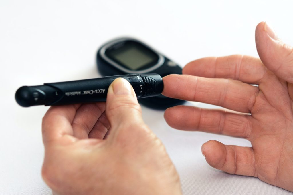
Probing an Outdated Diabetes Drug’s Insulin Resistance Lowering Abilities

Photo by Photomix Company on Pexels

Thiazolidinediones (TZDs) are a class of drug that can be used to treat type 2 diabetes by reversing insulin resistance, one of the main hallmarks of the disease. While TZDs were extremely popular in the 1990s and early 2000s, they have fallen out of use among physicians in recent decades because unwanted side effects emerged, including weight gain and excess fluid accumulation in body tissues.

Now, researchers at University of California San Diego School of Medicine are exploring how to isolate the positive effects of these drugs, which could help yield new treatments that don’t come with the old side effects.

In a new study published in Nature Metabolism, the researchers discovered how one of the most well-known TZD drugs works at the molecular level and were able to replicate its positive effects in mice without giving them the drug itself.

“For decades, TZDs have been the only drugs we have that can reverse insulin resistance, but we seldom use them anymore because of their side effects profile,” said Jerrold Olefsky, MD, a professor of medicine and assistant vice chancellor for integrative research at UC San Diego Health Sciences.

“Impaired insulin sensitivity is the root cause of type 2 diabetes, so any treatment we can develop to safely restore this would be a major step forward for patients.”

The main driver of insulin resistance in type 2 diabetes is obesity. Obesity-related inflammation causes macrophages to accumulate in adipose tissue, where they can comprise up to 40% of the total number of cells in the tissue.

When adipose tissue is inflamed, these macrophages release tiny nanoparticles containing instructions for surrounding cells in the form of microRNAs. These microRNA-containing capsules, called exosomes, are released into the circulation and can travel through the bloodstream to be absorbed by other tissues, such as the liver and muscles. This can then lead to the varied metabolic changes associated with obesity, including insulin resistance.

To understand how TZD drugs, which restore insulin resistance, affect this exosome system, the researchers treated a group of obese mice with the TZD drug rosiglitazone. Those mice became more sensitive to insulin, but they also gained weight and retained excess fluid, known side effects of rosiglitazone.

However, by isolating exosomes from the adipose tissue macrophages of the mice who had received the drug and injecting them into another group of obese mice that had not received it, the researchers were able to deliver the positive effects of rosiglitazone without transferring the negative effects.

“The exosomes were just as effective in reversing insulin resistance as the drug itself but without the same side effects,” said Olefsky.

“This indicates that exosomes can ultimately link obesity-related inflammation and insulin resistance to diabetes. It also tells us that we may be able to leverage this system to boost insulin sensitivity.”

The researchers were also able to identify the specific microRNA within the exosomes that was responsible for the beneficial metabolic effects of rosiglitazone. This molecule, called miR-690, could eventually be leveraged into new therapies for type 2 diabetes.

“It’s likely not practical to develop exosomes themselves as a treatment because it would be difficult to produce and administer them, but learning what drives the beneficial effects of exosomes at the molecular level makes it possible to develop drugs that can mimic these effects,” said Olefsky. “There’s also plenty of precedent for using microRNAs themselves as drugs, so that’s the possibility we’re most excited about exploring for miR-690 going forward.”

Source: University of California – San Diego

Certain Gut Bacteria Assist in Immunotherapy for Milk Allergy

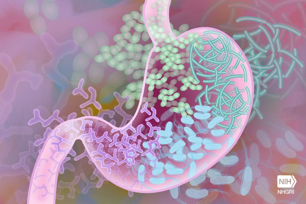
Gut Microbiome. Credit Darryl Leja National Human Genome Research Institute National Institutes Of Health

Researchers in Japan have discovered a link between gut bacteria and the success of milk-allergy oral immunotherapy. Published in the scientific journal Allergology International, the study found that Bifidobacterium – a genus of beneficial bacteria in the gut – was associated with a higher chance of successful treatment. The finding may help in the development of more effective oral immunotherapies, perhaps by combining them with probiotic supplements.

Many children have allergic reactions to certain milk proteins. Most grow out of it but some have to contend with lifelong allergy. Milk allergy can be improved by oral immunotherapy – taking small, increasing doses of milk. Unfortunately, while allergic reactions are controlled during treatment, in most cases, tolerance disappears soon after the treatment ends.

Gut bacteria are thought to help reduce allergic reactions to some foods, but little is known about the link between these bacteria and oral immunotherapy for milk allergy. Hiroshi Ohno led a team at the RIKEN Center for Integrative Medical Sciences to find out why. The researchers examined 32 children with cow’s milk allergy who received oral immunotherapy, with the first month being conducted in a hospital. “Oral immunotherapy is not without risk,” explains Ohno. “We closely monitored the children in the hospital, and in fact 4 children had such severe reactions to the milk that we could not allow them to continue the treatment.”

The remaining 28 children then completed an additional 12 months of treatment at home. Next, they avoided milk for two weeks, and were then tested on a double-blind, placebo-controlled food challenge to see if they could still tolerate milk without any allergic reactions. During the food challenge, children were initially given a tiny amount of placebo or milk (only 0.01mL) which was gradually increased every 20 minutes until they had an allergic reaction or until they could drink the final 30mL without a reaction.

The researchers focused their analyses on immunological and bacterial changes during the treatment and the relationship between gut bacteria and successful treatment—which was defined as showing milk tolerance that lasted beyond the treatment period by passing the food challenge. They found that during treatment, immunological markers for cow’s milk allergy improved, and bacteria in the gut changed. Nevertheless, after two weeks of avoiding milk, only 7 of the 28 children passed the food challenge, even though they had been able to drink milk safely at the end of the treatment.

To understand why the treatment worked for these seven children but not the others, the team looked for the clinical factors and types of gut bacteria that were related to successful treatment. Of the clinical factors, unsuccessful treatment was more likely in children who were being treated for eczema or asthma and in children who initially had higher levels of milk-protein antibodies. Among the gut bacteria, the presence of Bifidobacterium, a genus of beneficial bacteria in the Bifidobacteriaceae family was related to a higher chance of successful treatment. In fact, only children who passed the final food challenge showed an increasing trend in these bacteria over the course of treatment. When considering ways to improve oral immunotherapy, this is good news because while the first two factors are difficult to change, the types of bacteria in one’s gut are not set in stone.

“With this study, we have identified gut environmental factors that help establish immune tolerance against cow’s milk allergy via oral immunotherapy,” says Ohno. “The next step is to examine the mechanisms underlying this phenomenon and to develop ways to improve the effectiveness of oral immunotherapy, such as the addition of probiotic supplements.”

Source: RIKEN

Do More Mentally Challenging Jobs Protect against Cognitive Decline?

Source: Unsplash CC0

The harder your brain works at your job, the less likely you may be to have memory and thinking problems later in life, according to a new study published in Neurology®, the medical journal of the American Academy of Neurology. This study does not prove that stimulating work prevents mild cognitive impairment. It only shows an association.

“We examined the demands of various jobs and found that cognitive stimulation at work during different stages in life – during your 30s, 40s, 50s and 60s – was linked to a reduced risk of mild cognitive impairment after the age of 70,” said study author Trine Holt Edwin, MD, PhD, of Oslo University Hospital in Norway.

“Our findings highlight the value of having a job that requires more complex thinking as a way to possibly maintain memory and thinking in old age.”

The study looked at 7000 people and 305 occupations in Norway. Researchers measured the degree of cognitive stimulation that participants experienced while on the job. They measured the degree of routine manual, routine cognitive, non-routine analytical, and non-routine interpersonal tasks, which are skill sets that different jobs demand.

Routine manual tasks demand speed, control over equipment, and often involve repetitive motions, typical of factory work. Routine cognitive tasks demand precision and accuracy of repetitive tasks, such as in bookkeeping and filing.

Non-routine analytical tasks involve analysing information, engaging in creative thinking and interpreting information for others. Non-routine interpersonal tasks include establishing and maintaining personal relationships, motivating others and coaching. Non-routine cognitive jobs include public relations and computer programming.

Researchers divided participants into four groups based on the degree of cognitive stimulation that they experienced in their jobs. The most common job for the group with the highest cognitive demands was teaching. The most common jobs for the group with the lowest cognitive demands were mail carriers and custodians.

After age 70, participants completed memory and thinking tests to assess whether they had mild cognitive impairment. Of those with the lowest cognitive demands, 42% were diagnosed with mild cognitive impairment, compared to 27% for those with the highest cognitive demands.

After adjustment for age, sex, education, income and lifestyle factors, the group with the lowest cognitive demands at work had a 66% higher risk of mild cognitive impairment compared to the group with the highest cognitive demands at work.

“These results indicate that both education and doing work that challenges your brain during your career play a crucial role in lowering the risk of cognitive impairment later in life,” Edwin said. “Further research is required to pinpoint the specific cognitively challenging occupational tasks that are most beneficial for maintaining thinking and memory skills.”

A limitation of the study was that even within identical job titles, individuals might perform different tasks and experience different cognitive demands.

Source: American Academy of Neurology

Genetic Predisposition for Muscle Strength may Predict Longer Lifespan

Photo by Barbara Olsen on Pexels

A study conducted at the University of Jyväskylä showed that a genetic predisposition for higher muscle strength predicts a longer lifespan and a lower risk for developing common diseases. This study, published in The Journals of Gerontology: Series A, is the most comprehensive international study to date on hereditary muscle strength and its relationship to morbidity. The genome and health data of more than 340 000 Finns was used in the research.

Muscle strength, especially hand grip strength, can indicate an individual’s physiological resources to protect against age-related diseases and disabilities, as well as their ability to cope with them. Age-related loss of muscle strength is individual and influenced not only by lifestyle but also by genetics.

The study revealed that individuals with a genetic predisposition for higher muscle strength have a slightly lower risk for common noncommunicable diseases and premature mortality. It did not however predict better survival after acute adverse health events compared to the time before illness onset.  

“It seems that a genetic predisposition for higher muscle strength reflects more on an individual’s intrinsic ability to resist and protect oneself against pathological changes that occur during aging than the ability to recover or completely bounce back after severe adversity,” says doctoral researcher Päivi Herranen from the Faculty of Sport and Health Sciences. 

A unique study population  

Muscle strength is a multifactorial trait influenced by lifestyle and environmental factors but also by numerous genetic variants, each with a very small effect on muscle strength. In this study, the genetic predisposition for muscle strength was defined by constructing a polygenic score for muscle strength, which summarises the effects of hundreds of thousands of genetic variants into a single score. The polygenic score makes it possible to compare participants with an exceptionally high or low genetic predisposition for muscle strength, and to investigate associations with inherited muscle strength and other phenotypes, in this case, common diseases.  

“In this study, we were able to utilise both genetic information and health outcomes from over 340 000 Finnish men and women,” Herranen explains. “To our knowledge, this is the first study to investigate the association between a genetic predisposition for muscle strength and various diseases on this scale.” 

Further research on the effects of lifestyles is still needed 

Information about the genetic predisposition for muscle strength could be used alongside traditional risk assessment in identifying individuals who are at particularly high risk of common diseases and health adversities. However, further research on the topic is still needed. 

“Based on these results, we cannot say how lifestyle factors, such as physical activity, modify an individual’s intrinsic ability to resist diseases and whether their impact on health differs among individuals due to genetics,” Herranen notes. 

The study utilised the internationally unique FinnGen dataset, compiled through the collaboration of Finnish biobanks. The dataset consisted of 342 443 Finns who had given their consent and provided a biobank sample. The participants were aged 40 to 108 years, and 53% of them were women. The diagnoses selected for the study were based on the leading causes of death and the most significant noncommunicable diseases in Finland. Selected diagnoses included the most common cardiometabolic and pulmonary diseases, musculoskeletal and connective tissue diseases, falls and fractures, mental health and cognitive disorders, cancers, as well as overall mortality and mortality from cardiovascular diseases. 

Source: University of Jyväskylä

New Drug Shows Promise for Treating Rare and Aggressive Gliomas

MRI scan showing brain cancer. Credit: Michelle Monje, MD, PhD, Stanford University

An experimental drug may provide a new treatment option for some patients with rare incurable brain tumours, according to an analysis published in the Journal of Clinical Oncology.

Diffuse midline gliomas are diagnosed in about 800 people per year in the U.S., according to the Centers for Disease Control and Prevention.

A subset of particularly aggressive diffuse midline gliomas are caused by a H3 K27M mutation and the only effective treatment is radiation, as the location of the tumour in the brain makes surgery difficult. Even with radiation, relapse is virtually inevitable and more than 70% of patients with this subtype of brain tumour die from the cancer, according to the National Institutes of Health.

In the study, investigators analysed the results of five previous clinical trials testing the effectiveness of dordaviprone, an experimental drug which works by blocking a certain protein in tumours with the mutation.

The study included results from 50 patients (including four children) with H3 K27M–mutant diffuse midline gliomas and found that 30% of patients responded well to the drug. The most common side effect reported was fatigue, according to the study.

Now, the researchers are launching a trial at Northwestern Medicine hospitals to investigate the drug’s effectiveness in newly diagnosed patients.

Source: Northwestern University