Month: April 2024

Useless Antibiotic Prescriptions are Getting out of Hand

Photo from Pixabay CCO

According to a massive new medical insurance database study, the U.S. is going the wrong way with antibiotic stewardship, with 1 in 4 prescriptions going to patients who have conditions that the drugs simply won’t work on. In fact, the percentage of all antibiotic prescriptions given to treat conditions they’re useless against was even higher in December 2021 than it was before the pandemic began, the study shows – increasing the rate of antibiotic resistance development.

The percentage inappropriate prescriptions actually fell slightly in the early months of the pandemic, when far fewer people sought medical care for infectious or non-infectious reasons, the new research shows. But this trend was soon reversed.

The study, published in the journal Clinical Infectious Diseases by a team from the University of Michigan, Northwestern University and Boston Medical Center, is based on data from more than 37.5 million children and adults covered by private insurance or Medicare Advantage plans from 2017 to 2021. Patients received antibiotic prescriptions from both in-person and telehealth visits.

The team looked back at any new diagnosis given to each patient on the day they received a prescribed antibiotic or in the three days before getting the prescription. If none of these diagnoses justified the use of antibiotics, they classified the prescription as inappropriate.

Key findings:

  • In all, 60.6 million antibiotic prescriptions were dispensed in the five years of the study period from January 2017 to December 2021. The share that were inappropriate rose from 25.5% to 27.1% during this period.
  • The proportion of people getting inappropriate antibiotics was 1.7% in December 2019, dipped to 0.9% in April 2020 – largely because fewer people get antibiotics in general – and returned to 1.7% by December 2021.
  • Some groups of people were more likely to receive inappropriate antibiotics. At the end of 2021, 30% of antibiotics for older adults with Medicare Advantage coverage were inappropriate, compared with 26% of antibiotics for adults with private health insurance and 17% of antibiotics for children with private insurance.
  • Among the diagnoses listed for people who received antibiotics for inappropriate reasons, “contact with and suspected exposure to COVID-19” was one of top two most common reasons from March 2020 through December 2021. There is no evidence that taking antibiotics after an exposure can reduce risk of developing COVID-19.
  • Of all the inappropriately prescribed antibiotics dispensed in the last half of 2021, 15% were for a COVID-19 infection. And COVID-19 infections accounted for 2% of all antibiotic prescribing – regardless of appropriateness – from March 2020 through December 2021.
  • Telehealth appointments accounted for 9% of all inappropriate antibiotic prescriptions in the latter half of 2021, down somewhat from 2020. There were almost no telehealth-based antibiotic prescriptions before March 2020.
  • For 28% to 32% of the antibiotic prescriptions filled by patients in the study period, there was no diagnosis available to judge appropriateness, potentially because the patient received the prescription at an appointment that didn’t get billed to their insurance, or it was a refill of a past prescription. The percentage was especially high in the first months of the pandemic.
  • 45% of all the patients in the study received antibiotics at least once in the five years, and 13% received them four or more times.

Source: University of Michigan

Essential Tremor Increases Cognitive Impairment Risks over Time

Photo by Matthias Zomer on Pexels

Essential tremor, a nervous system disorder that causes rhythmic shaking, is one of the most common movement disorders. A new study published in the Annals of Neurology reveals details on the increased risk of mild cognitive impairment (MCI) and dementia that individuals with essential tremor may face.

The research represents the longest available longitudinal prospective study of rates of MCI and dementia in people with essential tremor. The study enrolled 222 patients, 177 of whom participated in periodic evaluations over an average follow-up of 5 years.

Investigators observed a cumulative prevalence of 26.6% and 18.5% for MCI and dementia, respectively. They also noted a cumulative incidence of 18.2% and 11.2% for MCI and dementia, respectively. Each year, 3.9% of patients with normal cognition “converted” to having MCI, and 12.2% of those with MCI “converted” to having dementia.

“We know from related research that the presence of cognitive impairment in patients with essential tremor has meaningful clinical consequences. For example, patients with essential tremor who are diagnosed with dementia are more likely to need to use a walker or wheelchair, to employ a home health aide, and to reside in non-independent living arrangements than are patients with essential tremor without dementia,” said corresponding author Elan D. Louis, MD, MS, of the University of Texas Southwestern Medical Center. “With this in mind, the findings of the present study highlight the importance of cognitive screening and monitoring in patients with essential tremor. Early detection of impairment may provide opportunities for interventions that may slow further cognitive decline and improve the quality of life of patients and their families.”

Source: Wiley

Study Links Emulsifiers and Type 2 Diabetes Risk

Photo by Amit Lahav on Unsplash

Emulsifiers, commonly used additives for improving the texture of food products and extending their shelf life, may be associated with the onset of type 2 diabetes, according to a large cohort study of over 100 000 people in France.

Researchers from Inserm, INRAE, Université Sorbonne Paris Nord, Université Paris Cité and Cnam, as part of the Nutritional Epidemiology Research Team (CRESS-EREN), studied the possible links between the dietary intake of food additive emulsifiers and the onset of type 2 diabetes between 2009 and 2023. They analysed the dietary and health data of 104 139 adults participating in the French NutriNet-Santé cohort study, specifically evaluating their consumption of this type of food additive using dietary surveys conducted every six months. The findings suggest an association between the chronic consumption of certain emulsifier additives and a higher risk of diabetes. The study is published in Lancet Diabetes & Endocrinology.

In Europe and North America, 30 to 60% of dietary energy intake in adults comes from ultra-processed foods. An increasing number of epidemiological studies suggest a link between higher consumption levels of ultra-processed foods with higher risks of diabetes and other metabolic disorders.

Emulsifiers are among the most commonly used additives. They are often added to processed and packaged foods such as certain industrial cakes, biscuits and desserts, as well as yoghurts, ice creams, chocolate bars, industrial breads, margarines and ready-to-eat or ready-to-heat meals, in order to improve their appearance, taste and texture and lengthen shelf life. These emulsifiers include for instance mono- and diglycerides of fatty acids, carrageenans, modified starches, lecithins, phosphates, celluloses, gums and pectins.

As with all food additives, the safety of emulsifiers had been previously evaluated by food safety and health agencies based on the scientific evidence that was available at the time of their evaluation.
However, some recent studies suggest that emulsifiers may disrupt the gut microbiota and increase the risk of inflammation and metabolic disruption, potentially leading to insulin resistance and the development of diabetes.

For the first time worldwide, a team of researchers in France has studied the relationships between the dietary intakes of emulsifiers, assessed over a follow-up period of maximum 14 years, and the risk of developing type 2 diabetes in a large study in the general population.

The results are based on the analysis of data from 104 139 adults in France (average age 43 years; 79% women) who participated in the NutriNet-Santé web-cohort study (see box below) between 2009 and 2023.

The participants completed at least two days of dietary records, collecting detailed information on all foods and drinks consumed and their commercial brands (in the case of industrial products). These dietary records were repeated every six months for 14 years, and were matched against databases in order to identify the presence and amount of food additives (including emulsifiers) in the products consumed. Laboratory assays were also performed in order to provide quantitative data. This allowed a measurement of chronic exposure to these emulsifiers over time.

During follow-up, participants reported the development of diabetes (1056 cases diagnosed), and reports were validated using a multi-source strategy (including data on diabetes medication use). Several well-known risk factors for diabetes, including age, sex, weight (BMI), educational level, family history, smoking, alcohol and levels of physical activity, as well as the overall nutritional quality of the diet (including sugar intake) were taken into account in the analysis.

After an average follow-up of seven years, the researchers observed that chronic exposure – evaluated by repeated data – to the following emulsifiers was associated with an increased risk of type 2 diabetes:

  • carrageenans (total carrageenans and E407; 3% increased risk per increment of 100 mg per day)
  • tripotassium phosphate (E340; 15% increased risk per increment of 500 mg per day)
  • mono- and diacetyltartaric acid esters of mono- and diglycerides of fatty acids (E472e; 4% increased risk per increment of 100 mg per day)
  • sodium citrate (E331; 4% increased risk per increment of 500 mg per day)
  • guar gum (E412; 11% increased risk per increment of 500 mg per day)
  • gum arabic (E414; 3% increased risk per increment of 1000 mg per day)
  • xanthan gum (E415; 8% increased risk per increment of 500 mg per day)

This study constitutes an initial exploration of these relationships, and further investigations are now needed to establish causal links. The researchers mentioned several limitations of their study, such as the predominance of women in the sample, a higher level of education than the general population, and generally more health-promoting behaviours among the NutriNet-Santé study participants. Therefore caution is needed when extrapolating the conclusions to the entire French population.

The study is nevertheless based on a large sample size, and the researchers have accounted for a large number of factors that could have led to confounding bias. They also used unique, detailed data on exposure to food additives, down to the commercial brand name of the industrial products consumed. In addition, the results remain consistent through various sensitivity analyses, which reinforces their reliability.

“These findings are issued from a single observational study for the moment, and cannot be used on their own to establish a causal relationship,”explain Mathilde Touvier, Research Director at Inserm, and Bernard Srour, Junior Professor at INRAE, lead authors of the study. “They need to be replicated in other epidemiological studies worldwide, and supplemented with toxicological and interventional experimental studies, to further inform the mechanisms linking these food additive emulsifiers and the onset of type 2 diabetes. However, our results represent key elements to enrich the debate on re-evaluating the regulations around the use of additives in the food industry, in order to better protect consumers.”

Among the next steps, the research team will be looking at variations in certain blood markers and the gut microbiota linked to the consumption of these additives, to better understand the underlying mechanisms. The researchers will also look at the health impact of additive mixtures and their potential ‘cocktail effects.’

They will also work in collaboration with toxicologists to test the impact of these exposures in in vitro and in vivo experiments, to gather more arguments in favour of a causal link.

Refurbished School for Paediatric Patients Bridges Critical Learning Gaps

Photo by Mary Taylor on Pexels

April 16 2024 – The Chris Hani Baragwanath Hospital School officially opened in its new location today, marking a key milestone in the partnership between Wits University and the academic hospital. The school caters for all learners in need of longer-term and chronic treatment for various paediatric conditions. Learners between Grades R and 12 are taught.  

“Sick children have multiple needs, and it’s our duty to ensure that they don’t miss out on any schooling. Everyone deserves the right to be educated and to contribute meaningfully to their communities as adults,” said Professor Shabir Madhi, Dean of the Wits Faculty of Health Sciences. 

Professor Madhi noted that the previous school building will be used as a campus for medical students and to grow the university’s teaching and learning footprint at Chris Hani Baragwanath Academic Hospital.  

The school district representative for Johannesburg Central, Ronica Ramdath, said that often sick children forfeit their education, which can be mitigated through the correct teaching approach and through supportive facilities. “When I first came to the school some years back, I was amazed at the teachers’ dedication. I remember seeing a teacher load all their educational resources in a bag and walk to the paediatric ward to teach sick children. Today, these children all benefit from such support,” she said.  

The Wits Faculty of Health Sciences heads of schools were present, together with hospital and teaching representatives.  

Meanwhile, Professor Madhi said that the university’s wifi is available at Chris Hani Baragwanath Hospital, underpinning Wits’ commitment to invest in a world-class academic hospital facility. “We are very proud of our footprint at the hospital and hope to continue to add value through research and clinical work,” he said. 

Source: University of the Witwatersrand – Faculty of Health Sciences

How Glucocorticoids Reprogram Immune Cells to Slow them Down

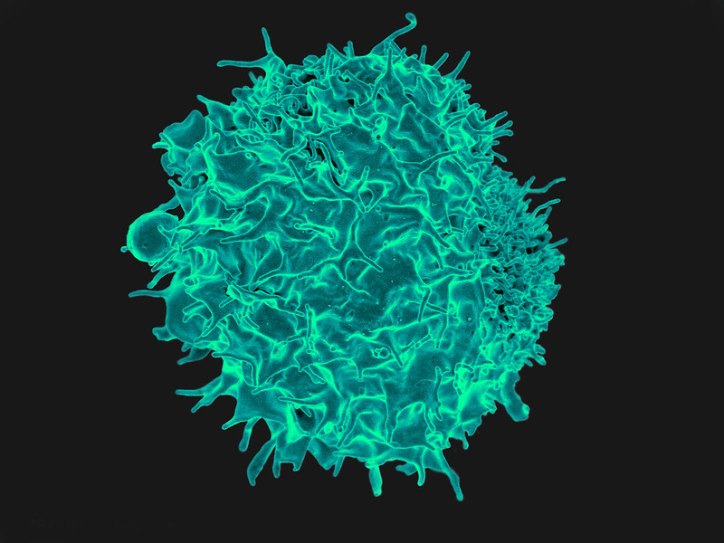
Scanning electron micrograph of a T cell lymphocyte. Credit: NIH / NIAID

Cortisone and other related glucocorticoids are extremely effective at curbing excessive immune reactions. But previously, astonishingly little was known about how they exactly do that. A team of researchers have now explored the molecular mechanism of action in greater detail. As the researchers report in the journal Nature, glucocorticoids reprogram the metabolism of immune cells, activating the body’s natural “brakes” on inflammation. These findings lay the groundwork for development of anti-inflammatory agents with fewer and less severe side effects.

The glucocorticoid cortisone is naturally present in the body as the stress hormone cortisol, which is released to improve the body’s responses to stress. Cortisol intervenes in sugar and fat metabolism and affects other parameters, including blood pressure and respiratory and heart rate. At higher doses, it also inhibits immune system activity, making it it useful for medical purposes. Due to their excellent efficacy, synthetic glucocorticoid derivates that inhibit inflammation even more strongly are used to treat a wide range of immune-mediated inflammatory diseases.

Glucocorticoids affect not only genes, but also cellular energy sources

Glucocorticoid-based medications come with side effects, especially at higher doses and when administered for longer periods. These side effects are related to the other effects of the body’s own cortisol, and include hypertension, osteoporosis, diabetes, and weight gain. With the aim of developing anti-inflammatory agents with fewer and less severe side effects, a team of researchers from from Charité – Universitätsmedizin Berlin, Uniklinikum Erlangen and Ulm University has now conducted a closer study of how the immunosuppressive effects of glucocorticoids exactly works.

Lead researcher Prof Gerhard Krönke, director of the Department of Rheumatology and Clinical Immunology at Charité, explains: “It was previously known that glucocorticoids activate a number of genes in different cells of the body. But through this mechanism, they mainly activate the resources present in the body. This does not adequately explains its strong immunosuppressive effect. In our study, we have now been able to show that glucocorticoids affect more than just the gene expression in immune cells. It also affects the cell´s powerhouses, the mitochondria. And that this effect on cell metabolism is in turn crucial to the anti-inflammatory effects exerted by glucocorticoids.”

Swords to plowshares

For the study, the research team focused on macrophages, a type of immune cell responsible for eliminating intruders such as viruses and bacteria. These cells can also play a role in the emergence of immune-mediated inflammatory diseases. In a mouse model, the researchers studied how these immune cells responded to inflammatory stimuli in a laboratory setting and what effects additional administration of a glucocorticoid had. The researchers observed that in addition to its effect on gene expression, glucocorticoids had a major effect in reversing changes in the cell metabolism that had been initiated by the inflammatory stimuli.

“When macrophages are put into ‘fight’ mode, they redirect their cellular energy into arming for a fight. Instead of supplying energy, their mitochondria produce the components needed to fight intruders,” Krönke says, describing the processes involved. “Glucocorticoids reverse the process, switching the ‘fight’ mode back off and turning swords into plowshares, so to speak. A tiny molecule called itaconate plays an especially important role in this.”

Itaconate mediates anti-inflammatory effect of glucocorticoids

Itaconate is an anti-inflammatory substance that the body naturally produces inside its mitochondria. Macrophages produce it early on when they are activated so that the inflammatory reaction will subside after a certain period. Generation of this natural immune “brake,” however, requires sufficient fuel. When the cell´s powerhouses are arming up for a fight, that is no longer the case, so itaconate production dwindles to a halt after a while. With normal, short-term inflammation, this timing is effective because the immune response has also subsided in the meantime.

“With a persistent inflammatory stimulus, the drop-off in itaconate production is an issue because there is then no immune ‘brake’ even though the immune system is still running on all cylinders, eventually contributing to chronic inflammation,” explains Dr Jean-Philippe Auger, a scientist at the Department of Medicine 3 – Rheumatology and Immunology at Uniklinikum Erlangen and the first author of the study. “This is where glucocorticoids intervenes. By reprogramming the mitochondrial function, they ramp up the formation of itaconate in the macrophages, restoring its anti-inflammatory effect.”

The search for new active substances

Using animal models for asthma and rheumatoid arthritis, the researchers showed how much the anti-inflammatory effect of glucocorticoids depends on itaconate. Glucocorticoids had no effect in animals unable to produce itaconate. So, if itaconate mediates the immunosuppressant effect of cortisone, what about administering itaconate directly, instead of glucocorticoids?

“Unfortunately, itaconate isn’t a particularly good candidate as an anti-inflammatory drug, because it’s unstable, and due to its high reactivity, it could cause side effects if administered systemically,” Krönke explains. “Aside from that, we assume the processes in humans to be a bit more complex than those in mice. So our plan is to look for new synthetic compounds that are just as effective as glucocorticoids at reprogramming the mitochondrial metabolism inside immune cells, but have fewer and less severe side effects.”

Source: Charité – Universitätsmedizin Berlin

Metabolic Health before Flu Vaccination Determines Its Effectiveness

A four-week healthy diet improved the effectiveness of a flu vaccine given to obese mice

Photo by Gustavo Fring on Pexels

Scientists at St. Jude Children’s Research Hospital have shown that improving metabolic health in obese mice before vaccination, but not after, protects against influenza virus.

Metabolic health (normal blood pressure, blood sugar and cholesterol levels, among other factors) influences the effectiveness of influenza vaccinations. Vaccination is known to be less effective in people with obesity compared to those with a healthier body mass index (BMI), but St. Jude Children’s Research Hospital scientists have found that the difference is attributable not to obesity itself, but rather metabolic dysfunction. In a study published in Nature Microbiology, the researchers found that switching obese mice to a healthy diet before flu vaccination, but not after, completely protected the models from a lethal dose of flu, despite BMI.

“We found that the vaccines worked effectively if at the time of vaccination an animal is metabolically healthy,” said corresponding author Stacey Schultz-Cherry, PhD, St. Jude Department of Host-Microbe Interactions and Center of Excellence for Influenza Research and Response co-director. “And the opposite was also true: regardless of what the mice looked like on the outside, if they had metabolic dysfunction, the vaccines did not work as well.”

Prior research has shown that 100% of obese mice succumbed to influenza after exposure, even after vaccination. Contrary to the scientists’ original expectations, when mice who were vaccinated while obese returned to a healthy weight, outcomes did not improve. These now outwardly healthy mice still all succumbed to disease when exposed to the real virus. Only switching to a healthy diet four weeks before vaccination improved survival, with drastic effect, despite high BMI.

“We were excited to see this effect because mice with obesity are so susceptible to severe disease and succumbing to the infection,” Schultz-Cherry said. “Getting 100% survival with the vaccine where we had only seen 0% survival was impressive.” The improved survival suggests the researchers have discovered a greater underlying principle determining influenza vaccine efficacy.

Metabolic dysfunction hinders the immune system

While studying how metabolic function influences influenza vaccine responses, the scientists found that poor metabolic health causes immune system dysfunction. T cells, the primary immune cells involved in anti-viral responses, failed to act in animals that had been in an unhealthy metabolic state at the time of vaccination, even during later viral exposure. Even when the animals ate a healthy diet after vaccination and maintained a normal BMI, the anti-flu T cells were “frozen” in that dysfunctional state.

However, a healthy diet before vaccination improved T-cell function, which resulted in a robust anti-flu response during later exposure.

“The T cells were better able to do their job in the metabolically healthy mice at the time of vaccination,” Schultz-Cherry said. “It wasn’t a matter of the numbers of them or the types of them. It was their functional activity. There were plenty of them in the lungs, not working. The healthy diet switched them from not working to functioning properly, but only if the switch occurred before vaccination.”

The earlier healthy diet also improved inflammation. Pro-inflammatory cytokines are upregulated in obese animals. Schultz-Cherry’s team found that models also returned to a lower basal cytokine level when switched to a healthy diet before vaccination.

“A healthy diet lowered some of the systemic meta-inflammation in these animals, and they regained some of the epithelial innate immune responses,” said Schultz-Cherry. “We started seeing better signalling of things like interferons, which we know is problematic in obesity and in general saw the immune system starting to function the way that it should.”

Improving metabolic health may improve influenza vaccine effectiveness

“What we found and are emphasising is that it’s not the phenotype of obesity that matters; it’s really about metabolic health,” Schultz-Cherry said. “It’s metabolic health at that moment of vaccination that really makes a difference.”

The study was restricted to mice, but it does open research opportunities to improve influenza vaccine efficacy in humans. The findings suggest methods of improving metabolic health may also improve subsequent influenza vaccinations. Given the recent introduction of metabolic improvement drugs, especially glucagon-like peptide 1 (GLP-1) agonists, there may be potential for a cooperative effect.

“We don’t know for sure, but if the outcome of using GLP-1 drugs is weight loss and improved metabolic health, we would hypothesise that it will help,” Schultz-Cherry said. “But we do know that we can do better protecting our vulnerable populations, and this study is a start for understanding how.”

Source: St. Jude Children’s Research Hospital

56% Stem Cell Donor Dropout Rate Puts Blood Cancer Patient Survival at Risk

Twenty-five-year-old Amahle is a proud stem cell donor – despite her fear of needles. Photo: supplied.

South Africans in need of life-saving stem cell transplants face an uphill battle due to a high rate of donor attrition. While more than 30 000 South Africans registered as stem cell donors in 2023, a 43% increase year-on-year, more than half of matched donors changed their minds when they received the call.

Palesa Mokomele, Head of Community Engagement and Communications at DKMS Africa, says that donor attrition reduces the chances of survival for many patients. “Unlike other medical donations, such as donating blood, which take place immediately, stem cell donation is a multi-stage process, meaning that those who have registered may be contacted weeks, months, or even years after they have signed up if they are a possible match for a patient. “The uncertainty around whether and when they will be called on to donate therefore impacts donor availability.”

“If notified, they will be asked to undergo confirmatory typing to determine whether they are the best match for the patient and healthy enough to donate. It is usually at this point that they decide whether to proceed with the donation or not,” shares Mokomele.

“Finding a matching stem cell donor is already like searching for a needle in a haystack, so when they choose not to follow through, it further delays the process of locating suitable donors while also increasing wait times for transplants – putting patient survival at risk,” she points out.

To prevent this and help give patients a second chance at life as fast as possible, Mokomele urges those who have registered to regularly update their donor profile to reflect their current health status and availability. “Although it can take some time between registering and receiving the call that you’re a match, it is well worth the wait.”

Twenty-five-year-old Amahle, who recently answered the call, concurs, saying, “I couldn’t believe I was going to give another person a chance to live a healthy life.”

After receiving the news, she underwent confirmatory typing and a preliminary health check. She was also given a detailed briefing call on what to expect.

As with most cases, a Peripheral Stem Cell Donation was required, which is similar to a blood donation in 90% of cases. Blood is drawn and passes through a machine (apheresis machine) that collects the stem cells after which the rest of the blood is returned back into the body. This procedure does not require anaesthetic or admission to hospital and is normally completed within four to six hours. To help Amahle generate sufficient stem cell quantities, she was injected with a hormone-like substance called G-CSF in the lead up to the donation so her body can produce more stem cells for her genetic twin. G-CSF is safe and is a significant part of the process.

Finally, the big day arrived and although she was a ball of nerves (especially given her fear of needles), she pushed through. “I was strong. I knew I needed to continue because soon I was going to save a life.”

“We applaud Amahle’s selfless act. It is moments like these that remind us of the profound impact each individual can have on another’s life. At the same time, however, we are forced to acknowledge the sobering reality that 56% of registered donors drop out. With every registration, there is hope. But hope alone is not enough. Action is what truly makes a difference. We, therefore, urge South Africans between the ages of 17 and 55 who are in good health to not only register as a stem cell donor but to act when the call for donation comes,” concludes Mokomele.

Register today at https://www.dkms-africa.org/register-now.

Vaccinologists Keith Klugman and Shabir Madhi awarded Sabin’s Prestigious Gold Medal

Professor Shabir Madhi of Wits University. Photo: supplied.

The Sabin Vaccine Institute presented the Albert B. Sabin Gold Medal to physician-researchers Keith Klugman and Shabir Madhi.

Nicole Basta, an associate professor at Canada’s McGill University and Canada Research Chair in Infectious Disease Prevention, received Sabin’s 2024 Rising Star Award.

The awards were made on 18 April 2024 at a ceremony in the National Academy of Sciences building in Washington D.C.

Formidable Wits alumni are world leaders in vaccinology

Klugman and Madhi received the Sabin Gold Medal, one of the highest recognitions for vaccinologists globally, for their seminal combined contributions to the development of vaccines against pneumonia and diarrhoeal disease – major causes of death in children in low- and middle-income countries (LMICs).

Klugman is a Wits University alumnus who received an honorary doctorate from his alma mater in 2023.

Madhi, also a Wits alumnus, is currently Professor of Vaccinology and Dean of the Faculty of Health Sciences at  Wits University.

The Gold Medal is Sabin’s highest scientific honour. It has been given annually for more than three decades to a distinguished member of the global health community who has made exceptional contributions to vaccinology or a complementary field. 

Klugman first met his then-graduate student Madhi at Wits University, where Klugman established, and Madhi expanded, a now globally renowned infectious diseases research institute. Apart from pneumonia, their work focused on maternal and children’s vaccines including influenza, respiratory syncytial virus (RSV), typhoid, and Group B streptococcus (GBS).

The evidence produced by these two awardees has and continues to inform the World Health Organization’s recommendations for vaccines. Klugman and Madhi’s research has helped pave the way for the introduction of lifesaving vaccines in public immunization programs – including the pneumococcal conjugate vaccine where their findings were pivotal in influencing vaccination policy in many low- and middle-income countries (LMICs).

Klugman’s efforts help prevent babies from dying of pneumonia

Fuelled by an early interest in science as a child in South Africa – in part due to a physician father – Klugman holds both a medical as well as a science doctorate degree from Wits University and was the first student in the school’s history to obtain them simultaneously.

He began his research career nearly five decades ago investigating the typhoid vaccine and has since distinguished himself as a formidable infectious diseases’ scientist.

Klugman is widely known for his work on pneumonia, which still kills a child under five every 43 seconds, many in the world’s poorest countries.

As the director of the pneumonia programme at the Seattle-based Bill & Melinda Gates Foundation, Klugman orchestrates strategic initiatives aimed at reducing deaths from pneumonia, RSV, neonatal sepsis, and meningitis.

He has authored hundreds of publications that have been cited over 50 000 times to date and has been elected to the National Academy of Medicine in the United States. He is also a professor emeritus of global health at Atlanta’s Emory University.

His scientific achievements aside, Klugman has long championed the need for the world’s poorest children to have equitable access to vaccines. While in South Africa he joined in Wits University’s struggle to allow access to the institution for all students.

“It is absolutely wonderful to be receiving this award, especially together with Shabir,” he says. “When I look down the list of previous awardees, I recognize the great majority of them, and it is extraordinary to now be numbered among them.”

Past award recipients include leaders of vaccinology and vaccine advocacy such as Drs. Barney Graham, Carol Baker, Bill Foege, Anne Gershon, Stanley Plotkin, and Kathrin Jansen.

Madhi’s research informed WHO recommendations on universal rotavirus vaccination

With a career spanning more than 25 years, Madhi, also from South Africa, is a trained paediatrician whose research continues to be instrumental in prioritising the rollout of vital vaccines and guiding global public health policies. At Wits University, he led clinical trials focused on respiratory and meningeal pathogens, including vaccines targeted at pregnant women and their unborn babies.

Madhi led the first study showing that a rotavirus vaccine could significantly prevent severe diarrhoea during the first year of life in African infants. That research served as a key piece of evidence for the WHO’s recommendation of universal rotavirus vaccination. In addition, he also led the first two COVID-19 vaccine trials in Africa, and a number of COVID-19 epidemiology studies which led to the first evidence suggesting that infection-induced immunity and vaccinations played a role in reducing severity of disease.

In addition to serving as Professor of Vaccinology and Dean of Health Sciences at Wits University, Madhi heads South Africa’s widely respected South African Medical Research Council (SAMRC) Vaccines and Infectious Diseases Analytics Research Unit (Wits VIDA). He is also the co-founder and co-Director of the African Leadership Initiative for Vaccinology Expertise (ALIVE).

He has co-authored hundreds of publications which have been cited over 59 000 times. Madhi is a recipient of numerous lifetime achievement awards in South Africa, as well being bestowed an Honorary Commander of the Order of the British Empire (CBE) from the British Government for his services to science and public health in a global pandemic.

“It is really humbling for me to be acknowledged for my contributions in the field of vaccinology along with those who have received the Gold Medal award,” says Madhi. “It makes me realise that the work my team and I have done is acknowledged by my peers as being of substance. Most significantly, we contributed to protecting lives in those settings where a majority of death and suffering occurs, and that is in LMICs.”

Amy Finan, Sabin’s chief executive officer, says, “I am honoured to award the Sabin Gold Medal to Dr Klugman and D. Madhi for their extraordinary work on vaccines that have saved lives in communities most in need of these interventions. Their pneumonia research has been particularly transformative in shaping our understanding of the disease and strengthening global health strategies to protect children from this vaccine-preventable disease.”

Source: Wits University

A Single Gene Variant that Gave Rise to Humans’ Unique Skull Base

Source: CC0

One of the unique features that Homo sapiens have compared with other closely related hominin species and primates is the shape of the base of the skull, which enabled larger brains to evolve. Now, in a study recently published in the American Journal of Human Genetics, a team from Tokyo Medical and Dental University (TMDU), the University of Helsinki, and the University of Barcelona has analysed a genomic variant responsible for this unique human skull base morphology.

Most of the genomic changes that occurred during human evolution did not occur directly to genes themselves, but in regions responsible for controlling and regulating the expression of genes. Variants in these same regions are often involved in genetic conditions, causing aberrant gene expression throughout development. Identifying and characterising such genomic changes is therefore crucial for understanding human development and disease.

The development of the basicranial region, the base of the skull where it joins the vertebra, was key in the evolution of Homo sapiens, as we developed a highly flexed skull base that allowed our increased brain size. Therefore, variants that affect the development of this region are likely to have been highly significant in our evolution.

First, the team searched for variants in just a single letter of the DNA code, called single nucleotide polymorphisms (SNPs), that caused different regulation of genes in the basicranial region in Homo sapiens compared with other extinct hominins. One of these SNPs stood out, located in a gene called TBX1.

They then used cell lines to show that the SNP, called “rs41298798,” is located in a region that regulates the expression levels of the TBX1 gene, and that the “ancestral” form of the SNP, found in extinct hominins, is associated with lower TBX1 expression, while the form found in Homo sapiens gives us higher levels of TBX1.

“We then employed a mouse model with lower TBX1 expression,” explains lead author Noriko Funato, “which resulted in distinct alterations to the morphology at the base of the skull and premature hardening of a cartilage joint where the bones fuse together, restricting the growth ability of the skull.” The changes in the Tbx1-knockout mice were reminiscent of the known basicranial morphology of Neanderthals.

These morphological changes are also reflected in human genetic conditions associated with lower TBX1 gene dosage, such as DiGeorge syndrome and velocardiofacial syndrome, further indicating the significance of this genetic variant in the evolution of our unique skull base morphology.

The identification of this genomic variant sheds light on human evolution, as well as providing insight into common genetic conditions associated with lower expression of the TBX1 gene, paving the way for greater understanding and management of these conditions.

Source: Tokyo Medical and Dental University

Dietary Intervention Trumps Drugs for IBS Treatment

Irritable bowel syndrome. Credit: Scientific Animations CC4.0

Dietary treatment is more effective than medications in irritable bowel syndrome (IBS), according to the results of a study conducted at the University of Gothenburg. With dietary adjustments, more than seven out of ten patients had significantly reduced symptoms.

Irritable bowel syndrome (IBS) is a common diagnosis that causes abdominal pain, gas and abdominal bloating, diarrhoea, and constipation, in various combinations and with varying degrees of severity.

Treatment often consists of dietary advice such as eating small and frequent meals and avoiding excessive intake of food triggers such as coffee, alcohol and fizzy drinks. Patients may also be given medications to improve specific symptoms, such as gas or constipation, diarrhoea, bloating or abdominal pain. Antidepressants are sometimes used to improve symptoms in IBS.

The current study, published in The Lancet Gastroenterology & Hepatology, compared three treatments: two dietary and one medication-based. The participants were adult patients with severe or moderate IBS symptoms at Sahlgrenska University Hospital in Gothenburg.

More symptom relief after dietary adjustment

The first group was given traditional IBS dietary advice, focusing on eating behaviour combined with low intake of fermentable carbohydrates (FODMAPs). These include products with lactose, legumes, onions, and grains, which ferment in the colon and can cause pain in IBS.

The second group received a dietary treatment low in carbohydrates and proportionally high in protein and fat. In the third group, the best possible medication was given based on the patient’s most troublesome IBS symptoms.

Each group included around 100 participants in four-week treatment periods. Treatment response was measure with an established IBS symptom scoring scale.

Of those who received traditional IBS dietary advice and low content of FODMAPs, 76% had significantly reduced symptoms. In the group receiving low carbohydrates and high protein and fat, the proportion was 71%, and in the medication group 58%.

All groups reported significantly better quality of life, less physical symptoms and less symptoms of anxiety and depression.

The importance of personalisation

At a six-month follow-up, when participants in the dietary groups had partially returned to their previous eating habits, a large proportion still had clinically significant symptom relief; 68% in the traditional dietary advice and low FODMAP group, and 60% in the low-carbohydrate diet group.

The study was led by Sanna Nybacka, Researcher and Dietician, Stine Störsrud, Associate Professor, and Magnus Simrén, Professor and Senior Consultant, all at Sahlgrenska Academy, University of Gothenburg.

“With this study, we can show that diet plays a central role in the treatment of IBS, but that there are several alternative treatments that are effective,” says Sanna Nybacka.

“We need more knowledge about how to best personalise the treatment of IBS in the future and we will further investigate whether there are certain factors that can predict whether individuals will respond better to different treatment options,” she concludes.

Source: University of Gothenburg