Day: April 10, 2024

At-home Nasal Spray for Episodes of Arrhythmia

Photo by CDC on Unsplash

A clinical trial led by Weill Cornell Medicine investigators showed that a nasal spray that patients administer at home, without a physician, successfully and safely treated recurrent episodes of a condition that causes arrythmia. The study, published in the Journal of the American College of Cardiology, provides real-world evidence that a wide range of patients can safely and effectively use the experimental drug etripamil to treat recurrent paroxysmal supraventricular tachycardia (PSVT) episodes at home. For many, this could spare the need for repeated hospital trips for more invasive treatments.

The study is the latest in a series of studies by lead author Dr James Ip, professor of clinical medicine at Weill Cornell Medicine and a cardiologist at New York-Presbyterian/Weill Cornell Medical Center, and colleagues to demonstrate the potential of nasal spray calcium-channel blocker etripamil as an at-home treatment for PSVT.

Patients with PSVT experience sudden and recurrent rapid heart rhythms triggered by abnormal electrical activity in the upper chambers of the heart.

Though the episodes are not commonly life-threatening, they can be frightening and cause shortness of breath, chest pain, dizziness or fainting and lead to frequent emergency department visits. Treatment for PSVT often requires hospitalisation to receive intravenous medication. Some patients undergo cardiac ablation.

Dr Ip and colleagues previously showed that almost two-thirds of patients with PSVT who took one or more doses of the calcium channel blocker etripamil without a physician present experienced symptom relief on average in 17 minutes.

The latest study builds on those findings, showing that etripamil is safe and effective under more real-world circumstances in a larger patient population, and could be safely used to treat multiple episodes of PSVT.

The new study enrolled 1116 patients at 148 sites in the United States, Canada and South America. It did not require a pretest dose supervised by a physician as the previous studies did. It also included patients with a history of atrial fibrillation or atrial flutter, who were excluded from the previous studies. Patients monitored their heart for one hour with a home electrocardiogram monitor after self-administering the first dose, took an additional dose if necessary, and were allowed to self-treat up to four PSVT episodes with etripamil. Two-thirds of the patients experienced relief within an hour, and the average time needed for symptom relief was 17 minutes. Mild, temporary nasal symptoms such as runny nose, nasal congestion or discomfort, and bloody nose were common after the first use of etripamil but became less common with subsequent use.

Source: Weill Cornell Medicine

In Alzheimer’s, Bungled Instructions are Carried between Neurons

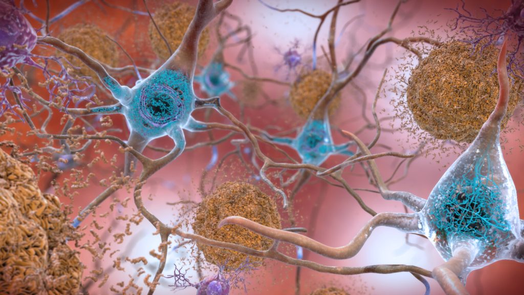
Neurons in the brain of an Alzheimer’s patient, with plaques caused by tau proteins. Credit: NIH

In findings published in Cell Reports, researchers discovered that the biological instructions within vesicles that communicate between cells differed significantly in postmortem brain samples donated from patients suffering from Alzheimer’s disease.

Small extracellular vesicles (sEVs) are tiny containers are produced by most cells in the body to ferry a wide variety of proteins, lipids and byproducts of cellular metabolism, as well as RNA nucleic acid codes used by recipient cells to construct new proteins.

Because this biologically active cargo can easily elicit changes in other cells, scientists are interested in brain sEVs as a medium for passing along normal as well as bungled instructions for misfolded proteins that accumulate in the brain as neurodegenerative diseases such as Alzheimer’s disease progress.

To be a potential contributor to the buildup of unwanted proteins, sEVs would have to carry blueprints with sufficient information to enable other cells to produce the problematic proteins. Most previous research had indicated that the messenger RNA (mRNA) carrying plans for proteins were chopped into too many shorter fragments to allow recipient cells to change their construction patterns.

“We found quite the opposite to be true in our study,” says senior author Jerold Chun, MD, PhD, professor in the Center for Genetic Disorders and Aging Research at Sanford Burnham Prebys. “We identified more than 10 000 full-length mRNAs through the use of a relatively newer DNA sequencing technique called PacBio long-read sequencing.”

The team isolated sEVs from the prefrontal cortex of 12 postmortem brain samples donated from patients diagnosed with Alzheimer’s disease and 12 from donors without Alzheimer’s disease (or any other known neurological disease). Nearly 80% of identified mRNAs were full-length, allowing them to be transcribed by recipient cells into viable proteins.

“To corroborate the results of long-read sequencing in the human samples, we also looked at vesicles isolated from mouse cells,” says first author Linnea Ransom, PhD, postdoctoral fellow. “We found similar averages of between 78% and 86% full-length transcripts in three brain cell types: astrocytes, microglia and neurons.”

The researchers also compared the sequence of genes reflected in the sEV mRNA transcriptome. In Alzheimer’s disease samples, 700 genes showed increased expression whereas nearly 1500 were found to have reduced activity.

The scientists determined that the 700 upregulated genes are associated with inflammation and immune system activation, which fits within known patterns of brain inflammation present in neurodegenerative diseases such as Alzheimer’s disease. The researchers also found many genes associated with Alzheimer’s disease in prior genome-wide association studies also were present in Alzheimer’s disease sEVs.

“The changes in gene expression contained in these vesicles reveal an inflammatory signature that may serve as a window into disease processes occurring in the brain as Alzheimer’s disease progresses,” says Chun.

Following this study, Chun and team will dig deeper into how cells package sEVs and how the enclosed mRNA codes lead to functional changes in other brain cells affected in Alzheimer’s disease. Better understanding of sEVs and their mRNA contents may enable the discovery of biomarkers that could be used to improve early detection of Alzheimer’s disease and potentially other neurological conditions, while identifying new disease mechanisms to provide new therapeutic targets.

“Additionally, sEVs naturally occur as a vehicle for transporting biologically active cargo between cells, so it also may be possible to leverage them as a targeted delivery system for future brain therapies” says Chun.

Source: Sanford-Burnham Prebys

Study Shows no Thyroid Cancer Risk from GLP-1 Agonists

By HualinXMN – Own work, CC BY-SA 4.0, https://commons.wikimedia.org/w/index.php?curid=133759262

GLP-1 analogues have become increasingly popular to treat diabetes and obesity, but there have been concerns that they might increase the risk of thyroid cancer. Now an extensive Scandinavian study led by researchers at Karolinska Institutet has found no evidence of such a link. The study is published in The BMJ.

GLP-1 receptor agonists, also known as GLP-1 analogues, reduce blood sugar levels and appetite. They are widely used in the treatment of type 2 diabetes and obesity, with their clinical use steadily increasing. Earlier studies and adverse event data have suggested that these drugs could be associated with an increased risk of thyroid tumours. However, due to limitations in data and methodology, clear conclusions could not be drawn, leading to uncertainty about this potential side effect.

“Many people take these medicines, so it is important to study potential risks associated with them,” says Björn Pasternak, principal researcher at the Department of Medicine, Solna, at Karolinska Institutet in Sweden. “Our study covers a broad group of patients and provides strong support that GLP-1 analogues are not associated with an increased risk of thyroid cancer.” 

The researchers analysed national register data from Denmark, Norway, and Sweden of about 145 000 patients treated with GLP-1 analogues, mainly liraglutide or semaglutide, and 290 000 patients treated with another diabetes drug (DPP4 inhibitors). The risk of thyroid cancer was compared between the groups over an average follow-up period of just under four years. 

GLP-1 treatment was not associated with an increased risk of thyroid cancer. The results were consistent also when compared to a third diabetes medication group (SGLT2 inhibitors).

“We cannot rule out that the risk of certain subtypes of thyroid cancer is increased in smaller patient groups that we could not study here, for example in people with a high congenital risk of medullary thyroid cancer who are advised against using these drugs,” says Peter Ueda, assistant professor at the Department of Medicine, Solna, at Karolinska Institutet.

The ongoing research program at Karolinska Institutet investigates the effects and potential side effects of newer diabetes medications such as GLP-1 analogues and SGLT2 inhibitors. These medications are now being used to treat broader patient groups, including those with obesity, heart failure, and kidney failure.

“We know from randomised clinical trials that they have positive effects, but clinical reality is different with patients varying in disease severity, comorbidities, and adherence to treatment recommendations,” says Björn Pasternak. “It’s therefore essential to investigate how these medicines perform in everyday clinical settings.”

Source: Karolinska Institutet

Virtual Reality Sessions can Lessen Cancer Pain, Trial Shows

Photo by Bradley Hook on Pexels

Hospitalised cancer patients who engaged in a 10-minute virtual reality (VR) session experienced significantly lessened pain in a trial published in CANCER, a peer-reviewed journal of the American Cancer Society. Participants still experienced sustained benefits a day later.

Most cancer patients experience pain, and treatment usually involves medications including opioids. VR sessions that immerse the user in new environments have been shown to be a noninvasive and nonpharmacologic way to lessen pain in different patient populations, but data are lacking in individuals with cancer. To investigate, Hunter Groninger, MD, of Georgetown University School of Medicine and MedStar Health and his colleagues randomized 128 adults with cancer with moderate or severe pain to a 10-minute immersive VR intervention involving calm, pleasant environments or to a 10-minute two-dimensional guided imagery experience on an iPad tablet.

The investigators found that both interventions lessened pain, but VR sessions had a greater impact. Based on patient-reported scores from 0 to 10, patients in the guided imagery group reported an average decrease of 0.7 in pain scores, whereas those in the VR group reported an average drop of 1.4. Twenty four hours after the assigned intervention, participants in the VR group reported sustained improvement in pain severity (1.7 points lower than baseline before the VR intervention) compared with participants in the guided imagery group (only 0.3 points lower than baseline before the active control intervention).

Participants assigned to the VR intervention also reported improvements related to pain “bothersomeness” (how much the pain bothered them, regardless of the severity of the pain) and general distress, and they expressed satisfaction with the intervention. 

“Results from this trial suggest that immersive VR may be a useful non-medication strategy to improve the cancer pain experience,” said Dr Groninger. “While this study was conducted among hospitalized patients, future studies should also evaluate VR pain therapies in outpatient settings and explore the impact of different VR content to improve different types of cancer-related pain in different patient populations. Perhaps one day, patients living with cancer pain will be prescribed a VR therapy to use at home to improve their pain experience, in addition to usual cancer pain management strategies like pain medications.”

Source: Wiley

Are Brain Organoids Derived from Foetal Tissue Ethical?

Image from Pixabay.

Brain organoids (BOs), though often referred to as “mini brains,” are not truly human brains. But the concerns over these lab-grown brain tissues, especially when they are developed from human foetal tissues, can be very human indeed.

In a paper published in EMBO Reports, researchers from Hiroshima University offer valuable insights into the complexities inherent in brain organoid research, highlighting often-overlooked ethical dilemmas for better decision-making, especially for foetal brain organoids (FeBOs).

Brain organoids are three-dimensional human brain tissues derived from stem cells. They replicate the complexity of the human brain in vitro, allowing researchers to study brain development and diseases.

Traditionally, brain organoids (BOs) are grown from pluripotent stem cells, an especially potent sub-type that is typical of early embryonic development, but new technologies now make it possible to generate these organoids from human foetal brain cells.

The research comes amid increasingly heated debates over human BOs. Central concerns are that lab-grown BOs might achieve consciousness and the ethical implications of transplanting them into animal models. The discourse includes matters of consent, commercialisation, integration with computational technologies, and legal ramifications. In addition, the public perception of BOs, often shaped by inaccurate media depictions.

Issues of consciousness arising and transplantation into animal models are particularly morally sensitive for tissue donors, and so rigorous informed consent is needed. With FeBOs, these become even more important. FeBOs, for example, can grow past the developmental stage of the initial foetal donor tissue.

“Our research seeks to illuminate previously often-overlooked ethical dilemmas and legal complexities that arise at the intersection of advanced organoid research and the use of foetal tissue, which is predominantly obtained through elective abortions,” said Tsutomu Sawai, an associate professor at Hiroshima University and lead author of the study.

The study highlights the urgent need for a sophisticated and globally harmonised regulatory framework tailored to navigate the complex ethical and legal landscape of FeBO research. One example is the 14-day rule used in embryo research, as neurogenesis does not occur in embryos prior to 14 days post-fertilisation. Using FeBOs derived from 12-15 week old foetuses therefore raises significant ethical questions, especially as there is a proposed 20-week ethical boundary.

The paper emphasises the importance of informed consent protocols, ethical considerations surrounding organoid consciousness, transplantation of organoids into animals, integration with computational systems, and broader debates related to embryo research and the ethics of abortion.

“Our plan is to vigorously advocate for the development of thorough ethical and regulatory frameworks for brain organoid research, including FeBO research, at both national and international levels,” said Masanori Kataoka, a fellow researcher at Hiroshima University.

“Rather than being limited to issues of consciousness, it’s imperative, now more than ever, to systematically advance the ethical and regulatory discussion in order to responsibly and ethically advance scientific and medical progress,” Sawai said.

Moving forward, the research duo plans to continue supporting the advancement of ethical and regulatory discussions surrounding brain organoid research. By promoting responsible and ethical progress in science and medicine, they aim to ensure that all research involving brain organoids, including FeBOs, is conducted within a framework that prioritises human dignity and ethical integrity.

Source: Hiroshima University