Month: March 2024

A New Treatment for Retinal Detachment – Based on Seaweed

Photoreceptor cells in the retina. Credit: Scientific Animations

In Korea, it is taboo to consume seaweed soup before exams since it there is a belief that the slippery nature of seaweed can lead to slipping up in the exam and failing. The slick surface of seaweeds is attributed to alginate, a mucilaginous carbohydrate substance. Now, Korean researchers have made use of this substance in the treatment of retinal detachment.

The result is an artificial vitreous body for treating retinal detachment, based on alginate from seaweed.

The research findings were recently published in Biomaterials, an international journal of biomaterials published by Elsevier. The work was a collaborative effort between Professor Hyung Joon Cha from the Department of Chemical Engineering and the School of Convergence Science and Technology and Dr. Geunho Choi from the Department of Chemical Engineering at Pohang University of Science and Technology (POSTECH), and Professor Woo Jin Jeong, Professor Woo Chan Park, and Professor Seoung Hyun An from the Dong-A University Hospital’s Department of Ophthalmology.

The vitreous body is a gel-like substance that occupies the space between the lens and retina, contributing to the eye’s structural integrity. Retinal detachment occurs when the retina separates from the inner wall of the eye and moves into the vitreous cavity, leading to detachment and potentially resulting in blindness in severe cases.

While a common approach involves removing the vitreous body and substituting it with medical intraocular fillers like expandable gas or silicone oil, these fillers have been associated with various side effects. To address these concerns, the research team employed a modified form of alginate, a natural carbohydrate sourced from algae.

Alginate, also known as alginic acid, is widely utilised in various industries, including food and medicine, for its ability to create viscous products. In this research, the team crafted a medical composite hydrogel based on alginate, offering a potential alternative for vitreous replacement.

The hydrogel has high biocompatibility and optical properties akin to authentic vitreous body, preserving the vision of patients post-surgery. Its distinctive viscoelasticity effectively regulates fluid dynamics within the eye, contributing to retinal stabilisation and the elimination of air bubbles.

To validate the hydrogel’s stability and effectiveness, the team conducted experiments using rabbit eyes, which closely resemble human eyes in structure, size, and physiological response.

Implanting the hydrogel into rabbit eyes demonstrated its success in preventing the recurrence of retinal detachment, maintaining stability, and functioning well over an extended period without any adverse effects.

Professor Hyung Joon Cha of the POSTECH who led the study remarked, “There is a correlation between retinal detachment and severe myopia and the prevalence of retinal detachment is increasing, particularly in young people. The incidence of retinal detachment cases in Korea rose by 50% in 2022 compared to 2017.” He expressed the team’s commitment by saying, “Our team will enhance and progress the technology to make the hydrogel suitable for practical use in real-world eye care through ongoing research.”

Professor Woo Jin Jeong from the Dong-A University Hospital stated, “The worldwide market for intraocular fillers is expanding at a rate of 3% per year.” He added, “We anticipate that the hydrogel we’ve created will prove beneficial in upcoming vitreoretinal surgeries.”

Source: Pohang University of Science & Technology (POSTECH)

Study Finds Multiple Unsafe Sleep Practices in Most Sudden Infant Deaths

Photo by William Fortunato on Pexels

There were multiple unsafe sleep practices at play in more than three-quarters of Sudden Unexpected Infant Deaths reported in 23 U.S. jurisdictions between 2011 and 2020, according to a new study published in Pediatrics. The researchers say the findings underscore the need for more comprehensive safe-sleep education for new parents, including from healthcare providers.

Of 7595 infant deaths reviewed, almost 60% of the infants were sharing a sleep surface, such as a bed, when they died.

This practice is strongly discouraged by sleep experts, who warn that a parent or other bed partner could unintentionally roll over and suffocate the baby.

Infants who died while sharing a sleep surface were typically younger (less than 3 months old), non-Hispanic Black, publicly insured, and either in the care of a parent at the time of death or being supervised by someone impaired by drugs or alcohol.

These infants were typically found in an adult bed, chair or couch instead of the crib or bassinet recommended by sleep experts.

“The large number of hazardous sleep practices for both infants who were sharing a sleep surface and sleeping alone at the time of death is alarming,” said researcher Fern Hauck, MD, MS, a safe-sleep expert at UVA Health and the University of Virginia School of Medicine.

“These are known risk factors for SUID [Sudden Unexpected Infant Death], and tells us that we need to do a better job of working with families to increase acceptance of the recommendations to create safer sleep spaces for their infants.”

Sudden Unexpected Infant Deaths

To better understand the factors contributing to SUID and improve safe-sleep messaging, Hauck and her collaborators analysed data from the federal Centers for Disease Control and Prevention’s SUID Case Registry.

The researchers obtained important insights on the prevalence of practices such as prenatal smoking, a known risk factor for SUID, and breastfeeding, which is thought to have a protective benefit.

More than 36% of mothers of infants who died had smoked while pregnant. This percentage was higher among moms who bed shared than those who didn’t, 41.4% to 30.5%. Both bed sharers and non-bed sharers had breastfed at similar rates.

The researchers note that it was rare for bedsharing to be the only risk factor present during a child’s death.

The findings highlight the need for better public education about safe-sleep practices, and for care providers to take a more active role in teaching new parents about the practices, the researchers say.

“Our findings support comprehensive safe sleep counselling for every family at every encounter beyond just asking where an infant is sleeping,” the researchers wrote

In addition to helping parents understand safe-sleep practices, care providers should take steps to ensure parents can follow those practices once they leave the hospital.

For example, some families may not have the means to purchase a crib or bassinet; a hospital might direct them to resources to help with that.

“SUID deaths in the U.S. are still higher than in most other countries, and this is unacceptable,” Hauck said.

“Clinicians and others caring for infants need to have thoughtful conversations with families at risk to understand the barriers to following safe-sleep guidelines and find ways to work together to overcome them.”

Source: University of Virginia Health System

Promising Drug Combination for Multiple Myeloma Treatment

Depiction of multiple myeloma. Credit: Scientific Animations

Researchers in Ireland have found that venetoclax, a medication currently approved for leukaemia, has benefits for patients with multiple myeloma when used in combination with another drug. This discovery offers a new avenue of treatment options for the currently incurable disease.

A type of blood cancer, multiple myeloma (MM) is still incurable despite treatment recent advances. The search for innovative treatment strategies is crucial, particularly for patients whose cancer is resistant to standard care.

In the new study published in Haematologica, researchers at the RCSI Department of Physiology and Medical Physics and the Beaumont RCSI Cancer Centre set out to identify complementary drugs that would enhance the efficiency of venetoclax, a drug approved for use in leukaemia, for MM treatment.

Although previously tested in MM, venetoclax, which blocks the function of a protein called BCL-2, was only found to be effective for a small proportion of patients.

The researchers discovered that combining venetoclax with a drug called 5-azacytidine significantly increased its effectiveness across many MM cell lines, indicating a broader potential patient population that could be treated with the new combination.

“This research is a significant step in identifying more effective treatment options for multiple myeloma. By combining venetoclax and 5-azacytidine we’ve seen enhanced efficacy across a wide range of patient samples. It shows the benefits of re-evaluating existing treatments in new contexts to expand their potential.” said Professor Tríona Ní Chonghaile, Associate Professor and research lead, Department of Physiology and Medical Physics.

Professor Siobhán Glavey, Chair, RCSI Department of Pathology and Clinician Scientist, Beaumont RCSI Cancer Centre commented: “Discovering the potential of this new drug combination is a promising development. Our next goal is to test for efficacy and safety for multiple myeloma in a clinical trial setting to bring us closer to offering a new treatment strategy for patients.”

The mechanism of how the two drugs work efficiently together was also investigated and it was shown that the combination of the two therapies was effective in patient samples from different stages of cancer, even if that patient had been previously treated with chemotherapy drugs.

The research was conducted in collaboration with the Department of Haematology, Beaumont Hospital, Dublin; Department of Medical Oncology, Dana-Farber Cancer Institute, Boston; and the Department of Medicine/Haematology, University of Galway, Galway.

This study was supported by funding from Leukemia Research Foundation, Breakthrough Cancer Research and AbbVie.

Source: RCSI

Trial of Minocycline for Dry Age-related Macular Degeneration Flops

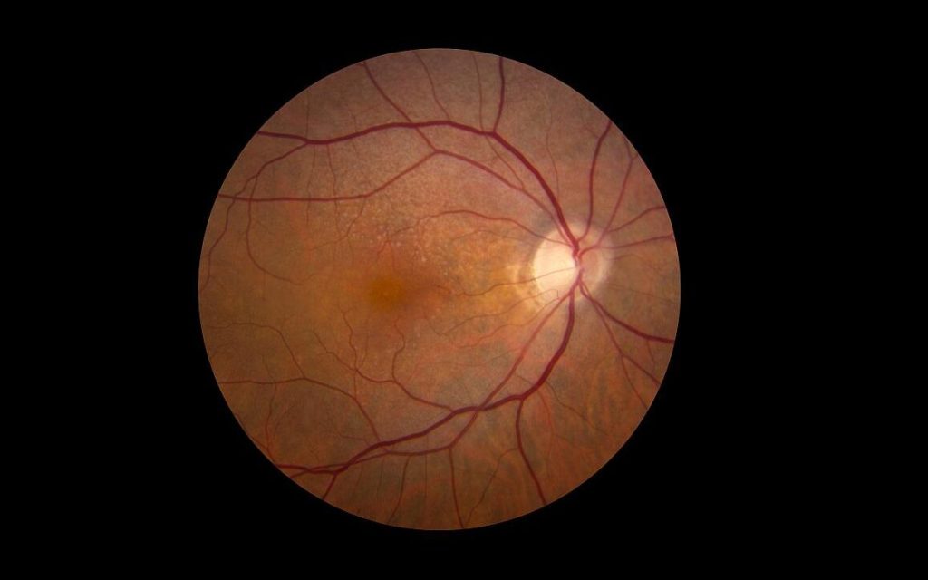
Retina showing reticular pseudodrusen. Although they can infrequently appear in individuals with no other apparent pathology, their highest rates of occurrence are in association with age-related macular degeneration (AMD), for which they hold clinical significance by being highly correlated with end-stage disease sub-types, choroidal neovascularisation and geographic atrophy. Credit: National Eye Institute

The drug minocycline, an antibiotic that also decreases inflammation, failed to slow vision loss or expansion of geographic atrophy in people with dry age-related macular degeneration (AMD), according to a phase II clinical study at the National Eye Institute (NEI), part of the National Institutes of Health.

Dry AMD affects the macula, the part of the retina that allows for clear central vision. In people with dry AMD, patches of photoreceptors and their nearby support cells begin to die off, leaving regions known as geographic atrophy. Over time, these regions expand, causing people to lose more and more of their central vision.

Microglia, immune cells that help maintain tissue and clear up debris, are present at higher levels around damaged retinal regions in people with dry AMD than in people without AMD. Scientists have suggested that inflammation – and particularly microglia – may be driving the expansion of geographic atrophy regions.

This study, led by Tiarnan Keenan, MD, PhD, a Stadtman Tenure-Track Investigator at the NEI’s Division of Epidemiology and Clinical Applications, tested whether inhibiting microglia with minocycline might help slow geographic atrophy expansion and its corresponding vision loss.

The trial enrolled 37 participants at the NIH Clinical Center in Bethesda, Maryland, and at the Bristol Eye Hospital, United Kingdom.

After a nine-month period where the researchers tracked each participant’s rate of geographic atrophy expansion, the participants took twice-daily doses of minocycline for two years.

The researchers compared each participant’s rate of geographic atrophy expansion while taking minocycline to their baseline rate, and found there was no difference in geographic atrophy expansion rate or vision loss with minocycline.

Previous studies have shown that minocycline can help reduce inflammation and microglial activity in the eye, including the retina.

The drug has shown beneficial effects for conditions such as diabetic retinopathy, but has not previously been tested for dry AMD.

Source: NIH/National Eye Institute

Adcock Ingram Critical Care Partners with Convatec to Supply Advanced Medical Products

Convatec’s Esteem stoma care system

Adcock Ingram Critical Care (AICC), a leading manufacturer and supplier of hospital and critical care products in Southern Africa, is expanding its reach in Ostomy and Advanced Wound Care. On 1 February 2024, AICC and Convatec signed a sales, marketing and distribution agreement covering South Africa and neighbouring countries.

Convatec is a globally renowned medical products and technologies company focused on therapies for managing chronic conditions, with leading positions in advanced wound care, ostomy care, continence care, and infusion care.

Colin Sheen, MD at AICC says: “This strategic agreement will add an important new pillar to AICC’s business. As a key pharmaceutical company in Southern Africa, AICC takes its responsibility towards healthcare professionals and patients seriously. As part of our commitment and responsibility to healthcare providers and patients, this agreement between AICC and Convatec is aligned with our mission to provide quality products that improve the health and lives of people in the markets we serve.”

The agreement extends throughout South Africa and neighbouring countries, and includes the import and distribution of a range of finished products in Advanced Wound Care, Ostomy Care and Continence Care. Convatec’s solutions provide various clinical and economic benefits that include infection prevention, protection of at-risk skin, and improved patient outcomes.

Sameer Singla, Vice President – Asia, Middle-East, Africa (AMEA) at Convatec says: “Convatec is pleased to partner with AICC to extend the reach of our products and solutions for patients. Convatec is committed to supporting people living with challenging medical conditions, and to addressing the care gap between the support patients need and what healthcare professionals can provide, which underpins our ‘forever caring’ promise. We look forward to partnering with AICC to meet the needs of patients and healthcare professionals.”

Rise in Global Fungal Drug-resistant Infections

In a recent study published in Pathogens and Immunity, researchers issue a call to action over how rising antifungal resistance is worsening the problem of invasive fungal infections.

Fungal infections have become more than just Epidemiological data published in Microbial Cell indicates that a rise in severe fungal infections has resulted in over 150 million cases annually and almost 1.7 million fatalities globally.

Skin contact with microorganisms found in soil or on hard surfaces, such as common shower facilities, or exposure to infected pets, can result in fungal infections known as dermatomycoses. Rashes, itching, burning and skin irritation are among the symptoms of fungal infection.

Thomas McCormick and Mahmoud Ghannoum, professors of dermatology at the Case Western Reserve University School of Medicine and affiliated with University Hospitals Cleveland Medical Center, explained extent of the problem. “This is not just an issue that affects individual patients,” McCormick said.

“The World Health Organization has recognised it as a widespread threat that has the potential to impact entire healthcare systems if left unchecked.”

Based on their findings, the researchers issued precautions and a “call to action” for the medical community to help protect people from multidrug-resistant fungi, starting with awareness and education.

“Healthcare providers must prioritise the use of diagnostic tests when faced with an unknown fungal infection,” Ghannoum said.

“Early detection can make all the difference in improving patient outcomes.”

Patients treated with medications to protect the immune system after cancer and transplant procedures are more vulnerable to fungal infections – making them especially more vulnerable to infections from drug-resistant fungi, the researchers said.

The emergence of multidrug-resistant fungal species, such as Candida auris and Trichophyton indotineae, is especially troubling and requires urgent attention, they reported.

In a study recently published in Emerging Infectious Diseases, Ghannoum’s research team and the Centers for Disease Control and Prevention (CDC), detailed a case that demonstrated Trichophyton indotineae, in addition to becoming drug-resistant, was also sexually transmissible.

To address the growing health concern, McCormick and Ghannoum suggest several measures:

  • Increased awareness and education: Raising awareness in the general healthcare setting to obtain a more accurate understanding of the rise of antifungal-resistant infections.
  • Diagnostic Testing: Routine use of diagnostic tests can guide appropriate treatment strategies.
  • Antifungal Susceptibility Testing (AST): Improving insurance reimbursement rates for AST and increasing the number of qualified laboratories with the capacity to perform these tests.
  • Call to Action: Addressing the emerging challenge of antifungal resistance involves concerted efforts from healthcare professionals, researchers, policymakers and the pharmaceutical industry to develop and implement strategies for managing and preventing antifungal resistance.

“The ultimate goal of these measures,” Ghannoum said, “is to improve the quality of patient care by ensuring effective treatment and preventing further escalation of the problem.”

Source: Case Western Reserve University

Social Bonding Gets People on the Same Wavelength

Forming social bonds facilitates effective communication and neural synchronisation across individuals of different social status within a group

When small hierarchical groups bond, neural activity between leaders and followers aligns, promoting quicker and more frequent communication, according to a study published on March 19th in the open-access journal PLOS Biology by Jun Ni from Beijing Normal University, China, and colleagues.

Social groups are often organised hierarchically, where status differences and bonds between members shape the group’s dynamic. To better understand how bonding influences communication within hierarchical groups and which brain regions are involved in these processes, the researchers recorded 176 three-person groups of human participants (who had never met before) while they communicated with each other, sitting face-to-face in a triangle. Participants wore caps with fNIRS (functional near-infrared spectroscopy) electrodes to non-invasively measure brain activity while they communicated with their group members. Each group democratically selected a leader, so each group of three ultimately included one leader and two followers. After strategising together, groups played two economic games designed to test their willingness to make sacrifices to benefit their group (or harm other groups).

Experimenters assigned some triads to go through a bonding session, where they were grouped according to colour preferences, given uniforms, and led through an introductory chat session to build familiarity. Bonded groups spoke more freely and bounced between speakers more frequently and rapidly, relative to groups that didn’t experience this bonding session. This bonding effect was stronger between leaders and followers than between two followers. Neural activity in two brain regions linked to social interaction, the right dorsolateral prefrontal cortex (rDLPFC) and the right temporoparietal junction (rTPJ), aligned between leaders and followers if they had bonded. The authors state that this neural synchronisation suggests that leaders may be anticipating followers’ mental states during group decision-making, though they acknowledge that their findings are restricted to East Asian Chinese individuals communicating via text (without non-verbal cues), whose culture emphasises group cohesion and commitment towards group leaders.

The authors add, “Social bonding increases information exchange and prefrontal neural synchronisation selectively among individuals with different social statuses, providing a potential neurocognitive explanation for how social bonding facilitates the hierarchical structure of human groups.”

Source: PLOS

Human Brains are Getting Larger, which may Protect against Dementia

Image: Pixabay CC0

A new study by researchers at UC Davis Health found human brains are getting larger. Study participants born in the 1970s had 6.6% larger brain volumes and almost 15% larger brain surface area than those born in the 1930s. The researchers hypothesise that the increased brain size may lead to an increased brain reserve, potentially reducing the overall risk of age-related dementias.

The findings were published in JAMA Neurology.

“The decade someone is born appears to impact brain size and potentially long-term brain health,” said first author Charles DeCarli, a distinguished professor of neurology and director of the UC Davis Alzheimer’s Disease Research Center.

“Genetics plays a major role in determining brain size, but our findings indicate external influences – such as health, social, cultural and educational factors – may also play a role.”

75-year study reveals brain changes between generations

The researchers used brain magnetic resonance imaging (MRIs) from participants in the Framingham Heart Study (FHS). The community-based study was launched in 1948 in Framingham, Massachusetts, to analyse patterns of cardiovascular and other diseases.

The original cohort consisted of 5209 men and women between the ages of 30 and 62. The research has continued for 75 years and now includes second and third generations of participants.

The MRIs were conducted between 1999 and 2019 with FHS participants born during the 1930s through the 1970s.

The brain study consisted of 3226 participants (53% female, 47% male) with an average age of about 57 at the time of the MRI.

The research led by UC Davis compared the MRIs of people born in the 1930s to those born in the 1970s.

It found gradual but consistent increases in several brain structures.

For example, a measure that looked at brain volume (intracranial volume) showed steady increases decade by decade.

For participants born in the 1930s, the average volume was 1234mL, but for those born in the 1970s, the volume was 1321 mL, or about 6.6% greater volume.

Cortical surface area showed an even greater increase over the decades.

Participants born in the 1970s had an average surface area of 2104cm2 compared to 2056cm2 for participants born in the 1930s — almost a 15% increase in volume.

The researchers found brain structures such as white matter, gray matter and hippocampus (a brain region involved in learning and memory) also increased in size when comparing participants born in the 1930s to those born in the 1970s.

Larger brains may mean lower incidence of dementia

Although the numbers are rising with America’s aging population, the incidence of Alzheimer’s – the percentage of the population affected by the disease – is decreasing.

A previous study found a 20% reduction in the incidence of dementia per decade since the 1970s.

Improved brain health and size may be one reason why.

“Larger brain structures like those observed in our study may reflect improved brain development and improved brain health,” DeCarli said.

“A larger brain structure represents a larger brain reserve and may buffer the late-life effects of age-related brain diseases like Alzheimer’s and related dementias.”

One of the study’s strengths is the design of the FHS study, which allows the researchers to examine brain imaging of three generations of participants with birthdates spanning almost 80 years.

A limitation is that non-Hispanic white participants make up the majority of the FHS cohort, which is not representative of the U.S. population.

Source: University of California – Davis Health

Is it Time for the International Definition of Triple-negative Breast Cancer to be Revised?

Photo by National Cancer Institute on Unsplash

An analysis of Swedish data, where the definition of triple negative breast cancer (TNBC) differs from that used internationally, brings additional insights to on ongoing discussion in the scientific community. The study was presented at the 2023 European Society for Medical Oncology (ESMO) meeting and is now published in Lancet Regional Health – Europe.

The Swedish definition of TNBC differs from the international version in that it also includes tumours with low expression of the Oestrogen Receptor (ER) biomarker, ie in 1–9% of tumour cells. Internationally, ER-low breast cancer is classified as hormone-sensitive and treated differently from TNBC patients. This is despite previous studies demonstrating that the majority of ER-low tumours are molecularly similar to ER-zero, the latter completely without expression of ER, and meta-analyses that show no survival benefit from endocrine therapy in ER-low tumours.

The Swedish population-based study included all women diagnosed with TNBC in Sweden during 2008–2020 using the National Quality Register for Breast Cancer. Patient and tumour characteristics, treatment and survival in patients with low ER expression was compared to patients with no ER tumour expression.

The study identified and included 5655, and 560 patients (10%) were defined as ER-low and 5095 (90%) as ER-zero. The data demonstrated there are only small differences in tumour characteristics, no differences in response to neoadjuvant chemotherapy and no significant differences in prognosis.

“The international cut-off for ER-positivity and thus the definition of TNBC as only completely ER-negative is now increasingly questioned. ER-low tumours behave like ER-zero tumours and should be treated as such. On the basis of real-world data, the Swedish cutoff for hormone receptor positivity appears to be more clinically relevant. A changed international definition would give patients with ER-low expressing breast cancer the same treatment options as in TNBC, within studies and in clinical routine,” says study leader Dr Irma Fredriksson.

The study was carried out in collaboration with the pharmaceutical company MSD.

Source: Karolinska Institutet

Scientists may have Found the Specific Neurons Behind Anorexia Nervosa

Anorexia nervosa, a mental health disorder in which people dangerously restrict their eating or purge their stomachs soon after a meal, is one of the deadliest psychological diseases. Yet, the neural mechanisms behind this have remained unclear, and therapies are limited.

Scientists have been tailing a lead for years, though. They’ve known that the disorder is often associated with anxiety and depression, hinting that the biological basis for anorexia could be regulated by neurons somewhere in the brain region that controls emotion – the amygdala.

That’s exactly where Haijiang Cai, a University of Arizona associate professor in the Department of Neuroscience and BIO5 Institute member, and his team found it: Anorexia is caused by a combination of two subregions in the amygdala, according to new research published in Cell Reports.

One knot of neurons in the central nucleus of the amygdala curbs appetite when a person gets full, feels nauseous or tastes something bitter. The other is in the oval region of the bed nucleus of the stria terminalis, which also halts eating due to inflammation and sickness.

Cai and his research team found that when they destroyed a certain type of brain cell, called PKC-delta neurons, in both of these regions, they could prevent anorexia development.

They also found that PKC-delta neurons become more active in response to eating during the anorexia development. What’s more, when they artificially activated these neurons, they caused a suppression in eating habits and increased exercise.

“This study suggests two important insights to treat anorexia,” Cai said. “One is that we need to target multiple brain regions to develop therapies. We also need to treat multiple conditions. For example, maybe one drug will target nausea and another drug target will target inflammation, and you have to combine them, like a cocktail therapy, to have better therapeutic effects.”

The team relied on mice models for their research.

“There’s no animal model that can mimic human disease completely, but this is as close as we can get,” Cai said. “For example, there are multiple common features, including a warped body image, a very low body weight, limited food intake and excessive exercise. We can’t know if an animal has a warped body image, but we can measure the other three features.”

One future step – since researchers cannot destroy neurons for human treatment – is to develop a method to silence the neurons temporarily, using drugs or some other method to test if that can prevent anorexia development or speed up recovery for people who have already developed the disorder.

Source: University of Arizona