Day: February 26, 2024

Metabolic Diseases may be Driven by Gut Microbiome, Loss of Ovarian Hormones

Photo by Ravi Patel on Unsplash

The gut microbiome interacts with the loss of female sex hormones to exacerbate metabolic disease, including weight gain, fat in the liver and the expression of genes linked with inflammation, researchers report in the journal Gut Microbes.

The findings, using rodent models, may shed light on why women are at significantly greater risk of metabolic diseases such as obesity and Type 2 diabetes after menopause, when ovarian production of female sex hormones diminishes.

“Collectively, the findings demonstrate that removal of the ovaries and female hormones led to increased permeability and inflammation of the gut and metabolic organs, and the high-fat diet exacerbated these conditions,” said Kelly S. Swanson, the director of the Division of Nutritional Sciences and a professor in nutrition at the University of Illinois Urbana-Champaign who is a corresponding author of the paper. “The results indicated that the gut microbiome responds to changes in female hormones and worsens metabolic dysfunction.”

“This is the first time it has been shown that the response of microbiome to the loss of ovarian hormone production can increase metabolic dysfunction,” said first author Tzu-Wen L. Cross, a professor of nutrition science and the director of the Gnotobiotic Animal Facility at Purdue University. Cross was a doctoral student at the U. of I. when she began the research.

“The gut microbiome is sensitive to sex hormone changes and can further impact the risk of disease development.”

Cross said early microbiome research, beginning around 2005, looked at how the microbiome contributes to obesity development, but most of those studies focused on males.

“Metabolic dysfunction that is driven by the loss of ovarian-function in menopausal women – and how much the gut microbiome contributes to that – has not been studied. The aetiology is clearly very complex, but those gut-microbiome related factors are certainly components that we speculated play a role,” she said.

The scientists created diet-induced obesity in female mice and simulated the loss of female sex hormones by removing the ovaries in half of the population to examine any metabolic and inflammatory changes, including those to enzymes in the gut. The diets for both groups of mice were identical except for the proportion of fat, which constituted 60% or 10% of calories for those in the high-fat and low-fat groups, respectively.

In the second leg of the study, faecal samples were harvested from mice with or without ovaries and implanted in germ-free mice to study the impact on weight gain and metabolic and inflammatory activity in the gut, liver and fat tissue.

“The mice that were recipients of the gut microbiome of ovariectomized mice gained more weight and fat mass, and they had greater expression of genes in the liver associated with inflammation, obesity, Type 2 diabetes, fatty liver disease and atherosclerosis compared with those in the control group,” Swanson said.

Assessing the severity of fatty tissue and triglyceride concentrations in the liver, the scientists found that the triglyceride levels were significantly higher and fatty deposits in the liver and groin were greater in the mice that consumed the high-fat diet compared with all other treatment groups.

Those on the high-fat diet and those without ovaries had significantly larger fat cells, which are associated with cell death and the infiltration of macrophages. Along with elevated expression of the genes associated with inflammation and macrophage markers, these mice had lower expression of genes that are involved with glucose and lipid metabolism.

In the donor mice without ovaries that consumed the low-fat diet, the scientists found increased levels of beta-glucuronidase, an enzyme produced by the colon and some intestinal bacteria that breaks down and recycles steroidal metabolites such as oestrogen and various toxins, including carcinogens.

The scientists also examined the expression of genes coding for tight-junction proteins, which affect cell membranes’ permeability. They found that the mice without ovaries and those fed the high-fat diet had lower levels of these proteins in the liver and colon, which suggested their gut barriers were more permeable, compromised by either their diet or the absence of female hormones.

In the livers of the recipient mice that received transplants from donors without ovaries, the scientists found elevated expression levels of the gene for arginase-1, which plays a critical role in the elimination of nitrogenous waste. High levels of this protein have been associated with cardiovascular problems such as hypertension and atherosclerosis.

Source: University of Illinois at Urbana-Champaign, News Bureau

Researchers Map out Protein Pathways of MND Development

Spinal neuron. Image by Scientific Animations CC4.0

For the first time, researchers from The University of Queensland (UQ) have mapped out the proteins implicated in the early stages of motor neurone disease (MND). This paper was published in Nature Communications.

Dr Rebecca San Gil, the study’s first author, has developed a longitudinal map of the proteins involved in MND across the trajectory of the disease, identifying potential therapeutic pathways for further investigation. This includes one protein, TDP-43, implicated in a number of MNDs.

“The map is a springboard for many more projects exploring the proteins activated and repressed during the onset, early and late stages of MND,” Dr San Gil said. “These proteins are biological factors that drive disease onset and progress its development over time.

“We measured differences in protein levels in the brain across the trajectory of the disease and collated this information into a longitudinal map.”

The map is now available for scientists worldwide and will accelerate investigations into MND.

Dr San Gil, in the lab of Associate Professor Adam Walker, has been working in mouse models of MND to understand the mechanisms driving TDP-43 pathology in the brain, which accounts for 95% of amyotrophic lateral sclerosis (ALS) cases and 50% of frontotemporal lobar degeneration (FTLD).

Building on the mapping project, Dr San Gil chose to focus on a protein-folding factor called DNAJB5.

“Before the onset of MND in mouse models, we observed a marked increase in protein groups responsible for physically assisting in the protein folding process. “One of these ‘chaperone’ proteins, DNAJB5, was particularly abundant early on, sparking our curiosity about its role in disease progression.

“In human brain tissue, we found DNAJB5 enriched in areas where TDP-43 aggregates. The short-term elevation of DNAJB5 is likely a protective mechanism by neurons in an attempt to control TDP-43 as it begins to dysfunction.

“This protective response to TDP-43 needs further investigation because it may help us identify preventative and therapeutic approaches to MND.”

A/ Prof Walker envisions that the lab will continue to follow other identified protein pathways, using gene therapy and repurposing medicine, to see if they can alter or prevent the disease.

Compiling the TDP map was a collaborative project with researchers from Macquarie University, the University of Auckland, and the Children’s Medical Research Institute.

Source: University of Queensland

New, More Accurate Approach to Blood Tests for Determining Diabetes Risks

Photo by National Cancer Institute on Unsplash

A new approach to blood tests could potentially be used to estimate a patient’s risk of type 2 diabetes, according to a new study appearing in BMC’s Journal of Translational Medicine. Currently, the most commonly used inflammatory biomarker currently used to predict the risk of type 2 diabetes is high-sensitivity C-reactive protein (CRP). But new research has suggested that jointly assessing of biomarkers, rather than assessing each individually, would improve the chances of predicting diabetes risk and diabetic complications.

A study by Edith Cowan University (ECU) researcher Dan Wu investigated the connection between systematic inflammation, assessed by joint cumulative high-sensitivity CRP and another biomarker called monocyte to high-density lipoprotein ratio (MHR), and incident type 2 diabetes.

The study followed more than 40 800 non-diabetic participants over a near ten-year period, with more than 4800 of the participants developing diabetes over this period.

Wu said that of those patients presenting with type 2 diabetes, significant interaction between MHR and CRP was observed.

“Specifically, increases in the MHR in each CRP stratum increased the risk of type 2 diabetes; concomitant increases in MHR and CRP presented significantly higher incidence rates and risks of diabetes.

“Furthermore, the association between chronic inflammation (reflected by the joint cumulative MHR and CRP exposure) and incident diabetes was highly age- and sex-specific and influenced by hypertension, high cholesterol, or prediabetes. The addition of the MHR and CRP to the clinical risk model significantly improved the prediction of incident diabetes,” said Wu.

Biological sex a risk factor

The study found that females had a greater risk of type 2 diabetes conferred by joint increases in CRP and MHR, with Wu stating that sex hormones could account for these differences.

Wu said that the research findings corroborated the involvement of chronic inflammation in causing early-onset diabetes and merited specific attention.

“Epidemiological evidence indicates a consistent increase in early-onset diabetes, especially in developing countries. Leveraging this age-specific association between chronic inflammation and type 2 diabetes may be a promising method for achieving early identification of at-risk young adults and developing personalised interventions,” she added.

Wu noted that the chronic progressive nature of diabetes and the enormous burden of subsequent comorbidities further highlighted the urgent need to address this critical health issue.

Although aging and genetics are non-modifiable risk factors, other risk factors could be modified through lifestyle changes.

Inflammation is strongly influenced by life activities and metabolic conditions such as diet, sleep disruptions, chronic stress, and glucose and cholesterol dysregulation, thereby indicating the potential benefits of monitoring risk-related metabolic conditions.

Wu said that the dual advantages of cost effectiveness and the wide availability of cumulative MHR and CRP in current clinical settings, potentiated the widespread use of these measures as a convenient tool for predicting the risk of diabetes.

Source: Edith Cowan University

Terahertz Biosensor can Accurately Detect Skin Cancer

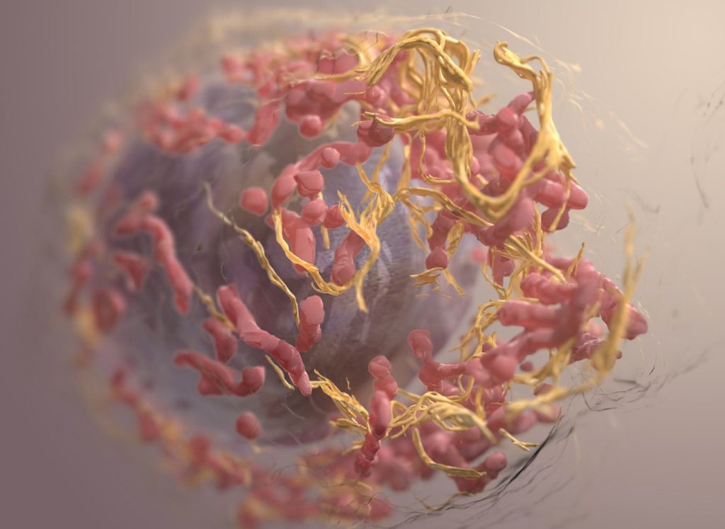
3D structure of a melanoma cell derived by ion abrasion scanning electron microscopy. Credit: Sriram Subramaniam/ National Cancer Institute

Researchers have developed a revolutionary biosensor using terahertz (THz) waves that can detect skin cancer with exceptional sensitivity, potentially paving the way for earlier and easier diagnoses. Published in the journal IEEE Transactions on Biomedical Engineering, the study presents a significant advancement in early cancer detection, thanks to a multidisciplinary collaboration of teams from Queen Mary University of London and the University of Glasgow.

“Traditional methods for detecting skin cancer often involve expensive, time-consuming, CT, PET scans and invasive higher frequencies technologies,” explains Dr Shohreh Nourinovin, Postdoctoral Research Associate at Queen Mary’s School of Electronic Engineering and Computer Science, and the study’s first author.

“Our biosensor offers a non-invasive and highly efficient solution, leveraging the unique properties of THz waves – a type of radiation with lower energy than X-rays, thus safe for humans – to detect subtle changes in cell characteristics.”

The key innovation lies in the biosensor’s design. Featuring tiny, asymmetric resonators on a flexible substrate, it can detect subtle changes in the properties of cells.

Unlike traditional methods that rely solely on refractive index, this device analyses a combination of parameters, including resonance frequency, transmission magnitude, and a value called “Full Width at Half Maximum” (FWHM). This comprehensive approach provides a richer picture of the tissue, allowing for more accurate differentiation between healthy and cancerous cells and to measure malignancy degree of the tissue.

In tests, the biosensor successfully differentiated between normal skin cells and basal cell carcinoma (BCC) cells, even at different concentrations. This ability to detect early-stage cancer holds immense potential for improving patient outcomes.

“The implications of this study extend far beyond skin cancer detection,” says Dr Nourinovin.

“This technology could be used for early detection of various cancers and other diseases, like Alzheimer’s, with potential applications in resource-limited settings due to its portability and affordability.”

Dr Nourinovin’s research journey wasn’t without its challenges.

Initially focusing on THz spectroscopy for cancer analysis, her project was temporarily halted due to the COVID pandemic. However, this setback led her to explore the potential of THz metasurfaces, a novel approach that sparked a new chapter in her research.

Source: Queen Mary University of London

Experimental Model Identifies New Drug–drug Interactions

Photo by Myriam Zilles on Unsplash

When taking oral drugs, transporter proteins found on cells that line the gastrointestinal tract facilitate their entry into the bloodstream. But for many drugs, it is not known which of those transporters they use to exit the digestive tract.

Identifying the transporters used by specific drugs could help to improve patient treatment because if two drugs rely on the same transporter, they can interfere with each other and should not be prescribed together.

Researchers at MIT, Brigham and Women’s Hospital, and Duke University have developed a multipronged strategy to identify the transporters used by different drugs, which appears in Nature Biomedical Engineering. Their approach, which makes use of both tissue models and machine-learning algorithms, has already revealed that a commonly prescribed antibiotic and a blood thinner can interfere with each other.

“One of the challenges in modelling absorption is that drugs are subject to different transporters. This study is all about how we can model those interactions, which could help us make drugs safer and more efficacious, and predict potential toxicities that may have been difficult to predict until now,” says Giovanni Traverso, an associate professor of mechanical engineering at MIT, a gastroenterologist at Brigham and Women’s Hospital, and the senior author of the study.

Learning more about which transporters help drugs pass through the digestive tract could also help drug developers improve the absorbability of new drugs by adding excipients that enhance their interactions with transporters.

Former MIT postdocs Yunhua Shi and Daniel Reker are the lead authors of the study.

Drug transport

Previous studies have identified several transporters in the GI tract that help drugs pass through the intestinal lining. Three of the most commonly used, which were the focus of the new study, are BCRP, MRP2, and PgP.

For this study, Traverso and his colleagues adapted a tissue model they had developed in 2020 to measure a given drug’s absorbability. This experimental setup, based on pig intestinal tissue grown in the laboratory, can be used to systematically expose tissue to different drug formulations and measure how well they are absorbed.

To study the role of individual transporters within the tissue, the researchers used short strands of RNA called siRNA to knock down the expression of each transporter. In each section of tissue, they knocked down different combinations of transporters, which enabled them to study how each transporter interacts with many different drugs.

“There are a few roads that drugs can take through tissue, but you don’t know which road. We can close the roads separately to figure out, if we close this road, does the drug still go through? If the answer is yes, then it’s not using that road,” Traverso says.

The researchers tested 23 commonly used drugs using this system, allowing them to identify transporters used by each of those drugs. Then, they trained a machine-learning model on that data, as well as data from several drug databases. The model learned to make predictions of which drugs would interact with which transporters, based on similarities between the chemical structures of the drugs.

Using this model, the researchers analysed a new set of 28 currently used drugs, as well as 1595 experimental drugs. This screen yielded nearly 2 million predictions of potential drug interactions. Among them was the prediction that doxycycline, an antibiotic, could interact with warfarin, a commonly prescribed blood-thinner. Doxycycline was also predicted to interact with digoxin, which is used to treat heart failure, levetiracetam, an antiseizure medication, and tacrolimus, an immunosuppressant.

Identifying interactions

To test those predictions, the researchers looked at data from about 50 patients who had been taking one of those three drugs when they were prescribed doxycycline. This data, which came from a patient database at Massachusetts General Hospital and Brigham and Women’s Hospital, showed that when doxycycline was given to patients already taking warfarin, the level of warfarin in the patients’ bloodstream went up, then went back down again after they stopped taking doxycycline.

That data also confirmed the model’s predictions that the absorption of doxycycline is affected by digoxin, levetiracetam, and tacrolimus. Only one of those drugs, tacrolimus, had been previously suspected to interact with doxycycline.

“These are drugs that are commonly used, and we are the first to predict this interaction using this accelerated in silico and in vitro model,” Traverso says. “This kind of approach gives you the ability to understand the potential safety implications of giving these drugs together.”

Source: Massachusetts Institute of Technology