Day: February 20, 2024

Anabolic Steroid Use can Increase Atrial Fibrillation Risk, Study Finds

Photo by Jonathan Borba on Unsplash

People using anabolic steroids could be increasing their underlying risk of atrial fibrillation, according to new research published in the Journal of Physiology

The team found that male sex hormones, such as testosterone, also called androgenic anabolic steroids (AAS), which are misused for muscle building particularly among in young men can increase the risk of atrial fibrillation in individuals genetically predisposed to heart diseases.

Dr Laura Sommerfeld, Postdoctoral Researcher at the UKE Hamburg, who completed her PhD at the Institute of Cardiovascular Sciences at the University of Birmingham focusing on this work is lead author of the study.

Dr Sommerfeld said: “Our study can significantly contribute to understanding the impact on the heart health of young men who misuse anabolic steroids to increase muscle mass. Recent reports have shown that young men in particular are being targeted on social media such as TikTok being sold testosterone products, but we have shown how the misuse of steroids carries a specific risk that many people will not be aware of.”

Professor Larissa Fabritz, Chair of Inherited Cardiac Conditions at UKE Hamburg and Honorary Chair in the Institute of Cardiovascular Sciences at the University of Birmingham added:

“Heart muscle diseases like ARVC affect young, athletic individuals and can lead to life-threatening heart rhythm disturbances. Atrial fibrillation is a common condition in the general population. Elevated testosterone levels can result in an earlier onset of these diseases.”

The scientists examined potential effects on a condition called arrhythmogenic right ventricular cardiomyopathy (ARVC), which is genetically determined and primarily attributed to disruptions in the formation of cell connections critical for heart muscle stability.

The scientists initially confirmed, based on clinical patient data from UHB and elsewhere, that ARVC occurs more frequently and severely in men than in women.

In laboratory experiments, they discovered that six weeks of AAS intake, combined with impaired cell connections, could lead to reduced sodium channel function in heart tissue and a slowing of signal conduction within the atria.

Dr Andrew Holmes, co-author and Assistant Professor in the Institute of Clinical Sciences at the University of Birmingham said:

“This work implies that young male individuals with key inherited genetic changes have a greater risk of developing electrical problems in the heart in response to anabolic steroid abuse.”

The research was conducted by an interdisciplinary consortium of clinicians and researchers led by University of Birmingham and collaborators in Germany.

Source: University of Birmingham

New Tool Predicts Burnout Risk

Photo by Mulyadi on Unsplash

It is not uncommon for people to “hit the wall” at work and experience burnout for short or long periods of time.

“We have found that approximately 13 per cent of Norwegian employees are at high risk of burnout,” says Leon De Beer, Associate Professor of Work and Organizational Psychology at the Norwegian University of Science and Technology (NTNU) Department of Psychology.

De Beer has contributed to a new study on burnout published in the Scandinavian Journal of Psychology with colleagues from the Healthy Workplaces research group.

They are working on a new tool that can identify people at risk of burnout. 

Signs that you might be at risk of burnout

If you are facing demands and stress at work that seem to be intractable, and you have frequently experienced the following symptoms in recent weeks, it might be a sign that you are on the verge of burning out:

  1. You feel mentally exhausted at work.
  2. You struggle to feel enthusiastic about your job.
  3. You have trouble concentrating when working.
  4. You sometimes overreact at work without meaning to.

Early intervention is key

It is important to identify the early signs of burnout in order to mitigate the harmful effects. The warning signs are often present before things have gone too far, as long as we manage to identify them.

“Not addressing the risk of employee burnout in time can have long-term consequences,” says De Beer.

The physical and psychological effects of burnout include cardiovascular disease, pain related to musculoskeletal injuries, sleeping problems, and depression. Organisations can also lose talented employees and experience an increase in sickness absence and lost productivity.

A new tool may become standard

De Beer’s research group has trialled a new measurement tool to identify the early warning signs of burnout. In the past, it has not always been that easy.

“Previously, we have not had a detailed enough measurement tool for use in both the field of practice and research that identifies workers who are at risk of burnout,” says De Beer.

There is currently no international standard for assessing burnout.

The new tool is called the Burnout Assessment Tool, or BAT among researchers who have a penchant for amusing abbreviations. The BAT consortium, of which the researchers are a part, is now testing the instrument in more than 30 countries.

https://burnoutassessmenttool.be/start_eng/

“Our studies show that BAT is a good tool for identifying the risk of burnout,” says De Beer.

Burnout is the body’s response to stress

BAT measures four main groups of risk factors: exhaustion, mental distancing, cognitive impairment and emotional impairment.

Burnout is not really an illness, but a feeling of being mentally or physically exhausted — the body’s response to a lasting, demanding situation.

Burnout is normally defined as a work-related syndrome, but there is evidence that work-life balance also plays a role. Stress and burnout don’t necessarily stop when you go home at the end of the day, as these effects often extend into other areas of life and vice versa.

Some may experience years of burnout

For some people, burnout can be stopped in its tracks and solutions found to improve their situation. For others, however, burnout can last for years if the problem isn’t addressed.

“We can deal with burnout through individual treatment, but it is of little use if people return to a workplace where the demands are too high and there are few resources. It is then highly likely that the employee will become ill again. Therefore, it is important to create good working conditions and structures that safeguard the health of employees,” says Professor Marit Christensen at NTNU’s Department of Psychology.

Culturally independent

The researchers studied a representative sample of 500 Norwegian workers. Norway is roughly on par with the EU average when it comes to mental health, but somewhat better when it comes to work-related matters.

A lower percentage of the Norwegian population struggles with exhaustion in connection with work. Somewhat fewer people than the EU average report health hazards at work, and we experience a better work-life balance.

“Using a recognised method, we found that around 13 per cent of the 500 surveyed workers were at high risk of burnout,” says Professor Christensen.

The tool can help identify who requires the most urgent follow up so that the risk of burnout can be reduced.

We do not yet know whether the prevalence of burnout in Norway is high in an international context. The Norwegian study is among several BAT studies that are currently taking place, so these answers will be available at a later date.

The tool is intended to be culturally independent, and it certainly works well in Norway. The researchers also found that the tool works regardless of gender.

“For entertainment and educational purposes, interested parties can use our online tool to test if they are at risk of burnout,” says Professor Christensen.

“Please note that the tool only gives an indication of risk and does not provide any type of formal diagnosis or medical advice. If you are concerned about your levels of work-related stress, we encourage you to visit a health care provider to discuss the matter,” says Professor Christensen.

Source: Norwegian University of Science and Technology

New Neural Prosthetic Device Can Help Restore Memory in Humans

Source: CC0

Scientists have demonstrated the first successful use of a neural prosthetic device to recall specific memories. The findings appear online in Frontiers in Computational Neuroscience.

This groundbreaking research was derived from a 2018 study led by Robert Hampson, PhD, professor of regenerative medicine, translational neuroscience and neurology at Wake Forest University School of Medicine. That study demonstrated the successful implementation of a prosthetic system that uses a person’s own memory patterns to facilitate the brain’s ability to encode and recall memory, improving recall by as much as 37%.

In the previous study, the team’s electronic prosthetic system was based on a multi-input multi-output (MIMO) nonlinear mathematical model, and the researchers influenced the firing patterns of multiple neurons in the hippocampus, a part of the brain involved in making new memories.

In this study, researchers from Wake Forest and University of Southern California (USC) built a new model of processes that assists the hippocampus in helping people remember specific information.

When the brain tries to store or recall information such as, “I turned off the stove” or “Where did I put my car keys?” groups of cells work together in neural ensembles that activate so that the information is stored or recalled.

Using recordings of the activity of these brain cells, the researchers created a memory decoding model (MDM) which let them decode what neural activity is used to store different pieces of specific information.

The neural activity decoded by the MDM was then used to create a pattern, or code, which was used to apply neurostimulation to the hippocampus when the brain was trying to store that information.

“Here, we not only highlight an innovative technique for neurostimulation to enhance memory, but we also demonstrate that stimulating memory isn’t just limited to a general approach but can also be applied to specific information that is critical to a person,” said Brent Roeder, Ph.D., a research fellow in the department of translational neuroscience at Wake Forest University School of Medicine and the study’s corresponding author.

The team enrolled 14 adults with epilepsy who were participating in a diagnostic brain-mapping procedure that used surgically implanted electrodes placed in various parts of the brain to pinpoint the origin of their seizures.

Participants underwent all surgical procedures, post-operative monitoring and neurocognitive testing at one of the three sites participating in this study including Atrium Health Wake Forest Baptist Medical Center, Keck Hospital of USC in Los Angeles and Rancho Los Amigo National Rehabilitation Center in Downey, California.

The team delivered MDM electrical stimulation during visual recognition memory tasks to see if the stimulation could help people remember images better.

They found that when they used this electrical stimulation, there were significant changes in how well people remembered things. In about 22% of cases, there was a noticeable difference in performance.

When they looked specifically at participants with impaired memory function, who were given the stimulation on both sides of their brain, almost 40% of them showed significant changes in memory performance.

“Our goal is to create an intervention that can restore memory function that’s lost because of Alzheimer’s disease, stroke or head injury,” Roeder said.

“We found the most pronounced change occurred in people who had impaired memory.”

Roeder said he hopes the technology can be refined to help people live independently by helping them recall critical information such as whether medication has been taken or whether a door is locked.

“While much more research is needed, we know that MDM-based stimulation has the potential to be used to significantly modify memory,” Roeder said.

Source: Atrium Health Wake Forest Baptist

Scientists Peer into a Transporter Protein for Inflammatory Signals

In the human body, a protein carrier called SPNS2 transports S1P molecules from endothelial cells to rally immune cell response in infected organs and tissues, resulting in inflammation. By enlarging the entire SPNS2 structure using nanoparticles, the S1P molecules contained within can be viewed via cryogenic electron microscopy. Using this information, small molecules can be developed to inhibit this signalling pathway and treat inflammatory diseases.

Scientists at the National University of Singapore and colleagues in China have analysed the structure of the SPNS2 protein at an atomic level that could provide greater insights into how S1P signalling molecules are released to communicate with the immune cells to regulate inflammatory responses. Their findings are published in Cell Research.

“Seeing is believing. This work shows that SPNS2 is directly exporting S1P for signalling and it is possible to inhibit its transport function with small molecules. This work provides the foundation for understanding of how S1P is released by SPNS2 and how this protein function is inhibited by small molecules for treatment of inflammatory diseases,” said team leader Dr Nguyen Nam Long.

The SPNS2 protein allows the binding of the S1P signalling molecules to trigger the immune cells to leave the lymph nodes and induce inflammation in different parts of the body when needed.

Made up of amino acids, the SPNS2 protein is malleable enough to change its shape and structure to release the S1P signalling molecules through small cavities found within the protein.

Through the discovery of how the SPNS2 protein releases S1P molecules, the SPNS2 structure can be exploited for future drug development.

Similar to discovering how the shape of the lock looks like before the key can be designed, this finding sheds more light into how future drugs can be designed to better target the protein to increase drug efficacy.

This finding builds on previous research which found that deleting SPNS2 protein from a pre-clinical model effectively blocks the S1P signalling pathway so that the S1P signalling molecules are unable to be transported to prompt immune cells to leave the lymph node to induce inflammation.

Both SPNS2 protein and S1P signalling molecule are required for immune cell recruitment to inflammatory organs, which goes towards treating various inflammatory diseases.

“Using pre-clinical models, we have shown that targeting SPNS2 proteins in the body blocks inflammatory responses in disease conditions, such as multiple sclerosis. This work has provided us a possibility to inhibit its transport function with small molecules that will go a long way to treating inflammatory diseases more efficiently and effectively,” said Dr Nguyen.

Source: National University of Singapore, Yong Loo Lin School of Medicine

Removing a Protein Lets Glioblastoma Chemo Remain Effective for Longer

Photo by Anna Shvets on Pexels

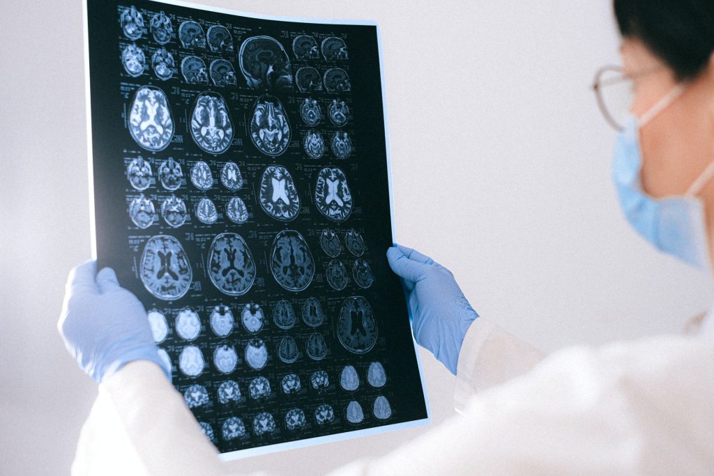
New research by the University of Sussex could help to increase life expectancy and improve treatment for glioblastoma. In the study, published in the Journal of Advanced Science, researchers have discovered that an understudied protein, called PANK4, is able to block cancer cells from responding to chemotherapeutic treatment for the highly intrusive brain cancer, glioblastoma.

Scientists at Sussex have demonstrated that if the protein is removed, cancer cells respond better to temozolomide, the main chemotherapy drug for the treatment of glioblastoma.

Prof Georgios Giamas, Professor of Cancer Cell Signalling at the University of Sussex explains: “Glioblastoma is a devastating brain cancer, and researchers are working hard to identify ways to delay progression of the disease, and tackle cell resistance to treatment. As this is the first time that PANK4 has been linked to glioblastoma, the next step is to develop a drug targeting this protein to try to reverse chemo-resistance and restore sensitivity, ensuring that patients receive the best treatment and have better outcomes.”

Glioblastoma is one of the most aggressive forms of brain cancer. Approximately 250 000 – 300 000 globally are diagnosed with it annually, with a best-case survival rate of just one to 18 months after diagnosis.

Following surgery to remove the tumour, glioblastoma patients are typically treated with radiation and the chemotherapeutic drug, temozolomide. Although patients initially respond well to the drug, the cancer cells quickly develop resistance to this treatment.

The University of Sussex scientists led an international research team to understand the possible reasons for this resistance, helping to guide future therapies to improve quality of life and increase life expectancy for those with glioblastoma.

The team identified a protein called PANK4 which, when removed from the cancer cells, can lead to the cell’s death, and saw patients better responding to temozolomide. Linked to this, the researchers found that patients expressing high levels of the PANK4 protein had lower survival rates.

Dr Viviana Vella, research fellow at the University of Sussex explains: “There are a multitude of under-investigated proteins that may hold great potential for therapeutic intervention. Our study sheds light on this understudied protein, PANK4, unveiling a protective role in temozolomide-resistant cancer cells. Ultimately, PANK4 depletion represents a vulnerability that can now be exploited to restore sensitivity to the drug and improve treatment.”

Source: University of Sussex