Day: February 13, 2024

How Lung Cancer Transforms from One Type to Another
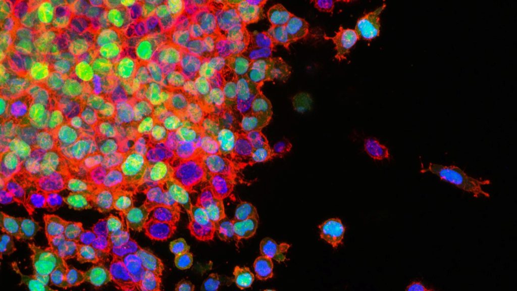
Lung cancer metastasis. Credit: National Cancer Institute

Adenocarcinomas sometimes respond to initially effective treatments by transforming into a much more aggressive small cell lung cancer (SCLC) that spreads rapidly and has few options for treatment. Researchers at Weill Cornell Medicine have developed a mouse model that illuminates this problematic process, known as histological transformation.

The researchers, whose results were published in Science, discovered that during the transition from lung adenocarcinoma to small cell lung cancer (SCLC), the mutated cells appeared to undergo a change in cell identity through an intermediate, stem cell-like state, which facilitated the transformation.

“It is very difficult to study this process in human patients. So my aim was to uncover the mechanism underlying the transformation of lung adenocarcinoma to small cell lung cancer in a mouse model,” said study lead Dr Eric Gardner, a postdoctoral fellow in the laboratory of Dr Harold Varmus.

The complex mouse model took several years to develop and characterise but has allowed the researchers to crack this difficult problem.

This study was in collaboration with Dr Ashley Laughney, assistant professor of physiology and biophysics and a member of the Meyer Cancer Center at Weill Cornell Medicine and Ethan Earlie, a graduate student in the Laughney lab and part of the Tri-Institutional Computational Biology and Medicine program.

“It is well known that cancer cells continue to evolve, especially to escape the pressure of effective treatments,” said Dr Varmus.

“This study shows how new technologies – including the detection of molecular features of single cancer cells, combined with computer-based analysis of the data – can portray dramatic, complex events in the evolution of lethal cancers, exposing new targets for therapeutic attack.”

Catching Transformation in the Act

SCLC most commonly occurs in heavy smokers, but this type of tumour also develops in a significant number of patients with lung adenocarcinomas, particularly after treatment with therapies that target a protein called Epidermal Growth Factor Receptor (EGFR), which promotes tumour growth.

The new SCLC-type tumours are resistant to anti-EGFR therapy because their growth is fuelled by a new cancer driver, high levels of Myc protein.

To unravel the interplay of these cancer pathways, the researchers engineered mice to develop a common form of lung adenocarcinoma, in which lung epithelial cells are driven by a mutated version of the EGFR gene.

They then turned the adenocarcinoma tumours into SCLC-type tumours, which generally arise from neuroendocrine cells.

They did this by shutting off EGFR in the presence of several other changes including losses of the tumour suppressor genes Rb1 and Trp53 as well as turning up the production of Myc,a known driver of SCLC.

Oncogenes, such as EGFR and Myc, are mutated forms of genes that normally control cell growth. They are known for their roles in driving the growth and spread of cancer. Tumor suppressor genes, on the other hand, normally inhibit cell proliferation and tumor development.

Context-dependent change

Surprisingly, this study showed that oncogenes act in a context-dependent manner.

While most lung cells are resistant to becoming cancerous by Myc, neuroendocrine cells, are very sensitive to the oncogenic effects of Myc. Conversely, epithelial cells, which line the air sacs of the lungs and are the precursors to lung adenocarcinomas, grow excessively in response to mutated EGFR.

“This shows that an ‘oncogene’ in the wrong cell type doesn’t act like an oncogene anymore,” Dr Laughney said.

“So, it fundamentally changes how we think about oncogenes.”

The researchers also discovered a stem cell-like intermediate that was neither adenocarcinoma nor SCLC.

Cells in this transitional state became neuroendocrine in nature only when mutations in the tumour suppressor genes RB1 and TP53 were present.

They observed that loss of another tumour suppressor called Pten accelerated this process.

At that stage, oncogenic Myc could drive these intermediate stem-like cells to form SCLC-type tumours.

This study further supports efforts seeking therapeutics that target Myc proteins, which are implicated in many types of cancers. The researchers now plan to use their new mouse model to further explore the adenocarcinoma-SCLC transition, detailing, for example, how the immune system normally responds to this transition.

Source: Weill Cornell Medicine

‘Junk Cells’ Actually Have a Powerful Role against Malaria

Red blood cell Infected with malaria parasites. Colourised scanning electron micrograph of red blood cell infected with malaria parasites (teal). The small bumps on the infected cell show how the parasite remodels its host cell by forming protrusions called ‘knobs’ on the surface, enabling it to avoid destruction and cause inflammation. Uninfected cells (red) have smoother surfaces. Credit: NIAID

Researchers from The Australian National University (ANU) have discovered a previously unknown ability of a group of immune system cells, known as Atypical B cells (ABCs), to fight infectious diseases such as malaria.

The discovery, published in Science Immunology, provides new insight into how the immune system fights infections and brings scientists a step closer to harnessing the body’s natural defences to combat malaria.

The scientists say ABCs could also be key to developing new treatments for chronic autoimmune conditions such as lupus. According to the researchers, ABCs have long been associated with malaria, as malaria patients have more of these cells in their system compared to the general population.

“In this study, we wanted to understand the mechanisms that drive the creation of ABCs in the immune system, but also find out whether these cells are good or bad for us when it comes to fighting infection,” lead author Dr Xin Gao, from ANU, said.

“Although ABCs are known to contribute to chronic inflammatory diseases and autoimmunity, we’ve discovered a previously unknown ability of these cells to fight disease. In this sense, ABCs are like a double-edged sword.

“Contrary to past belief, ABCs are not junk cells; they are more important than we thought.

“Our research found that ABCs are also instrumental in developing T follicular helper cells. These helper cells generate powerful antibodies that help the body fight malaria parasites.

“Antibodies can block parasites in the blood as they travel from the site of the infectious mosquito bite to the liver, where the infection is first established.”

In 2022, malaria killed more than 600 000 people worldwide. Although the disease is preventable and curable, scientists face an uphill battle to find long-lasting treatments as malaria parasites continue to find new ways to build resistance to current therapies.

Using gene-editing technology on mice, the ANU researchers discovered a gene called Zeb2 is crucial to the production of ABCs.

“We found that manipulating the Zeb2 gene disrupted the creation of ABCs in the immune system,” study co-author Professor Ian Cockburn, from The ANU John Curtin School of Medical Research, said.

“Importantly, we found that mice without the Zeb2 gene were unable to control malaria infection.

“Therefore, the findings show that ABCs play a crucial role in fighting malaria infections.”

The researchers say targeting ABCs could also pave the way for new treatments for certain autoimmune diseases such as lupus.

“ABCs also appear in large numbers in many autoimmune diseases, including lupus, which can be life-threating in severe cases,” Professor Cockburn said.

“By developing a better understanding of the role of ABCs in the immune system and the cells’ role in fighting disease, it could bring us a step closer to one day developing new and more effective therapies.”

Source: Australian National University

Unemployed Doctors March to Department of Health

They demand permanent jobs and no budget cuts to healthcare

Doctors marched to the Department of Health offices in Pietermaritzburg on Monday to demand jobs. Photo: Joseph Bracken.

Over 80 unemployed doctors marched from UNISA campus on Longmarket Road to the KwaZulu-Natal Department of Health’s offices in Langalibele Street, Pietermaritzburg, on Monday.

They went to hand over their CVs and a memorandum demanding that the healthcare budget be increased to accommodate over 700 qualifying medical practitioners. The department was given 14 days to respond.

Eighty-four unemployed doctors also signed a register handed to the department.

The doctors were met by Deputy Health Minister Sibongiseni Dhlomo who said health minister Joe Phaahla had another engagement. Dhlomo said the department was working to address the issue of unemployed doctors, and that the minister would raise it this week in Parliament and ask that healthcare be exempt from budget cuts.

Dr Siya Shozi, part of a “small committee” of unemployed doctors with no political affiliation mandated to liaise with the department, said the march was coordinated through a WhatsApp group. Shozi was happy with the turnout but said it did not represent the large number of unemployed doctors in KZN and its rural areas.

Busiziwe Mancotywa, a grade one medical officer who has been unemployed since completing her training at the end of last year, said, “You apply for some positions where you meet the minimum requirements but for whatever reason you are never contacted”.

Mancotywa was joined by her brother, Nqaba, who is finishing his internship at Greys Hospital. He said if action is not taken now, he won’t find a job in the future.

Nomfundo Mbanjwa, also a grade one medical officer, complained about the cost of applying for jobs, including printing applications and transport to interviews. Mbanjwa says she had to sell her car to cover these costs.

Representatives from the South African Medical Association Trade Union (SAMATU) and the Public Servants Association of South Africa (PSA) joined the march and pledged support for the doctors.

Republished from GroundUp under a Creative Commons Attribution-NoDerivatives 4.0 International License.

Source: GroundUp

Even Today, DDT Impacts Men’s Reproductive Health in South Africa and Elsewhere

Photo by Arjun Mj on Unsplash

In a study that signals potential reproductive and health complications in humans, now and for future generations, researchers from McGill University, the University of Pretoria, Université Laval, Aarhus University, and the University of Copenhagen, have concluded that fathers exposed to environmental toxins, notably DDT, may produce sperm with health consequences for their children.

The decade-long research project examined the impact of DDT on the sperm epigenome of South African Vhavenda and Greenlandic Inuit men, some of whom live in Canada’s North.

The study, published in the journal Environmental Health Perspectives, demonstrates a link between long-term exposure to DDT and changes in the sperm epigenome. These changes, particularly in genes vital for fertility, embryo development, neurodevelopment, and hormone regulation, correspond to increased rates of birth defects and diseases, including neurodevelopmental and metabolic disorders.

“We identified regions of the sperm epigenome that are associated with the serum levels of DDE (chemicals that form when DDT breaks down) and this association follows a dose-response trend. I think that’s quite striking, in that the more DDE you’re exposed to, the higher the chromatin, or DNA methylation defects are in the sperm,” said Ariane Lismer, PhD, the study’s lead author, who completed the work while pursuing her PhD at McGill’s Department of Pharmacology and Therapeutics.

“We demonstrate the sperm epigenome’s response to toxin exposures may be linked with disease in the next generation,” said Sarah Kimmins, PhD, who led the research as Professor of Pharmacology and Therapeutics at McGill and is also now a professor in the Department of Pathology and Cell Biology at Université de Montréal. “This is a critical new step for the field because while there are many studies of animals demonstrating toxin effects on the sperm epigenome, studies in humans have not comprehensively demonstrated this.”

Malaria, climate change, and the ‘grasshopper effect’

Despite a global ban on DDT to protect humans and the environment from its effects, the South African government has special permission to use it as an insecticide to control malaria. In some areas, home interiors are coated with the toxin. The study’s findings underscore the urgency to find alternative ways to control malaria and other vector-borne diseases.

“The reality is that people, especially young children and pregnant women, are still dying from malaria. We cannot afford for people in malaria-endemic regions to refuse spraying of their houses, as it will increase their risk of getting malaria,” says Tiaan de Jager, PhD, Dean of the Faculty of Health Sciences and Professor in Environmental Health at the School of Health Systems and Public Health at the University of Pretoria.

What’s more, the number of people and animals exposed to DDT is reportedly increasing due to climate change. DDT can travel vast distances through what is known as the ‘grasshopper effect,’ evaporating with warm air and returning to Earth with rain and snow in colder regions, where it persists in the Arctic food chain.

Rethinking fathers’ role in child development

The findings also highlight the importance of considering fathers in discussions about child health and development. While it’s commonly understood that women should avoid exposure to environmental contaminants during pregnancy, less attention has been given to how toxins affect fathers.

“We tend to think all fathers have to do is fertilise. But in fact, we forget that half of that genome and epigenome comes from the fathers, and half of it comes from the mothers. What that epigenome does in embryo development is critical for normal development,” says study co-author, Janice Bailey, PhD, formerly Professor of Animal Sciences at Université Laval and now the Scientific Director at Fonds de Recherche du Québec en Nature et Technologies (FRQNT).

Although the study focuses on DDT exposures, the researchers say it is not a leap to suggest that exposures to more common household endocrine disruptors such as those found in cosmetics and personal care items may act similarly.

Source: McGill University

For Neuropathic Pain, the More Capsaicin Patch Applications the Better

Photo by Sasun Bughdaryan on Unsplash

Capsaicin, derived from hot chili pepper plants, has been used to treat various types of pain, and a high concentration capsaicin patch (HCCP) is approved for the treatment of neuropathic pain. In a real-world study published in Pain Practice that included 97 outpatients in Germany diagnosed primarily with neuropathic back pain, postoperative/posttraumatic neuropathic pain, or postherpetic neuralgia (shingles pain), patients appeared to benefit from multiple HCCP applications.

Among the study participants, 38 received 2 HCCP treatments, and 59 received at least 3. Following HCCP treatments, most patients required significantly lower doses of opioids to manage their pain. Also, two-thirds of patients experienced a reduction in pain intensity after multiple HCCP treatments, and the proportion of patients experiencing a reduction in pain intensity was substantially higher among those who received at least 3 applications compared with those who received 2 applications.

“Consistent with the progressive response seen in prospective clinical trials involving repeated use of topical capsaicin, our research indicates that patients appear to benefit from multiple applications in terms of pain intensity and concomitant opioid use in real-world clinical practice,” said corresponding author Kai-Uwe Kern, MD, PhD, of the Institute for Pain Medicine/Pain Practice, in Wiesbaden, Germany.

Source: Wiley