Day: January 30, 2024

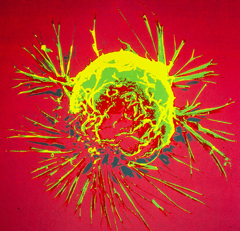
Destroying Tumour Cells with Calcium

Colourised scanning electron micrograph of a breast cancer cell. Credit: NIH

Calcium ions are essential for cells, but can be toxic in higher concentrations. A team of researchers has now designed and prepared a combination drug that kills tumour cells by modulating the calcium influx into the cell. An external calcium source is not necessary because only the calcium ions already present in the tumour tissue are used, according to the study published in the journal Angewandte Chemie.

Biological cells need calcium ions, among other things, for the proper functioning of the mitochondria, the powerhouses of the cells.

However, if there is too much calcium, the mitochondrial processes become unbalanced and the cell suffocates.

A research group led by Juyoung Yoon of Ewha Womens University in Seoul, South Korea, together with teams from China, has now taken advantage of this process and developed a synergistic antitumour drug that can open calcium channels and thus trigger a deadly calcium storm inside the tumour cell.

The researchers targeted two channels, the first one in the outer membrane, and the other was a calcium channel in the endoplasmic reticulum, a cell organelle that also stores calcium ions.

The channel located in the outer membrane opens when it is exposed to a large amount of reactive oxygen species (ROS), while the channel in the endoplasmic reticulum is activated by nitric oxide molecules.

To generate the ROS that open the outer membrane calcium channel, the researchers used the dye indocyanine green.

This bioactive agent can be activated by irradiation with near-infrared light, which not only triggers reactions that lead to ROS, but it also heats up the environment.

The team explains that the high local temperature activates the other active agent, BNN-6, to release nitric oxide molecules that open the channel in the endoplasmic reticulum.

Following successful trials in tumour cell lines, the team tested an injectable formulation in tumour-implanted mice.

To create a biocompatible combined drug, the researchers loaded the active ingredients into tiny modified porous silica beads that are not harmful to the body, but can be recognized by tumour cells and transported into the cell.

After injecting the beads into the bloodstream of the mice, the researchers observed that the drug accumulated in the tumour.

Exposure to near-infrared light successfully triggered the mechanism of action, and the tumour disappeared after a few days in mice that received the preparation.

The authors emphasise that this ion influx approach may also be useful in related biomedical research areas where a similar mechanism could activate ion channels different from calcium in order to find new therapeutic approaches.

Source: Wiley

Healthcare Workers among Those Who Conceal their Infectious Illness

Photo by Brittany Colette on Unsplash

A startling number of people – including healthcare workers – conceal an infectious illness to avoid missing work, travel, or social events, new research at the University of Michigan suggests. The findings however, reported in Psychological Science, exclude being ill with COVID.

Across a series of studies involving healthy and sick adults, 75% of the 4110 participants said they had either hidden an infectious illness from others at least once or might do so in the future.

Many participants reported boarding planes, going on dates, and engaging in other social interactions while secretly sick.

More than 61% of healthcare workers participating in the study also said they had concealed an infectious illness.

Interestingly, the researchers found a difference between how people believe they would act when ill and how they actually behave, said Wilson N. Merrell, a doctoral candidate and lead author on the study.

“Healthy people forecasted that they would be unlikely to hide harmful illnesses – those that spread easily and have severe symptoms – but actively sick people reported high levels of concealment regardless of how harmful their illness was to others,” Merrell said.

In the first study, Merrell and his colleagues, psychology professor Joshua M. Ackerman and PhD student Soyeon Choi, recruited 399 university healthcare employees and 505 students.

The participants reported the number of days they felt symptoms of an infectious illness, starting in March 2020, when the COVID pandemic began.

They then rated how often they actively covered up symptoms from others, came to campus or work without telling others they were feeling ill, or falsified mandatory symptom screeners that the university had required for anyone using campus facilities.

More than 70% of the participants reported covering up their symptoms.

Many said they hid their illness because it would conflict with social plans, while a small percentage of participants cited pressure from institutional policies (eg, lack of paid time off). Only five participants reported hiding a COVID infection.

In a second study, the researchers recruited 946 participants online and randomly assigned them to one of nine conditions in which they imagined being either moderately or severely sick while in a social situation.

In each condition, the risk of spreading the illness was designated as low, medium, or high.

(To control for the special stigma associated with COVID at the time, the researchers asked participants not to imagine being sick with that disease.) Participants were most likely to envision themselves hiding their sickness when symptom severity was low, and least likely to conceal when symptoms were severe and highly communicable.

Source: Association for Psychological Science

Hydroxyurea for Children with Sickle Cell Anaemia Significantly Reduces Infections

Sickle cell disease. Credit: National Institutes of Health

A clinical trial in Uganda has revealed that hydroxyurea significantly reduces infections in children with sickle cell anaemia. Their latest findings enhance strong evidence of hydroxyurea’s effectiveness and could ultimately reduce death in children in Africa, the continent most burdened by the disease.

The group’s research, appearing in the journal Blood, revealed that hydroxyurea treatment resulted in a remarkable 60% reduction in severe or invasive infections, including malaria, bacteraemia, respiratory tract infections and gastroenteritis, among Ugandan children with sickle cell anaemia.

“Our investigation provides powerful justifications for hydroxyurea’s use in children with sickle cell anaemia in Africa,” said Dr Chandy John, paediatrics professor at IU School of Medicine and co-lead investigator of the latest study.

“Given the high rates of infection in this region, we hope our evidence will encourage ministries of health to continue supporting and expanding access to hydroxyurea for young patients who can greatly benefit from the treatment.”

Sickle cell anaemia is a genetic blood disorder that alters the structure of red blood cells and affects oxygen distribution throughout the body, increasing susceptibility to serious health complications and life-threatening infections.

According to the World Health Organization, more than 300 000 children worldwide are born with sickle cell disease each year, with a high prevalence found in African countries.

While hydroxyurea has had U.S. Food and Drug Administration approval as a sickle cell disease treatment for children since 2017, its accessibility and acceptance in Africa have been comparatively limited.

As hydroxyurea has become more recognised in African countries for its effectiveness in treating sickle-cell-related complications, John and his colleagues noticed a knowledge gap about the treatment’s effect on infections.

This led the research group to incorporate hydroxyurea treatment and analysis into their established clinical trial, Zinc for Infection Prevention in Sickle Cell Anemia, led by Indiana University School of Medicine and collaborators in Uganda.

During the study, the researchers examined the effects of hydroxyurea on 117 children in Uganda and focused on a range of infections. After hydroxyurea treatment, results showed a substantial decrease in the incidence of these infections.

Additionally, eight of the nine deaths that occurred in the trial were children whose parents declined hydroxyurea treatment. The only death in a child on hydroxyurea treatment occurred four days after starting treatment, providing insufficient time for hydroxyurea to have an effect.

Of the five children for whom a cause of death was known, all five died of infectious causes.

The high death rate in the study, despite expert clinical care by study personnel, provides further evidence of the urgent need for additional interventions to decrease mortality in children with sickle cell disease in Africa.

“Infections commonly precede other complications related to sickle cell anaemia and often result in hospitalizations that can lead to death,” said Dr Ruth Namazzi, site principal investigator, first author and a lecturer in the Department of Pediatrics and Child Health at Makerere University in Uganda.

“We believe incorporating hydroxyurea treatment as the standard of care for sickle cell anaemia across Africa will not only reduce infections but will more importantly save countless lives.”

Source: Indiana University

Alzheimer’s Disease Cases Caused by Growth Hormone Treatment

Neurons in the brain of an Alzheimer’s patient, with plaques caused by tau proteins. Credit: NIH

Five cases of Alzheimer’s are believed to have arisen as a result of medical treatments decades earlier, according to a new paper published in Nature Medicine. Alzheimer’s disease is caused by the amyloid-beta protein, and is usually a sporadic condition of late adult life, or more rarely as an inherited condition from a faulty gene.

The study, by a team of UCL and UCLH researchers, provides the first evidence of Alzheimer’s disease in living people that appears to have been medically acquired and due to transmission of the amyloid-beta protein.

The people described in the paper had all been treated as children with a type of human growth hormone extracted from pituitary glands from deceased individuals (cadaver-derived human growth hormone or c-hGH). This was used to treat at least 1848 people in the UK between 1959 and 1985, and used for various causes of short stature.

It was withdrawn in 1985 after it was recognised that some c-hGH batches were contaminated with prions (infectious proteins) which had caused Creutzfeldt-Jakob disease (CJD) in some people.

c-hGH was then replaced with synthetic growth hormone that did not carry the risk of transmitting CJD.

These researchers previously reported that some patients with CJD due to c-hGH treatment (called iatrogenic CJD) also had prematurely developed deposits of the amyloid-beta protein in their brains.* The scientists went on to show in a 2018 paper that archived samples of c-hGH were contaminated with amyloid-beta protein and, despite having been stored for decades, transmitted amyloid-beta pathology to laboratory mice when it was injected.

They suggested that individuals exposed to contaminated c-hGH, who did not succumb to CJD and lived longer, might eventually develop Alzheimer’s disease.

This latest paper reports on eight people referred to UCLH’s National Prion Clinic at the National Hospital for Neurology and Neurosurgery in London, who had all been treated with c-hGH in childhood, often over several years.

Five of these people had symptoms of dementia, and either had already been diagnosed with Alzheimer’s disease or would otherwise meet the diagnostic criteria for this condition; another person met criteria for mild cognitive impairment. These people were between 38 and 55 years old when neurological symptoms started. Biomarker analyses supported the diagnoses of Alzheimer’s disease in two patients with the diagnosis, and was suggestive of Alzheimer’s in one other person; an autopsy analysis showed Alzheimer’s pathology in another patient.

The unusually young age at which these patients developed symptoms suggests they did not have the usual sporadic Alzheimer’s which is associated with old age. In the five patients in whom samples were available for genetic testing, the team ruled out inherited Alzheimer’s disease.

As c-hGH treatment is no longer used, there is no risk of any new transmission via this route. There have been no reported cases of Alzheimer’s acquired from any other medical or surgical procedures. There is no suggestion that amyloid-beta can be passed on in day-to-day life or during routine medical or social care.

However, the researchers caution that their findings highlight the importance of reviewing measures to ensure there is no risk of accidental transmission of amyloid-beta via other medical or surgical procedures which have been implicated in accidental transmission of CJD.

The lead author of the research, Professor John Collinge, Director of the UCL Institute of Prion Diseases and a consultant neurologist at UCLH, said: “There is no suggestion whatsoever that Alzheimer’s disease can be transmitted between individuals during activities of daily life or routine medical care. The patients we have described were given a specific and long-discontinued medical treatment which involved injecting patients with material now known to have been contaminated with disease-related proteins.

“However, the recognition of transmission of amyloid-beta pathology in these rare situations should lead us to review measures to prevent accidental transmission via other medical or surgical procedures, in order to prevent such cases occurring in future.

“Importantly, our findings also suggest that Alzheimer’s and some other neurological conditions share similar disease processes to CJD, and this may have important implications for understanding and treating Alzheimer’s disease in the future.”

Source: University College London

Expert Warns that Red Light Therapy for Myopia could Damage the Retina

Retina showing reticular pseudodrusen. Although they can infrequently appear in individuals with no other apparent pathology, their highest rates of occurrence are in association with age-related macular degeneration (AMD), for which they hold clinical significance by being highly correlated with end-stage disease sub-types, choroidal neovascularisation and geographic atrophy. Credit: National Eye Institute

A University of Houston optometry researcher is warning against the use of low-level red light (LLRL) therapy as a method to control myopia, or nearsightedness, especially in children. Over the last few years, LLRL has emerged as a viable myopia treatment after studies reported the treatment as effective and responsible for significant reduction in myopia progression. The company behind one of the devices reports that it is already being used to address myopia in over 100 000 paediatric patients.

But the excitement over its results as a myopia treatment may have come too soon, ahead of its proven safety.

“Based on measurements in our laboratory, it is recommended that clinicians strongly reconsider the use of LLRL therapy for myopia in children until safety standards can be confirmed,” reports Lisa Ostrin, associate professor at the UH College of Optometry in The College of Optometrists journal.

Ostrin reports the therapy can put the retina at risk of photochemical and thermal damage.

“The safety profiles of red-light laser devices for myopia have not been fully investigated,” she said.

For LLRL therapy, children are instructed to look into a red light-emitting instrument for three minutes, twice a day, five days a week, for the duration of the treatment period, which could last years.

“We found that the red-light instruments for myopia exceed safety limits,” said Ostrin, whose research characterises the laser output and determines the thermal and photochemical maximum permissible exposure (MPE) of LLRL devices.

“For both LLRL devices evaluated here, three minutes of continuous viewing approached or surpassed the luminance dose MPE, putting the retina at risk of photochemical damage.”

Ostrin examined two different LLRL devices, and while both instruments were confirmed to be Class-1 laser products, as defined by International Electrotechnical Commission standards, according to Ostrin they are unsafe to view continuously for the required treatment duration of three minutes.

Class-1 lasers are low-powered devices that are considered safe from all potential hazards when viewed accidentally and briefly.

Examples of Class-1 lasers are laser printers, CD players and digital video disc (DVD) devices.

Class-1 lasers are not meant to be viewed directly for extended periods.

“Thermal ocular injury from a laser can occur with exposures at any wavelength when the temperature change of the retina is greater than 10°C, resulting in the denaturation of proteins. With thermal damage, the lesion size is typically less than the size of the beam diameter, and the resultant scotomas are permanent.” said Ostrin.

Source: University of Houston