Day: January 26, 2024

Gene Therapy Restores Hearing in Children with Hereditary Deafness

Photo by jonas mohamadi

A new study co-led by investigators from Mass Eye and Ear, a member of Mass General Brigham, demonstrated the effectiveness of a gene therapy towards restoring hearing function for children suffering from hereditary deafness.

In a trial of six children taking place at the Eye & ENT Hospital of Fudan University in Shanghai, China, the researchers found the novel gene therapy to be an effective treatment for patients with a specific form of autosomal recessive deafness caused by mutations of the OTOF (otoferlin) gene, called DFNB9. With its first patient treated in December 2022, this research represents the first human clinical trial to administer gene therapy for treating this condition, with the most patients treated and longest follow-up to date. Their results are published in The Lancet.

“If children are unable to hear, their brains can develop abnormally without intervention,” said Zheng-Yi Chen, DPhil, an associate scientist in the Eaton-Peabody Laboratories at Mass Eye and Ear and associate professor of Otolaryngology–Head and Neck Surgery at Harvard Medical School. “The results from this study are truly remarkable. We saw the hearing ability of children improve dramatically week by week, as well as the regaining of their speech.”

Hearing loss affects more than 1.5 billion people worldwide, with congenital deafness making up about 26 million of those individuals. For hearing loss in children, more than 60% stem from genetic reasons. DFNB9 for example, is a hereditary disease caused by mutations of the OTOF gene and a failure to produce a functioning otoferlin protein, which is necessary for the transmission of the sound signals from the ear to the brain. There are currently no FDA-approved drugs to help with hereditary deafness, which has opened the door for new solutions like gene therapies.

In order to test this novel treatment, six children with DFNB9 were observed over a 26-week period at the Eye & ENT Hospital of Fudan University. The Mass Eye and Ear collaborators utilised an adeno-associated virus (AAV) carrying a version of the human OTOF gene to carefully introduce the gene into the inner ears of the patients through a special surgical procedure. Differing doses of the single injection of the viral vector were used.

All six children in the study had total deafness, as indicated by an average auditory brainstem response (ABR) threshold of over 95 decibels. After 26 weeks, five children demonstrated hearing recovery, showing a 40-57 decibel reduction in ABR testing, dramatic improvements in speech perception and the restored ability to conduct normal conversation. Overall, no dose-limiting toxicity was observed. While following up on the patients, 48 adverse events were observed, with a significant majority (96%) being low grade, and the rest being transitory with no long-term impact.

Trial findings will also be presented February 3rd at the Association for Research in Otolaryngology Annual Meeting.

This study provides evidence towards the safety and effectiveness of gene therapies in treating DFNB9, as well as their potential for other forms of genetic hearing loss. Moreover, the results contribute to an understanding of the safety of AAV insertion into the human inner ear. In regard to the usage of AAVs, the success of a dual-AAV vector carrying two pieces of the OTOF gene is notable. Typically, AAVs have a gene size limit, and so for a gene like OTOF that exceeds that limit, the achievement with a dual viral vector opens the door for AAV’s use with other large genes that are typically too big for the vector.

“We are the first to initiate the clinical trial of OTOF gene therapy. It is thrilling that our team translated the work from basic research in animal model of DFNB9 to hearing restoration in children with DFNB9,” said lead study author Yilai Shu, MD, of the Eye & ENT Hospital of Fudan University at Fudan University. Shu previously served as a postdoctoral fellow in Chen’s lab at Mass Eye and Ear. “I am truly excited about our future work on other forms of genetic hearing loss to bring treatments to more patients.”

The researchers plan to expand the trial to a larger sample size as well as track their outcomes over a longer timeline.

“Not since cochlear implants were invented 60 years ago, has there been an effective treatment for deafness,” said Chen. “This is a huge milestone that symbolises a new era in the fight against all types of hearing loss.”

Source: Massachusetts Eye and Ear Infirmary

Single Dose Typhoid Conjugate Vaccine (TCV) Provides Lasting Efficacy in Children

Healthy children in Malawi participating in study to test efficacy of typhoid conjugate vaccine. Credit: TyVAC/Madalitso Mvula

A single dose of the typhoid conjugate vaccine, Typbar TCV®, provides lasting efficacy in preventing typhoid fever in children ages 9 months to 12 years old, according to a new phase 3 clinical study published in The Lancet.

The study conducted by researchers at University of Maryland School of Medicine’s (UMSOM) Center for Vaccine Development and Global Health (CVD) and led by in-country partners at the Malawi-Liverpool Wellcome Trust (MLW) Clinical Research Programme.

The research team enrolled more than 28 000 healthy children in Malawi and randomly assigned about half the group to receive the TCV and the other half to receive a meningococcal capsular group A conjugate (MenA) control vaccine. During the more than four years of follow-up, 24 children in the TCV group and 110 in the MenA group developed typhoid fever, which was confirmed via blood culture. That resulted in an efficacy of 78.3% in the TCV group, with one case of typhoid prevented for every 163 children vaccinated. TCV was effective in all age groups and over the study period – which ended in 2022 – vaccine efficacy remained strong, decreasing by only 1.3% per year.

Typhoid fever causes more than 9 million illnesses and at least 110,000 deaths worldwide every year, mostly in sub-Saharan Africa and southeast Asia. It is a contagious bacterial infection that occurs from consuming contaminated food or beverages. Symptoms include nausea, fever, and abdominal pain that, if left untreated, can be deadly.

“These findings have significant implications for identification of the contribution of TCVs in the control and potential elimination of typhoid fever in endemic settings,” wrote the authors of a commentary published in The Lancet alongside the study.

In May 2023, the Malawi government launched a national rollout of the TCV in children under 15 years. Going forward, all children in Malawi will receive TCV at 9 months of age as part of the routine immunisation schedule.

“The newly published study supports the long-lasting impacts of a single shot of TCV, even in the youngest children, and offers hope of preventing typhoid in the most vulnerable children,” said Kathleen Neuzil, MD, MPH, CVD Director, the Myron M. Levine, MD, DTPH, Professor in Vaccinology at UMSOM and coauthor of the current study.  “We could not have had a better partner in this endeavor than MLW, whose long-standing excellence in typhoid research and strong surveillance infrastructure made this study possible.”

“The CVD’s outstanding record of generating data to accelerate public health decisions continues with this landmark study,” said UMSOM Dean Mark T. Gladwin, MD, Vice President for Medical Affairs, University of Maryland, Baltimore, and the John Z. and Akiko K. Bowers Distinguished Professor. “The research could not come at a more critical time when Malawi and other African countries are struggling with climate change, extreme weather events and increased urbanisation patterns, which are likely to contribute to increases in enteric diseases, including typhoid.”

Source: University of Maryland

Walking Fitness can Predict Fracture Risk in Older Adults

Photo by Teona Swift on Unsplash

The ability to walk one kilometre comfortably can help predict fracture risk, according to researchers at the Garvan Institute of Medical Research. The findings, published in JAMA Network Open, suggest that simply asking a patient about walking limitation could allow clinicians to identify those in need of further bone health screening and prescribe interventions that could prevent fractures from occurring.

“We’ve discovered that trouble walking even short distances appears closely tied to higher fracture risk over the following five years,” says lead author of the study, Professor Jacqueline Center, Head of Garvan’s Clinical Studies and Epidemiology Lab.

“Just a few simple questions about how far someone can walk could give doctors an early warning sign to check bone health.”

The researchers examined data on nearly 267 000 adults aged 45 and older from the Sax Institute’s 45 and Up Study, a major ongoing research initiative that has been tracking health outcomes in adults in the Australian state of New South Wales for more than 15 years.

Participants were asked if health issues limited their ability to walk various distances, with answer options of ‘not at all,’ ‘a little,’ or ‘a lot’. The group was then followed for five years to track fracture outcomes.

The researchers found that one in five adults reported some walking limitation at the beginning of the study.

Those with more difficulty walking were significantly more likely to experience a fracture during follow-up. For example, women who said they were limited ‘a lot’ in walking one kilometre had a 60% higher fracture risk than women with no limitation.

For men, the increased risk was over 100%.

“We saw a clear ‘dose-response’ pattern, where greater walking limitation meant higher fracture risk. This suggests a direct relationship between low walking ability and weaker bones,” says first author of the study Dr Dana Bliuc, Senior Research Officer at Garvan.

Approximately 60% of all fractures in the study were attributable to some level of walking limitation.

The link remained strong even after accounting for other factors like age, falls, prior fractures, and weight, and the findings were consistent across different fracture sites like hips, vertebrae, arms, and legs.

“In this generally healthy community-based population, we still found one in five people had trouble walking a kilometre,” says Professor Center.

“We think this simple assessment could help identify many more at-risk individuals who may benefit from bone density screening or preventative treatment.”

Osteoporosis medications, lifestyle changes, and other interventions are available to improve bone strength and avoid first or repeat fractures.

However, screening rates currently remain low, meaning many miss out on fracture risk assessments.

Finding easy but accurate ways to detect at-risk people is an important target for research.

“Fracture risk assessment generally relies on a bone density test, which many people have not had when seeing their doctor,” says Professor Center.

“Asking about walking ability takes just seconds and could be a free, non-invasive way to tell if someone needs their bones checked.”

The researchers stress that walking limitation may have many causes beyond weak bones, from heart disease to arthritis.

However, a difficulty in walking even short distances appears closely tied to fracture risk independently.

“We hope these findings will encourage clinicians to consider walking ability as a red flag for possible bone health issues. For patients, if you can’t walk a full kilometre comfortably, it may be wise to ask your doctor about getting your bones checked,” says Dr Bliuc.

Source: Garvan Institute of Medical Research

New Ultrasound Method can Predict Risk of Preterm Delivery

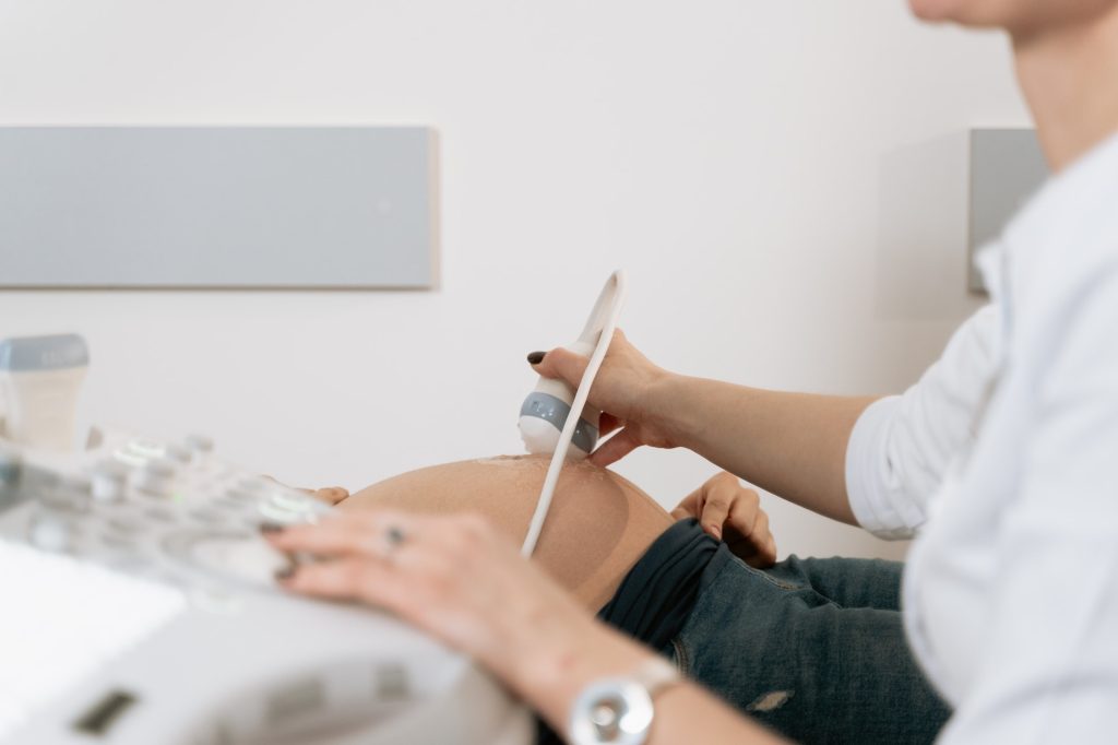
Photo by Mart Production on Pexels

Researchers have developed a way to use ultrasound to estimate the risk of delivering a baby preterm. The new method measures microstructural changes in a woman’s cervix using quantitative ultrasound. The method works as early as 23 weeks into a pregnancy, according to the research, which is published in the American Journal of Obstetrics & Gynecology Maternal Fetal Medicine.

The current method for assessing a woman’s risk of preterm birth is based solely on whether she has previously given birth prematurely. This means there has been no way to assess risk in a first-time pregnancy.

“Today, clinicians wait for signs and symptoms of a preterm birth,” such as a ruptured membrane, explained lead author Barbara McFarlin, a professor emeritus of nursing at University of Illinois Chicago.

“Our technique would be helpful in making decisions based on the tissue and not just on symptoms.”

The new method is the result of more than 20 years of collaboration between researchers in nursing and engineering at UIC and University of Illinois Urbana-Champaign. In a study of 429 women who gave birth without induction at the University of Illinois Hospital, the new method was effective at predicting the risk of preterm births during first-time pregnancies.

And for women who were having a subsequent pregnancy, combing the data from quantitative ultrasound with the woman’s delivery history was more effective at assessing risk than just using her history.

The new approach differs from a traditional ultrasound where a picture is produced from the data received.

In quantitative ultrasound, a traditional ultrasound is performed but the radio frequency data itself is read and analysed to determine tissue characteristics.

The study is the culmination of a research partnership that began in 2001 when McFarlin was a nursing PhD student at UIC. Having previously worked as a nurse midwife and sonographer, she had noticed that there were differences in the appearance of the cervix in women who went on to deliver preterm.

She was interested in quantifying this and discovered that “no one was looking at it.”

She was put in touch with Bill O’Brien, a UIUC professor of electrical and computer engineering, who was studying ways to use quantitative ultrasound data in health research.

Together, over the past 22 years, they established that quantitative ultrasound could detect changes in the cervix and, as McFarlin had suspected long ago, that those changes help predict the risk of preterm delivery.

The preterm birth rate hovers around 10-15% of pregnancies, O’Brien said.

“That’s a very, very high percentage to not know what is happening,” he said.

If a clinician could know at 23 weeks that there was a risk of preterm birth, they would likely conduct extra appointments to keep an eye on the foetus, the researchers said.

But since there had previously been no routine way to assess preterm birth risk this early, there have been no studies to show what sort of interventions would be helpful in delaying labour.

This study, O’Brien explains, will allow other researchers to “start studying processes by which you might be able to prevent or delay preterm birth.”

Source: University of Illinois Chicago

mRNA Technology Restores Tumour Suppressor Protein in Ovarian Cancer

Photo by Sangharsh Lohakare on Unsplash

Using mRNA technology developed and matured for certain COVID vaccines, researchers have successfully restored the tumour-suppressing p53 protein in mouse models of advanced human ovarian cancer, significantly extending their survival. They report their results in Cancer Communications.

Ovarian cancer is often only detected at an advanced stage and metastases have already formed — usually in the intestines, abdomen or lymph nodes. At such a late stage, only 20 to 30% of all those affected survive the next five years. “Unfortunately, this situation has hardly changed at all over the past two decades,” says Professor Klaus Strebhardt, Director of the Department of Molecular Gynecology and Obstetrics at University Hospital Frankfurt.

In 96% of all ovarian cancer (high-grade) patients, the tumour suppressor gene p53 has mutated and is now non-functional. The gene contains the building instructions for an important protein that normally recognises damage in each cell’s DNA. It then prevents these abnormal cells from proliferating and activates repair mechanisms that rectify the damage.

If this fails, it induces cell death. “In this way, p53 is very effective in preventing carcinogenesis,” explains Strebhardt. “But when it is mutated, this protective mechanism is eradicated.”

If a cell wants to produce a certain protein, it first makes a transcript of the gene containing the building instructions for it. Such transcripts are called mRNAs. In women with ovarian cancer, the p53 mRNAs are just as defective as the gene from which they were copied.

“We produced an mRNA in the laboratory that contained the blueprint for a normal, non-mutated p53 protein,” says Dr Monika Raab from the Department of Molecular Gynecology and Obstetrics, who conducted many of the key experiments in the study.

“We packed it into small lipid vesicles, known as liposomes, and then tested them first in cultures of various human cancer cell lines. The cells used the artificial mRNA to produce functional p53 protein.”

In the next step, the scientists cultivated ovarian tumours – organoids – from patient cells sourced by the team led by Professor Sven Becker, Director of the Women’s Clinic at University Hospital Frankfurt.

After treatment with the artificial mRNA, the organoids shrank and began to die.

To test whether the artificial mRNA is also effective in organisms and can combat metastases in the abdomen, the researchers implanted human ovarian tumour cells into the ovaries of mice and injected the mRNA liposomes into the animals some time later.

The result was very convincing, says Strebhardt: “With the help of the artificial mRNA, cells in the animals treated produced large quantities of the functional p53 protein, and as a result both the tumours in the ovaries and the metastases disappeared almost completely.”

That the method was so successful is partly due to recent advances in mRNA technology: Normally, mRNA transcripts are very sensitive and degraded by cells within minutes.

However, it is meanwhile possible to prevent this by specifically modifying the molecules.

This extends their lifespan substantially, in this study to up to two weeks.

In addition, the chemical composition of the artificial mRNA is slightly different to that of its natural counterpart.

This prevents the immune system from intervening after the molecule has been injected and from triggering inflammatory responses.

In 2023, the Hungarian scientist Katalin Karikó and her American colleague Drew Weissman were awarded the Nobel Prize in Physiology or Medicine for this discovery.

“Thanks to the development of mRNA vaccines such as those of BioNTech and Moderna, which went into action during the SARS-CoV-2 pandemic, we now also know how to make the molecules even more effective,” explains Strebhardt.

Strebhardt, Raab and Becker are now looking for partners to join the next step of the translational project: testing on patients with ovarian cancer. “What is crucial now is the question of whether we can implement the concept and the results in clinical reality and use our method to help cancer patients,” says Strebhardt. The latest results make him very optimistic that the tide could finally turn in the treatment of ovarian carcinomas. “p53 mRNA is not a normal therapeutic that targets a specific weak point in cancer cells. Instead, we are repairing a natural mechanism that the body normally uses very effectively to suppress carcinogenesis. This is a completely different quality of cancer therapy.”

Source: Goethe University Frankfurt