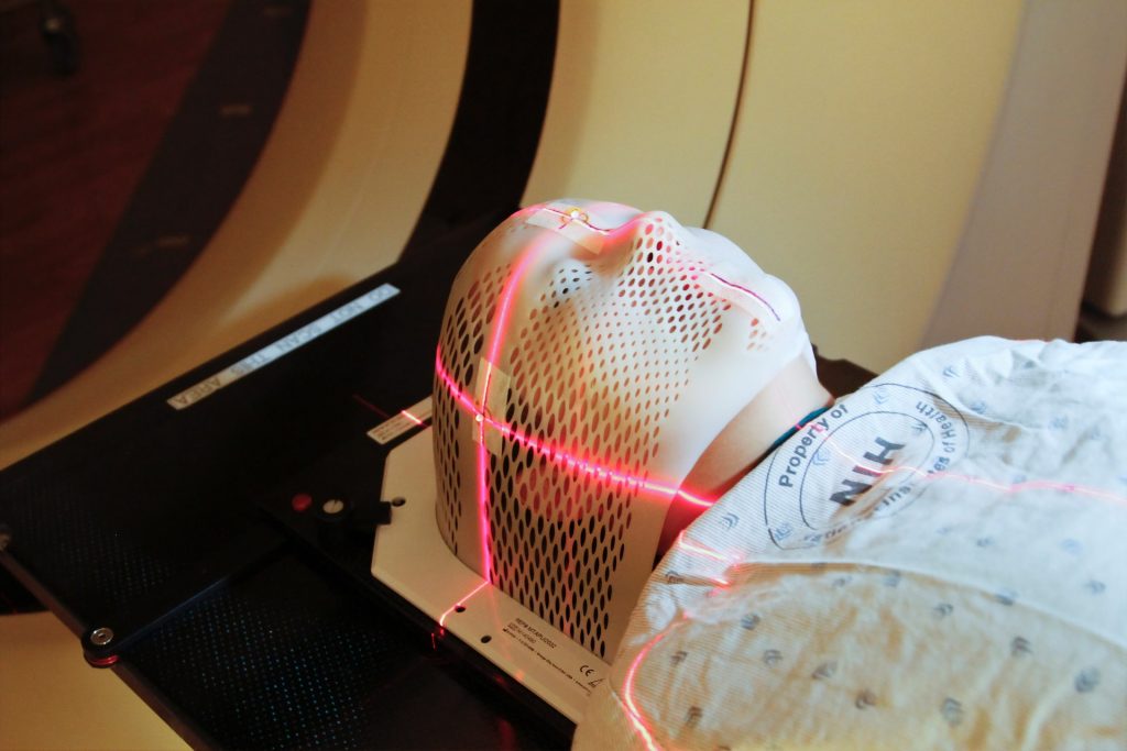
Microglia, the brain’s immune cells, can trigger cognitive deficits after radiation exposure and may be a key target for preventing these symptoms, University of Rochester researchers have found. Their work, published in the International Journal of Radiation Oncology Biology Biophysics, builds on previous research showing that after radiation exposure microglia damage synapses, the connections between neurons that are important for cognitive behaviour and memory.
“Cognitive deficits after radiation treatment are a major problem for cancer survivors,” M. Kerry O’Banion, MD, PhD, professor of Neuroscience, member of the Wilmot Cancer Institute, and senior author of the study said.
“This research gives us a possible target to develop therapies to prevent or mitigate against such deficits in people who need brain radiotherapy.”
Using several behavioural tests, researchers investigated the cognitive function of mice before and after radiation exposure.
Female mice performed the same throughout, indicating a resistance to radiation injury but Male mice could not remember or perform certain tasks after radiation exposure.
This cognitive decline correlates with the loss of synapses and evidence of potentially damaging microglial over-reactivity following the treatment.
Researchers then targeted the pathway in microglia important to synapse removal. Mice with these mutant microglia had no cognitive decline following radiation. And others that were given the drug, Leukadherin-1, which is known to block this same pathway, during radiation treatment, also had no cognitive decline.
“This could be the first step in substantially improving a patient’s quality of life and need for greater care,” said O’Banion. “Moving forward, we are particularly interested in understanding the signals that target synapses for removal and the fundamental signaling mechanisms that drive microglia to remove these synapses. We believe that both avenues of research offer additional targets for developing therapies to help individuals receiving brain radiotherapy.”
O’Banion also believes this work may have broader implications because some of these mechanisms are connected to Alzheimer’s and other neurodegenerative diseases.
A new study from UC Riverside has added more reasons to stick to New Year’s diet resolutions: it showed that that high-fat diets affect genes linked not only to obesity, colon cancer and irritable bowels, but also to the immune system, brain function, and potentially COVID risk.
While other studies have examined the effects of a high-fat diet, this one is unusual in its scope. UCR researchers fed mice three different diets over the course of 24 weeks where at least 40% of the calories came from fat. Then, they looked not only at the microbiome, but also at genetic changes in all four parts of the intestines.
One group of mice ate a diet based on saturated fat from coconut oil, another got a monounsaturated, modified soybean oil, a third got an unmodified soybean oil high in polyunsaturated fat. Compared to a low-fat control diet, all three groups experienced concerning changes in gene expression, the process that turns genetic information into a functional product, such as a protein.
Plant-based or not, high-fat is bad
“Word on the street is that plant-based diets are better for you, and in many cases that’s true. However, a diet high in fat, even from a plant, is one case where it’s just not true,” said Frances Sladek, a UCR cell biology professor and senior author of the new study.
The study, published in Scientific Reports, documents the many impacts of high-fat diets. Some of the intestinal changes did not surprise the researchers, such as major changes in genes related to fat metabolism and the composition of gut bacteria. For example, they observed an increase in pathogenic E. coli and a suppression of Bacteroides, which helps protect the body against pathogens.
Other observations were more surprising, such as changes in genes regulating susceptibility to infectious diseases. “We saw pattern recognition genes, ones that recognise infectious bacteria, take a hit. We saw cytokine signalling genes take a hit, which help the body control inflammation,” Sladek said. ‘So, it’s a double whammy. These diets impair immune system genes in the host, and they also create an environment in which harmful gut bacteria can thrive.”
The team’s previous work with soybean oil documents its link to obesity and diabetes, both major risk factors for COVID. This paper now shows that all three high-fat diets increase the expression of ACE2 and other host proteins that are used by COVID spike proteins to enter the body.
Additionally, the team observed that high-fat food increased signs of stem cells in the colon. “You’d think that would be a good thing, but actually they can be precursors to cancer,” Sladek said.
In terms of effects on gene expression, coconut oil showed the greatest number of changes, followed by the unmodified soybean oil. Differences between the two soybean oils suggest that polyunsaturated fatty acids in unmodified soybean oil, primarily linoleic acid, play a role in altering gene expression.
Negative changes to the microbiome in this study were more pronounced in mice fed the soybean oil diet. This was unsurprising, as the same research team previously documented other negative health effects of high soybean oil consumption.
Soybeans are fine, but watch the oil
In 2015, the team found that soybean oil induces obesity, diabetes, insulin resistance, and fatty liver in mice. In 2020, the researchers team demonstrated the oil could also affect genes in the brain related to conditions like autism, Alzheimer’s disease, anxiety, and depression.
Interestingly, in their current work they also found the expression of several neurotransmitter genes were changed by the high fat diets, reinforcing the notion of a gut-brain axis that can be impacted by diet.
The researchers have noted that these findings only apply to soybean oil, and not to other soy products, tofu, or soybeans themselves. “There are some really good things about soybeans. But too much of that oil is just not good for you,” said UCR microbiologist Poonamjot Deol, who was co-first author of the current study along with UCR postdoctoral researcher Jose Martinez-Lomeli.
Also, the studies were conducted using mice, and mouse studies do not always translate to the same results in humans. However, humans and mice share 97.5% of their working DNA. Therefore, the findings are concerning, as soybean oil is the most commonly consumed oil in the United States, and is increasingly being used in other countries, including Brazil, China, and India.
By some estimates, Americans tend to get nearly 40% of their calories from fat, which mirrors what the mice were fed in this study. “Some fat is necessary in the diet, perhaps 10 to 15%. Most people though, at least in this country, are getting at least three times the amount that they need,” Deol said.
Readers should not panic about a single meal. It is the long-term high-fat habit that caused the observed changes. Recall that the mice were fed these diets for 24 weeks. “In human terms, that is like starting from childhood and continuing until middle age. One night of indulgence is not what these mice ate. It’s more like a lifetime of the food,” Deol said.
That said, the researchers hope the study will cause people to closely examine their eating habits.
Coup and contrecoup brain injury. Credit: Scientific Animations CC4.0
Researchers have created a new brain imaging method that allows to be diagnosed, even when existing imaging techniques like magnetic resonance imaging (MRI) The technique involves loading gadolinium, a standard MRI contrast agent, into ‘backpacks’ that are attached macrophages. mTBIs cause inflammation, attracting macrophages there. Coupling the gadolinium contrast agent to these cells enables MRI to reveal brain inflammation and increase the number of correctly diagnosed mTBI cases, improving patient care. The method is described in a new paper in Science Translational Medicine.
“70-90% of reported TBI cases are categorised as ‘mild,’ yet as many as 90% of mTBI cases go undiagnosed, even though their effects can last for years and they are known to increase the risk of a host of neurological disorders including depression, dementia, and Parkinson’s disease,” said senior author Samir Mitragotri, PhD, in whose lab the research was performed. “Our cell-based imaging approach exploits immune cells’ innate ability to travel into the brain in response to inflammation, enabling us to identify mTBIs that standard MRI imaging would miss.”
Using immune cells to identify inflammation
Most of us know someone who has had a concussion (another name for an mTBI), sometimes even more than one. But the vast majority of people who experience an mTBI are never properly diagnosed. Without that diagnosis, they can exacerbate their injuries by returning to normal activity before they’re fully recovered, which can lead to further damage. Some studies even suggest that repeated mTBIs can lead to chronic traumatic encephalopathy (CTE), the neurodegenerative disease that has been found to afflict more than 90% of professional American football players.
Because the effects of mTBI are believed to be caused by “invisible” brain inflammation, members of the Mitragotri lab decided to leverage their experience with immune cells to create a better diagnostic. “Our previous projects have focused on controlling the behaviour of immune cells or using them to deliver drugs to a specific tissue. We wanted to exploit another innate ability of immune cells – homing to sites of inflammation in the body – to carry imaging agents into the brain, where they can provide a visible detection signal for mTBI,” said first author Lily Li-Wen Wang, Ph.D.. Wang is a former Research Fellow in the Mitragotri Lab at the Wyss Institute and SEAS who is now a scientist at Landmark Bio.
Gadolinium needs water to show up on MRI
The team planned to use their cellular backpack technology to attach gadolinium molecules to macrophages, known to infiltrate the brain in response to inflammation. But right away, they ran into a problem: in order to function as a contrast agent for MRI scans, gadolinium needs to interact with water. Their original backpack microparticles are made of a hydrophobic polymer called PLGA. So Wang and her co-authors started developing a new backpack made out of a hydrogel material that could be manufactured at a large scale in the lab.
After years of hard work, they finally created a new hydrogel backpack that could produce a strong gadolinium-mediated MRI signal, attach stably to both mouse and pig macrophages, and maintain their cargo for a sustained period of time in vitro. They named their new microparticles M-GLAMs, short for “macrophage-hitchhiking Gd(III)-Loaded Anisotropic Micropatches.” Now, it was time to test them in a more realistic setting, for which they partnered with researchers and clinicians at Boston Children’s Hospital.
First, they injected mouse M-GLAMs macrophages into mice to see if they could visualize them in vivo. They were especially interested to see if they accumulated in the kidney, as existing gadolinium-based contrast agents like Gadavist® can cause health risks for patients with kidney disease. Their M-GLAMs did not accumulate in the mice’s kidneys, but persisted in their bodies for over 24 hours with no negative side effects. In contrast, mice injected with Gadavist® showed substantial accumulation of the contrast agent in their kidneys within 15 minutes of injection, and the substance was fully cleared from their bodies within 24 hours.
Then, they tested porcine M-GLAMs in a pig model of mTBI. They injected the M-GLAMs into the animals’ blood two days after a mock mTBI, then used MRI to evaluate the concentration of gadolinium in the brain. They focused on a small region called the choroid plexus, which is known as a major conduit of immune cells into the brain. Pigs that received the M-GLAMs displayed a significant increase in the intensity of gadolinium present in the choroid plexus, while those injected with Gadavist® did not, despite confirmation of increased inflammation macrophage density in the brains of both groups. The animals showed no toxicity in any of their major organs following administration of the treatments.
“Another important aspect of our M-GLAMs is that we are able to achieve better imaging at a much lower dose of gadolinium than current contrast agents – 500-1000-fold lower in the case of Gadavist®,” said Wang. “This could allow the use of MRI for patients who are currently unable to tolerate existing contrast agents, including those who have existing kidney problems.”
Yale researchers report in the journal Nature that they have identified a drug target that may alleviate joint degeneration associated with osteoarthritis.
The most common therapies for the degenerative disease have been pain relievers and lifestyle changes, to reduce pain and stiffness, but there is a pressing need for therapies that can prevent joint breakdown that occurs in osteoarthritis, which occurs as a result of the breakdown of cartilage in the joints.
Sodium channels found in cell membranes produce electrical impulses in “excitable” cells within muscles, the nervous system, and the heart. And in previous research, Yale’s Stephen G. Waxman identified the key role of one particular sodium channel, called Nav1.7, in the transmission of pain signals.
Now, the labs of Chuan-Ju Liu, professor of orthopaedics, and Waxman, professor neurology, neuroscience and pharmacology, have found that the same Nav1.7 channels are also present in non-excitable cells that produce collagen and help maintain the joints in the body. These channels can be targeted by existing drugs to block them.
In the new study, the researchers deleted Nav1.7 genes from these collagen-producing cells and significantly reduced joint damage in two osteoarthritis models in mice.
They also demonstrated that drugs used to block Nav1.7 – including carbamazepine, a sodium channel blocker currently used to treat epilepsy and trigeminal neuralgia – also provided substantial protection from joint damage in the mice.
“The function of sodium channels in non-excitable cells has been a mystery,” Waxman said.
“This new study provides a window on how small numbers of sodium channels can powerfully regulate the behaviour of non-excitable cells.”
“The findings open new avenues for disease-modifying treatments,” added Wenyu Fu, a research scientist in the Liu laboratory and first author of the study.
A new study from researchers at the University of Colorado Anschutz Medical Campus finds that older adult drivers who are recently diagnosed with migraines are three times as likely to experience a motor vehicle crash. Older adult drivers who reported having ever had migraines in the past were no more likely to have a motor vehicle crash than those without migraines.
The study, published in the Journal of the American Geriatrics Society, also explored the relationships medications commonly prescribed for migraine management have with increased crash risk.
“Migraine headaches affect more than 7% of US adults over the age of 60,” says Carolyn DiGuiseppi, MPH, PhD, MD, professor with the Colorado School of Public Health and study lead author.
“The US population is aging, which means increasing numbers of older adult drivers could see their driving abilities affected by migraine symptoms previously not experienced. These symptoms include sleepiness, decreased concentration, dizziness, debilitating head pain and more.”
Researchers conducted a five-year longitudinal study of more than 2500 active drivers aged 65-79 in five sites across the United States.
Participants were categorised as having previously been diagnosed with migraine symptoms (12.5%), no previous diagnosis but experienced symptoms during the study timeframe (1.3%) or never migraine respondents.
Results indicate those with previous diagnosis did not have a different likelihood of having crashes after baseline, while those with new onset migraines were three times as likely to experience a crash within one year of diagnosis.
Previously diagnosed drivers nevertheless had experienced more hard braking events compared to adults who had never experienced a migraine.
Additionally, researchers examined the role medications commonly prescribed for migraines have in motor vehicle events and found that there was no impact on the relationship between migraines and either crashes or driving habits.
Few participants in the study sample were using acute migraine medications, however.
“These results have potential implications for the safety of older patients that should be addressed,” says DiGuiseppi. “Patients with a new migraine diagnosis would benefit from talking with their clinicians about driving safety, including being extra careful about other risks, such as distracted driving, alcohol, pain medication and other factors that affect driving.”