Month: January 2024

ER+ Tumours Driving Surge in Breast Cancer Diagnoses among Younger Women

Photo by National Cancer Institute on Unsplash

Diagnoses of breast cancer have increased steadily in women under age 50 over the past two decades, with steeper increases in more recent years, according to a study published in JAMA Network Open. The surge is driven largely by increases in the number of women diagnosed with oestrogen-receptor positive (ER+) tumours.

While overall trends show increases, however, some decreases have occurred in specific tumour types and among specific groups of women. Such changes in disease rates in young women observed over time – analysed by age, race, tumour type, tumour stage and other factors – may offer clues to possible prevention strategies.

“For most women, regular breast cancer screening does not begin until at least age 40, so younger women diagnosed with breast cancer tend to have later-stage tumours, when the disease is more advanced and more difficult to treat,” said senior author Adetunji T. Toriola, MD, PhD, a professor of surgery and co-leader of the Cancer Prevention and Control Program at Siteman Cancer Center, based at Barnes-Jewish Hospital and Washington University School of Medicine. “This research offers a way to begin identifying the factors driving these increasing rates, with the goal of finding ways to slow or reverse them. It also could help identify young women who are at high risk of developing early-onset breast cancer, so that we can design interventions to evaluate in clinical trials to see if we can lower that risk.”

The research team analysed data from over 217 000 U.S. women diagnosed with any type of breast cancer from 2000 through 2019. In 2000, the incidence of breast cancer among women ages 20 to 49 was about 64 cases per 100 000 people. Over the next 16 years, that rate slowly went up, increasing at about 0.24% per year. By 2016, the rate had reached about 66 cases per 100 000. But after 2016, for reasons researchers do not yet understand, the trend line made a steep uphill turn, suddenly increasing at 3.76% per year. By 2019 the rate had reached 74 cases per 100 000.

An additional intriguing aspect of the data is that the increase in breast cancer incidence is due almost entirely to an increase in tumours that are ER+ according to Toriola, who is also a William H. Danforth Washington University Physician-Scientist Scholar. These tumours have proteins on their surfaces that bind to oestrogen, which fuels tumour growth. In fact, the incidence of tumours without the oestrogen receptor decreased over the 20 years of data analysed in the study.

“We need to understand what is driving the specific increase in oestrogen-receptor positive tumours,” Toriola said. “We also hope to learn from the decrease in oestrogen-receptor negative tumours. If we can understand what is driving that rate down, perhaps we can apply it in efforts to reduce or prevent other breast tumour types.”

The researchers also found higher rates of breast cancer among Black women, especially among those ages 20 to 29. Black women in this age group have a 53% increased risk of breast cancer compared with white women of the same age group. A higher risk for Black women also continues from ages 30 to 39, but the increased risk is smaller, at about 15% greater risk compared with white women of the same age range. Then, from ages 40 to 49, the rate for Black women drops below that of white women.

Toriola said his group is evaluating breast tumour tissue from cancer patients of different ages and races to see if there are molecular differences that could shed light on what is driving cancer to develop more in young Black women. Of note, Hispanic women in the study had the lowest incidence of breast cancer of any group.

The researchers also showed an increase in diagnoses of stage 1 and stage 4 tumours, and a decrease in diagnoses of stage 2 and stage 3 tumours. Toriola said such data suggest that improvements in screening over the past two decades, and perhaps greater awareness of family history and genetic risk factors for breast cancer, have led to many tumours being caught earlier. But it also suggests that when stage 1 tumours are missed in younger women, the tumours tend not to be found until they reach stage 4.

The researchers also found differences in breast cancer risk by year of birth. Toriola said the most dramatic difference was a greater than 20% increased risk of breast cancer among women born in 1990 compared with women born in 1955.

“We are hopeful this study will offer clues to prevention strategies that will be effective in younger women, especially younger Black women, who are at particularly high risk of developing breast cancer before age 40,” Toriola said.

Source: Washington University School of Medicine in St. Louis

New Study Sheds Light on Placenta Accreta Spectrum Disorder

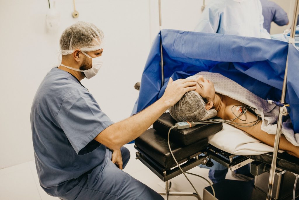
Photo by Jonathan Borba on Unsplash

A new UCLA-led study published in American Journal of Obstetrics & Gynecology may change the way clinicians and scientists understand, diagnose and treat placenta accreta spectrum disorder, a serious condition in which the placenta fails to separate from the uterus at birth. Researchers previously believed that certain overly invasive placental cells, called trophoblasts, were responsible for keeping the connection intact.

But this new research, which identifies genetic and cellular changes within single cells where the placenta and uterus join, shifts the focus to how the structural support of tissues, and the blood vessels of the uterus, can cause a “loss of normal boundary limits” between the placenta and the uterus.

“We utilized two new techniques in single-cell analysis to create an atlas of cells involved in placenta accreta to better understand this increasingly prevalent disorder that can have devastating implications for maternal and neonatal health,” said Dr Yalda Afshar, a maternal-foetal medicine specialist and researcher at the David Geffen School of Medicine at UCLA, and the first and corresponding author.

“This work revealed a subset of genes differentially expressed in placenta accreta spectrum disorder, which provides the basis for the ‘permissive environment’ for the placenta to attach to the uterine lining,” said Dr Deborah Krakow, a maternal-foetal medicine specialist and researcher, chair of the Department of Obstetrics and Gynecology at the David Geffen School of Medicine at UCLA, and the paper’s senior author.

The research showed that the decidua, the layer of the uterine lining that forms during pregnancy, and blood vessels, are sending different signals to the placenta when a pregnant person has placenta accreta.

In placenta accreta, the placenta is stuck on too tight, which becomes the reason for many of the maternal complications of placenta accreta.

“Our goal was to characterize the intimate relationship between the maternal and fetal tissue at the site of accreta or malfunction,” Afshar said.

“The genes and signaling pathways we identified go beyond providing a better understanding of the mechanism of the disease; they may be used as targets to help us refine diagnostic tests, track disease progression over time, and discover new, more effective therapies.”

The incidence of placenta accreta spectrum (PAS) disorders has increased dramatically in recent decades, the cause of which is not certain, though cesarean deliveries, is one of several risk factors.

Today, incidence is estimated at 1 in 272 births in the U.S., up from 1 in about 30 000 pregnancies in the 1960s, researchers say.

For this study, the research team performed multiple placental biopsies on 12 placentas, six with PAS disorder and six controls, conducting single-cell RNA analysis on 31 406 individual cells.

The researchers also applied spatial transcriptomics to 36 regions of interest: 12 in PAS-adherent, 12 in PAS-nonadherent, and 12 in controls.

Spatial transcriptomics allow researchers to precisely measure and map the gene activity within a single tissue sample.

“At the end of the day, understanding the biology of pregnancy and pregnancy-related diseases, like accreta, is inspired by only one thing – finding ways to improve the care we can provide to pregnant people and their families,” said Afshar, a physician-scientist who manages the care of many patients with placenta accreta spectrum disorders at UCLA Health.

Source: University of California – Los Angeles Health Sciences

Switching to Vegan or Keto Diets Impacts Immune System

Photo by Pixabay: https://www.pexels.com/photo/broccoli-161514/

Researchers at the National Institutes of Health observed rapid and distinct immune system changes in a small study of people who switched to a vegan or a ketogenic (“keto”) diet. They found that the vegan diet prompted responses linked to innate immunity while the keto diet prompted responses associated with adaptive immunity. Metabolic changes and shifts in the participants’ microbiomes were also observed. More research is needed to determine if these changes are beneficial or detrimental and what effect they could have on nutritional interventions for diseases such as cancer or inflammatory conditions.

Scientific understanding of how different diets impact the human immune system and microbiome is limited. Therapeutic nutritional interventions, which involve changing the diet to improve health, are not well understood, and few studies have directly compared the effects of more than one diet. The keto diet is a low-carbohydrate diet that is generally high in fat. The vegan diet eliminates animal products and tends to be high in fibre and low in fat.

The study was conducted by researchers from the NIH’s National Institute of Allergy and Infectious Diseases (NIAID) and National Institute of Diabetes and Digestive and Kidney Diseases (NIDDK) at the Metabolic Clinical Research Unit in the NIH Clinical Center.

The 20 participants were diverse with respect to ethnicity, race, gender, body mass index (BMI), and age. Participants sequentially ate vegan and keto diets for two weeks, in random order. Each person ate as much as desired of one diet (vegan or keto) for two weeks, followed by as much as desired of the other diet for two weeks. People on the vegan diet, which contained about 10% fat and 75% carbohydrates, chose to consume fewer calories than those on the keto diet, which contained about 76% fat and 10% carbohydrates. Throughout the study period, blood, urine, and stool were collected for analysis.

The effects of the diets were examined using a “multi-omics” approach that analysed multiple data sets to assess the body’s biochemical, cellular, metabolic, and immune responses, as well as changes to the microbiome.

Participants remained on site for the entire month-long study, allowing for careful control of the dietary interventions. Switching exclusively to the study diets caused notable changes in all participants.

The vegan diet significantly impacted pathways linked to the innate immune system, including antiviral responses. On the other hand, the keto diet led to significant increases in biochemical and cellular processes linked to adaptive immunity, such as pathways associated with T and B cells.

The keto diet affected levels of more proteins in the blood plasma than the vegan diet, as well as proteins from a wider range of tissues, such as the blood, brain and bone marrow. The vegan diet promoted more red blood cell-linked pathways, including those involved in heme metabolism, which could be due to the higher iron content of this diet.

Additionally, both diets produced changes in the microbiomes of the participants, causing shifts in the abundance of gut bacterial species that previously had been linked to the diets.

The keto diet was associated with changes in amino acid metabolism – an increase in human metabolic pathways for the production and degradation of amino acids and a reduction in microbial pathways for these processes – which might reflect the higher amounts of protein consumed by people on this diet.

The distinct metabolic and immune system changes caused by the two diets were observed despite the diversity of the participants, which shows that dietary changes consistently affect widespread and interconnected pathways in the body. More study is needed to examine how these nutritional interventions affect specific components of the immune system. According to the authors, the results of this study demonstrate that the immune system responds surprisingly rapidly to nutritional interventions. The authors suggest that it may be possible to tailor diets to prevent disease or complement disease treatments, such as by slowing processes associated with cancer or neurodegenerative disorders.

Source: NIH/National Institute of Allergy and Infectious Diseases

Gut Microbiome Composition Affects Sensitivity to Respiratory Viruses

Gut Microbiome. Credit Darryl Leja National Human Genome Research Institute National Institutes Of Health

The composition of microbiota found in the gut influences how susceptible mice are to respiratory virus infections and the severity of these infections, according to Georgia State University researchers. The findings, published in the journal Cell Host & Microbe, report that segmented filamentous bacteria, a bacterial species found in the intestines, protected mice against influenza virus infection when these bacteria were either naturally acquired or administered.

This protection against infection also applied to respiratory syncytial virus (RSV) and severe acute SARS-CoV-2. To maintain this protection, the study noted that segmented filamentous bacteria required immune cells in the lungs called basally resident alveolar macrophages.

In this study, the researchers investigated how differences in specific microbial species can impact outcomes of respiratory virus infections and how they might do so, which hasn’t been well defined previously.

They studied mice with discrete microbiome differences and mice differing in only the presence or absence of segmented filamentous bacteria.

Viral titers in the lung were measured several days after infection and varied significantly depending on the nature of the microbiome of the different animal groups.

“These findings uncover complex interactions that mechanistically link the intestinal microbiota with the functionality of basally resident alveolar macrophages and severity of respiratory virus infection,” said Dr. Andrew Gewirtz, co-senior author of the study and Regents’ Professor in the Institute for Biomedical Sciences at Georgia State.

The study found that in segmented filamentous bacteria-negative mice, basally resident alveolar macrophages were quickly depleted as respiratory virus infection progressed.

However, in segmented filamentous bacteria-colonised mice, basally resident alveolar macrophages were altered to resist influenza virus infection depletion and inflammatory signaling.

The basally resident alveolar macrophages disabled influenza virus, in large part by activating a component of the immune system referred to as the complement system.

“We find it remarkable that the presence of a single common commensal bacterial species, amidst the thousands of different microbial species that inhabit the mouse gut, had such strong impacts in respiratory virus infection models and that such impacts were largely attributable to reprogramming of basally resident alveolar macrophages,” said D. Richard Plemper, co-senior author of the study, Regents’ Professor and director of the Center for Translational Antiviral Research at Georgia State.

“If applicable to human infections, these findings will have major implications for the future risk assessment of a patient to advance to severe disease.”

“We find it highly unlikely that segmented filamentous bacteria is the only gut microbe capable of impacting the phenotype of alveolar macrophages, and consequently, proneness to respiratory virus infection,” Gewirtz said.

“Rather, we hypothesize that gut microbiota composition broadly influences proneness to respiratory virus infection. Microbiota mediated programming of basally resident alveolar macrophages may not only influence the severity of acute respiratory virus infection, but may also be a long-term post-respiratory virus infection health determinant.”

Source: Georgia State University

Vegan Diet in Pregnancy may Increase Preeclampsia and Low Birth Weight Risks

Photo by Anna Hecker on Unsplash

Women who follow vegan diets during pregnancy may face higher risks of developing preeclampsia and of giving birth to newborns with lower birth weight, suggests a recent study published in Acta Obstetricia et Gynecologica Scandinavica.

For the study, 65 872 women identified themselves as omnivorous, 666 as fish/poultry vegetarians, 183 as lacto/ovo vegetarians, and 18 as vegans. Based on a questionnaire completed mid-pregnancy, investigators found that protein intake was lower among lacto/ovo vegetarians (13.3%) and vegans (10.4%) compared with omnivorous participants (15.4%). Micronutrient intake was also much lower among vegans, but when dietary supplements were considered, no major differences were observed.

Compared with omnivorous mothers, vegan mothers had a higher prevalence of preeclampsia (a pregnancy complication characterised by high blood pressure), and their newborns weighed an average of 240 g less.

“Further research is needed regarding possible causality between plant-based diets and pregnancy and birth outcomes, to strengthen the basis for dietary recommendations,” the authors wrote.

Source: Wiley

Destroying Tumour Cells with Calcium

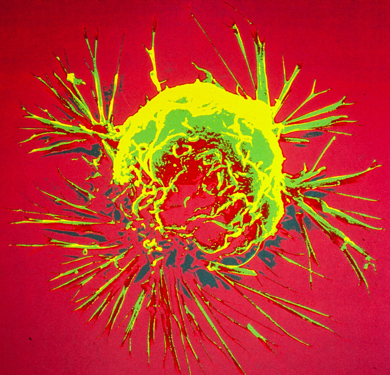
Colourised scanning electron micrograph of a breast cancer cell. Credit: NIH

Calcium ions are essential for cells, but can be toxic in higher concentrations. A team of researchers has now designed and prepared a combination drug that kills tumour cells by modulating the calcium influx into the cell. An external calcium source is not necessary because only the calcium ions already present in the tumour tissue are used, according to the study published in the journal Angewandte Chemie.

Biological cells need calcium ions, among other things, for the proper functioning of the mitochondria, the powerhouses of the cells.

However, if there is too much calcium, the mitochondrial processes become unbalanced and the cell suffocates.

A research group led by Juyoung Yoon of Ewha Womens University in Seoul, South Korea, together with teams from China, has now taken advantage of this process and developed a synergistic antitumour drug that can open calcium channels and thus trigger a deadly calcium storm inside the tumour cell.

The researchers targeted two channels, the first one in the outer membrane, and the other was a calcium channel in the endoplasmic reticulum, a cell organelle that also stores calcium ions.

The channel located in the outer membrane opens when it is exposed to a large amount of reactive oxygen species (ROS), while the channel in the endoplasmic reticulum is activated by nitric oxide molecules.

To generate the ROS that open the outer membrane calcium channel, the researchers used the dye indocyanine green.

This bioactive agent can be activated by irradiation with near-infrared light, which not only triggers reactions that lead to ROS, but it also heats up the environment.

The team explains that the high local temperature activates the other active agent, BNN-6, to release nitric oxide molecules that open the channel in the endoplasmic reticulum.

Following successful trials in tumour cell lines, the team tested an injectable formulation in tumour-implanted mice.

To create a biocompatible combined drug, the researchers loaded the active ingredients into tiny modified porous silica beads that are not harmful to the body, but can be recognized by tumour cells and transported into the cell.

After injecting the beads into the bloodstream of the mice, the researchers observed that the drug accumulated in the tumour.

Exposure to near-infrared light successfully triggered the mechanism of action, and the tumour disappeared after a few days in mice that received the preparation.

The authors emphasise that this ion influx approach may also be useful in related biomedical research areas where a similar mechanism could activate ion channels different from calcium in order to find new therapeutic approaches.

Source: Wiley

Healthcare Workers among Those Who Conceal their Infectious Illness

Photo by Brittany Colette on Unsplash

A startling number of people – including healthcare workers – conceal an infectious illness to avoid missing work, travel, or social events, new research at the University of Michigan suggests. The findings however, reported in Psychological Science, exclude being ill with COVID.

Across a series of studies involving healthy and sick adults, 75% of the 4110 participants said they had either hidden an infectious illness from others at least once or might do so in the future.

Many participants reported boarding planes, going on dates, and engaging in other social interactions while secretly sick.

More than 61% of healthcare workers participating in the study also said they had concealed an infectious illness.

Interestingly, the researchers found a difference between how people believe they would act when ill and how they actually behave, said Wilson N. Merrell, a doctoral candidate and lead author on the study.

“Healthy people forecasted that they would be unlikely to hide harmful illnesses – those that spread easily and have severe symptoms – but actively sick people reported high levels of concealment regardless of how harmful their illness was to others,” Merrell said.

In the first study, Merrell and his colleagues, psychology professor Joshua M. Ackerman and PhD student Soyeon Choi, recruited 399 university healthcare employees and 505 students.

The participants reported the number of days they felt symptoms of an infectious illness, starting in March 2020, when the COVID pandemic began.

They then rated how often they actively covered up symptoms from others, came to campus or work without telling others they were feeling ill, or falsified mandatory symptom screeners that the university had required for anyone using campus facilities.

More than 70% of the participants reported covering up their symptoms.

Many said they hid their illness because it would conflict with social plans, while a small percentage of participants cited pressure from institutional policies (eg, lack of paid time off). Only five participants reported hiding a COVID infection.

In a second study, the researchers recruited 946 participants online and randomly assigned them to one of nine conditions in which they imagined being either moderately or severely sick while in a social situation.

In each condition, the risk of spreading the illness was designated as low, medium, or high.

(To control for the special stigma associated with COVID at the time, the researchers asked participants not to imagine being sick with that disease.) Participants were most likely to envision themselves hiding their sickness when symptom severity was low, and least likely to conceal when symptoms were severe and highly communicable.

Source: Association for Psychological Science

Hydroxyurea for Children with Sickle Cell Anaemia Significantly Reduces Infections

Sickle cell disease. Credit: National Institutes of Health

A clinical trial in Uganda has revealed that hydroxyurea significantly reduces infections in children with sickle cell anaemia. Their latest findings enhance strong evidence of hydroxyurea’s effectiveness and could ultimately reduce death in children in Africa, the continent most burdened by the disease.

The group’s research, appearing in the journal Blood, revealed that hydroxyurea treatment resulted in a remarkable 60% reduction in severe or invasive infections, including malaria, bacteraemia, respiratory tract infections and gastroenteritis, among Ugandan children with sickle cell anaemia.

“Our investigation provides powerful justifications for hydroxyurea’s use in children with sickle cell anaemia in Africa,” said Dr Chandy John, paediatrics professor at IU School of Medicine and co-lead investigator of the latest study.

“Given the high rates of infection in this region, we hope our evidence will encourage ministries of health to continue supporting and expanding access to hydroxyurea for young patients who can greatly benefit from the treatment.”

Sickle cell anaemia is a genetic blood disorder that alters the structure of red blood cells and affects oxygen distribution throughout the body, increasing susceptibility to serious health complications and life-threatening infections.

According to the World Health Organization, more than 300 000 children worldwide are born with sickle cell disease each year, with a high prevalence found in African countries.

While hydroxyurea has had U.S. Food and Drug Administration approval as a sickle cell disease treatment for children since 2017, its accessibility and acceptance in Africa have been comparatively limited.

As hydroxyurea has become more recognised in African countries for its effectiveness in treating sickle-cell-related complications, John and his colleagues noticed a knowledge gap about the treatment’s effect on infections.

This led the research group to incorporate hydroxyurea treatment and analysis into their established clinical trial, Zinc for Infection Prevention in Sickle Cell Anemia, led by Indiana University School of Medicine and collaborators in Uganda.

During the study, the researchers examined the effects of hydroxyurea on 117 children in Uganda and focused on a range of infections. After hydroxyurea treatment, results showed a substantial decrease in the incidence of these infections.

Additionally, eight of the nine deaths that occurred in the trial were children whose parents declined hydroxyurea treatment. The only death in a child on hydroxyurea treatment occurred four days after starting treatment, providing insufficient time for hydroxyurea to have an effect.

Of the five children for whom a cause of death was known, all five died of infectious causes.

The high death rate in the study, despite expert clinical care by study personnel, provides further evidence of the urgent need for additional interventions to decrease mortality in children with sickle cell disease in Africa.

“Infections commonly precede other complications related to sickle cell anaemia and often result in hospitalizations that can lead to death,” said Dr Ruth Namazzi, site principal investigator, first author and a lecturer in the Department of Pediatrics and Child Health at Makerere University in Uganda.

“We believe incorporating hydroxyurea treatment as the standard of care for sickle cell anaemia across Africa will not only reduce infections but will more importantly save countless lives.”

Source: Indiana University

Alzheimer’s Disease Cases Caused by Growth Hormone Treatment

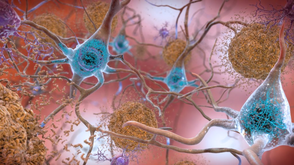
Neurons in the brain of an Alzheimer’s patient, with plaques caused by tau proteins. Credit: NIH

Five cases of Alzheimer’s are believed to have arisen as a result of medical treatments decades earlier, according to a new paper published in Nature Medicine. Alzheimer’s disease is caused by the amyloid-beta protein, and is usually a sporadic condition of late adult life, or more rarely as an inherited condition from a faulty gene.

The study, by a team of UCL and UCLH researchers, provides the first evidence of Alzheimer’s disease in living people that appears to have been medically acquired and due to transmission of the amyloid-beta protein.

The people described in the paper had all been treated as children with a type of human growth hormone extracted from pituitary glands from deceased individuals (cadaver-derived human growth hormone or c-hGH). This was used to treat at least 1848 people in the UK between 1959 and 1985, and used for various causes of short stature.

It was withdrawn in 1985 after it was recognised that some c-hGH batches were contaminated with prions (infectious proteins) which had caused Creutzfeldt-Jakob disease (CJD) in some people.

c-hGH was then replaced with synthetic growth hormone that did not carry the risk of transmitting CJD.

These researchers previously reported that some patients with CJD due to c-hGH treatment (called iatrogenic CJD) also had prematurely developed deposits of the amyloid-beta protein in their brains.* The scientists went on to show in a 2018 paper that archived samples of c-hGH were contaminated with amyloid-beta protein and, despite having been stored for decades, transmitted amyloid-beta pathology to laboratory mice when it was injected.

They suggested that individuals exposed to contaminated c-hGH, who did not succumb to CJD and lived longer, might eventually develop Alzheimer’s disease.

This latest paper reports on eight people referred to UCLH’s National Prion Clinic at the National Hospital for Neurology and Neurosurgery in London, who had all been treated with c-hGH in childhood, often over several years.

Five of these people had symptoms of dementia, and either had already been diagnosed with Alzheimer’s disease or would otherwise meet the diagnostic criteria for this condition; another person met criteria for mild cognitive impairment. These people were between 38 and 55 years old when neurological symptoms started. Biomarker analyses supported the diagnoses of Alzheimer’s disease in two patients with the diagnosis, and was suggestive of Alzheimer’s in one other person; an autopsy analysis showed Alzheimer’s pathology in another patient.

The unusually young age at which these patients developed symptoms suggests they did not have the usual sporadic Alzheimer’s which is associated with old age. In the five patients in whom samples were available for genetic testing, the team ruled out inherited Alzheimer’s disease.

As c-hGH treatment is no longer used, there is no risk of any new transmission via this route. There have been no reported cases of Alzheimer’s acquired from any other medical or surgical procedures. There is no suggestion that amyloid-beta can be passed on in day-to-day life or during routine medical or social care.

However, the researchers caution that their findings highlight the importance of reviewing measures to ensure there is no risk of accidental transmission of amyloid-beta via other medical or surgical procedures which have been implicated in accidental transmission of CJD.

The lead author of the research, Professor John Collinge, Director of the UCL Institute of Prion Diseases and a consultant neurologist at UCLH, said: “There is no suggestion whatsoever that Alzheimer’s disease can be transmitted between individuals during activities of daily life or routine medical care. The patients we have described were given a specific and long-discontinued medical treatment which involved injecting patients with material now known to have been contaminated with disease-related proteins.

“However, the recognition of transmission of amyloid-beta pathology in these rare situations should lead us to review measures to prevent accidental transmission via other medical or surgical procedures, in order to prevent such cases occurring in future.

“Importantly, our findings also suggest that Alzheimer’s and some other neurological conditions share similar disease processes to CJD, and this may have important implications for understanding and treating Alzheimer’s disease in the future.”

Source: University College London

Expert Warns that Red Light Therapy for Myopia could Damage the Retina

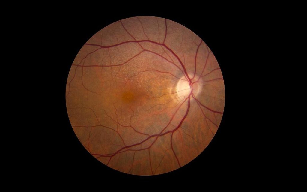
Retina showing reticular pseudodrusen. Although they can infrequently appear in individuals with no other apparent pathology, their highest rates of occurrence are in association with age-related macular degeneration (AMD), for which they hold clinical significance by being highly correlated with end-stage disease sub-types, choroidal neovascularisation and geographic atrophy. Credit: National Eye Institute

A University of Houston optometry researcher is warning against the use of low-level red light (LLRL) therapy as a method to control myopia, or nearsightedness, especially in children. Over the last few years, LLRL has emerged as a viable myopia treatment after studies reported the treatment as effective and responsible for significant reduction in myopia progression. The company behind one of the devices reports that it is already being used to address myopia in over 100 000 paediatric patients.

But the excitement over its results as a myopia treatment may have come too soon, ahead of its proven safety.

“Based on measurements in our laboratory, it is recommended that clinicians strongly reconsider the use of LLRL therapy for myopia in children until safety standards can be confirmed,” reports Lisa Ostrin, associate professor at the UH College of Optometry in The College of Optometrists journal.

Ostrin reports the therapy can put the retina at risk of photochemical and thermal damage.

“The safety profiles of red-light laser devices for myopia have not been fully investigated,” she said.

For LLRL therapy, children are instructed to look into a red light-emitting instrument for three minutes, twice a day, five days a week, for the duration of the treatment period, which could last years.

“We found that the red-light instruments for myopia exceed safety limits,” said Ostrin, whose research characterises the laser output and determines the thermal and photochemical maximum permissible exposure (MPE) of LLRL devices.

“For both LLRL devices evaluated here, three minutes of continuous viewing approached or surpassed the luminance dose MPE, putting the retina at risk of photochemical damage.”

Ostrin examined two different LLRL devices, and while both instruments were confirmed to be Class-1 laser products, as defined by International Electrotechnical Commission standards, according to Ostrin they are unsafe to view continuously for the required treatment duration of three minutes.

Class-1 lasers are low-powered devices that are considered safe from all potential hazards when viewed accidentally and briefly.

Examples of Class-1 lasers are laser printers, CD players and digital video disc (DVD) devices.

Class-1 lasers are not meant to be viewed directly for extended periods.

“Thermal ocular injury from a laser can occur with exposures at any wavelength when the temperature change of the retina is greater than 10°C, resulting in the denaturation of proteins. With thermal damage, the lesion size is typically less than the size of the beam diameter, and the resultant scotomas are permanent.” said Ostrin.

Source: University of Houston