Month: October 2023

Microplastics are a Danger to our Health. Here’s How to Reduce Our Exposure to Them

Photo by FLY:D on Unsplash

By Neil Thomas Stacey for GroundUp

About ten billion tonnes of plastic have been produced to date, of which around six billion tonnes have been discarded as waste. This is a severe threat to the environment, particularly oceans and lakes.

When plastics break down into particles smaller than five millimetres we call them microplastics. They are especially worrying.

Microplastics are an emerging threat to human health. They have been detected in organs in the human body and circulating in our bloodstreams. Studies have shown microplastics may deform red blood cells, inhibiting their ability to transport and transfer oxygen.

A study on mice exposed to microplastics found them in every tissue examined, and showed behavioural changes and heightened inflammation. While the exact effects on human health are not yet known, the risk is high enough that we should be very cautious about allowing them to pervade our atmosphere and food supply.

Microplastics have even been detected in high amounts in clouds, where they may affect rainfall patterns. They can also enter our food supply through rainfall.

A recent study of sediments in the Vaal river found an alarmingly high abundance of microplastics, which may enter the local food supply through crop irrigation. The sampling in this study was done in the region of the Vaal River Barrage, which is downstream of the Vaal Dam and fed by rivers that pass through heavily populated areas including Johannesburg.

Sampling at the Barrage gives direct insight into the rates at which we are producing microplastics in major population centres. And sampling at the Vaal Dam, which is the major drinking water supply for Gauteng, provides insight into the extent to which our drinking water is affected. Both these sampling points are needed as we track the levels of microplastics. Those levels are likely to rise dramatically; the microplastics we are seeing currently are only the tip of the iceberg, as there is a lag between the production of plastic, and it breaking down into microplastics.

Microplastic proliferation is not tied directly to accumulation of waste plastic. Examination of microplastics to ascertain their source is not an exact science, but it is reported that the main sources of microplastic pollution, at least for now, are car tyres and textiles and the pollution arises, not at the end-of-life when these are discarded as waste, but during their day-to-day use.

In other words, even if we solve the problem of waste plastic, we would still face the problem of microplastics that are emitted during the normal lifespan of products made of plastic.

There are, fortunately, some concrete steps that people can take to reduce personal exposure to microplastics. While microplastics are clearly able to travel throughout the atmosphere, their levels are concentrated around the sources releasing them. Microplastic concentrations are higher in indoor than outdoor air; old-fashioned fresh air and good ventilation are beneficial. So too is regularly wiping down surfaces, as they accumulate microplastic dust. Household air filters may also reduce microplastic concentrations.

Perhaps the most useful thing we as individuals can do is to have a different relationship with clothing. Synthetic fabrics are a prolific source of microplastics. These are released in our immediate surroundings, making our exposure to them disproportionately high.

Most microplastic release from textiles occurs within the first few washes after purchase, so purchasing long-lasting clothing rather than frequently replacing items of clothing can reduce your exposure, as can choosing natural fabrics such as cotton, where possible.

The other major source of microplastics is car tyres, which shed microplastics constantly as they wear down.

There are also activities which may seem environmentally-friendly but probably exacerbate microplastics pollution.

It is increasingly common to convert waste plastic into useable products from shoes and clothing to integration of waste plastic into road surfaces.

At first glance, this appears to be an environmental win-win. But recycled products tend to be more susceptible to the abrasion that causes microplastic release. Moreover, waste and recycled plastics tend to wear out more quickly and require replacement more frequently.

This is perhaps most harmful in the case of clothing made of waste or recycled plastic; the release of microplastics in early washes will be more severe because of the weaker polymer. This is particularly worth highlighting because recent research has shown that tumble-drying of synthetic textiles results in prolific microplastic release, much of which may be discharged into the indoor environment and breathed in or otherwise consumed.

Currently we have no practical way to remove microplastics from the environment; the particles are simply too small and widely dispersed. This means that we must exercise extreme caution to minimise emissions and our personal exposure to them.

Republished from GroundUp under a Creative Commons Attribution-NoDerivatives 4.0 International License.

Source: GroundUp

Hold the GLP-1 Agonists Before Surgery, New Advice Says

Photo by Natanael Melchor on Unsplash

Patients taking Glucagon-like peptide-1 (GLP-1) receptor agonists should stop taking them before they have surgery, due to the risk of aspirating while under general anaesthesia. This is the latest advice from the American Society of Anesthesiologists (ASA).

Initially approved by the Food and Drug Administration (FDA) for type 2 diabetes mellitus and cardiovascular risk reduction, GLP-1 agonists have shot up in popularity due to their effectiveness in weight loss. Despite having recent FDA approval, they have been used off-label for this purpose for quite some time.

When it comes to surgery, a number of organisations have recommended to hold these drugs either the day before or day of the procedure. For patients on weekly dosing, it is recommended to hold the dose for a week, the ASA notes.

GLP-1 agonists are associated with adverse gastrointestinal effects such as nausea, vomiting and delayed gastric emptying. The effects on gastric emptying are reported to be reduced with long-term use, most likely through rapid tachyphylaxis at the level of vagal nerve activation. Based on recent anecdotal reports, there are concerns that delayed gastric emptying from GLP-1 agonists can increase the risk of regurgitation and pulmonary aspiration of gastric contents during general anaesthesia and deep sedation. Patient taking GLP-1 agonists are more likely to have increased residual gastric contents as predicted by adverse gastrointestinal symptoms (nausea, vomiting, dyspepsia, abdominal distension).

The use of GLP-1 agonists in paediatrics has primarily been reported for the management of type 2 diabetes mellitus and obesity. The published literature on GLP-1 agonists in paediatrics is predominantly from paediatric patients 10 to 18 years old and concerns are similar to those reported in adults. During the conduct of general anaesthesia/deep sedation, children on GLP-1 agonists have similar gastrointestinal adverse events at a rate similar to adults.

In a review of the literature, the ASA Task Force on Preoperative Fasting found that, beyond a few case reports, there was little evidence for guidance on preoperative management of GLP-1 agonists. Nevertheless, they made recommendations for elective procedures. In the case of urgent or emergent procedures, they suggested treating the patient as ‘full stomach’.

If the patient’s GLP-1 agonists prescribed for diabetes management are held for longer than the dosing schedule, the guidelines urge surgeons to consider consulting an endocrinologist for bridging the antidiabetic therapy in order to avoid hyperglycaemia.

They further recommend that if gastrointestinal symptoms, such as severe nausea/vomiting/retching, abdominal bloating, or abdominal pain, are present, surgeons should consider delaying elective procedures. If the patient has no gastrointestinal symptoms and the GLP-1 agonists have been held as advised, the surgical team can carry on as normal.

Source: American Society of Anesthesiologists

Study Classifies Four Eating Eating Behaviours of Children

Photo by cottonbro studio

Children fall broadly into four eating categories, according to new research at Aston University, and parents feed their children differently depending on those categories.

The four categories identified by Dr Abigail Pickard and the team in the School of Psychology are ‘avid’, ‘happy’, ‘typical’, and ‘fussy’. The results, which showed specific temperaments and carer feeding patterns associated with overeating, are published in the journal Appetite.

In the UK, around a fifth of children are overweight or obese when they begin school, rising to around a third by the time they leave primary school at age 11. The team sought to identify eating behaviour patterns and how these are associated with temperament, feeding practices and food insecurity, as a way to predict which children are more at risk of becoming overweight.

Typical eaters made up 44% of the children in the study, while fussy eaters accounted for 16%. But of greatest interest to the team was that around one in five young children in the study were found to show “avid eating,” including greater enjoyment of food, faster eating speed, and weaker sensitivity to internal cues of ‘fullness’. The behaviours that distinguish children with avid eating from those who show ‘happy’ eating (17.7% of children in the study), who have similarly positive responses to food, are wanting to eat (or eating more) in response to the sight, smell or taste of palatable food, and a higher level of emotional overeating. In combination, these eating behaviours can lead to overeating and subsequent weight gain.

Dr Pickard and the team have also shown that there are significant differences in children’s temperament and caregivers’ feeding practices between each of the four eating behaviour patterns. Children with avid eating are more likely to be active and impulsive, and their caregivers are more likely to give them food to regulate their emotions or to restrict food for health reasons. Children with avid eating were also less food secure than children who showed happy or typical eating behaviours.

Principal investigator of the project, Professor Jackie Blissett, said: “Whilst feeding practices are key intervention targets to change children’s eating behaviour and child weight outcomes, there has been little evaluation of how feeding practices interact with children’s food approach behaviours to predict eating behaviour.”

She explained that despite the knowledge of the influence of feeding practices on children’s weight, current public health advice is generic and does not reflect variability in children’s appetites. Parents and caregivers can be left feeling frustrated when trying to manage their child’s food intake. By defining the four eating behaviour profiles, this research project, which is funded by the Economic and Social Research Council and co-developed by Professor Claire Farrow, Dr Clare Llewellyn, Dr Moritz Herle, Professor Emma Haycraft and Dr Helen Croker will make it easier to identify the best feeding practices for each eating pattern and provide tailored, effective advice for parents.

Dr Pickard said: “Parents can use this research to help them understand what type of eating pattern their child presents. Then based on the child’s eating profile the parent can adapt their feeding strategies to the child. For example, children in the avid eating profile may benefit more from covert restriction of food, i.e., not bringing snacks into the home or not having foods on display, to reduce the temptation to eat foods in the absence of hunger. Whereas, if a child shows fussy eating behaviour it would be more beneficial for the child to have a balanced and varied selection of foods on show to promote trying foods without pressure to eat.”

The team has planned further research investigating avid eating behaviour and will invite the caregivers and their children into the specialist eating behaviour lab at Aston University to get a better picture of what avid and typical eating behaviours look like in a real-life setting. All the findings will be integrated and the researchers will work with parents to develop feasible and helpful feeding guidelines to reduce children’s intake of palatable snack foods.

Source: Aston University

Surgery-free Deep Brain Stimulation Could be New Treatment for Dementia

A new form of deep brain stimulation offers hope for an alternative treatment option for dementia, without the need for surgery.

Researchers at Imperial College London are leading the development of the technique, known as temporal interference (TI). This non-invasive method works by delivering electrical fields to the brain through electrodes placed on the patient’s scalp and head. Their initial findings, which are published in the journal Nature Neuroscience, could lead to an alternative treatment for brain diseases such as Alzheimer’s, and its associated memory loss.

Temporal interference

By targeting the overlapping electrical fields researchers were able to stimulate an area deep in the brain called the hippocampus, without affecting the surrounding areas – a procedure that until now required surgery to implant electrodes into the brain.

The approach has been successfully trialled with 20 healthy volunteers for the first time by a team at the UK Dementia Research Institute (UK DRI) at Imperial and the University of Surrey.

Their initial results show that when healthy adults perform a memory task whilst receiving TI stimulation it helped to improve memory function.

The team is now conducting a clinical trial in people with early-stage Alzheimer’s disease, where they hope TI could be used to improve symptoms of memory loss.

Dr Nir Grossman, from the Department of Brain Sciences at Imperial College London, who led the work said: “Until now, if we wanted to electrically stimulate structures deep inside the brain, we needed to surgically implant electrodes which of course carries risk for the patient, and can lead to complications.

“With our new technique we have shown for the first time, that it is possible to remotely stimulate specific regions deep within the human brain without the need for surgery. This opens up an entirely new avenue of treatment for brain diseases like Alzheimer’s which affect deep brain structures.”

Reaching deep brain regions

TI was first described by the team at Imperial College London in 2017 and shown to work in principle in mice.

This latest work, funded and carried out through the UK Dementia Research Institute, shows for the first time that TI is effective at stimulating regions deep within the human brain.

According to the researchers, this could have broad applications and will enable scientists to stimulate different deep brain regions to discover more about their functional roles, accelerating the discovery of new therapeutic targets.

Source: Imperial College London

Combining Diagnosis and Treatment into One to Treat Pancreatic Cancer

Pancreatic cancer cells. Credit: NIH

Pancreatic ductal adenocarcinoma (PDAC) is one of the deadliest cancers worldwide, with a 5-year survival rate of less than 10%. Many PDAC tumours go undetected in early stages since they go undetected by conventional imaging methods such as fluorodeoxyglucose positron emission tomography (PET) scans. To tackle this problem, researchers in Japan are combining diagnostic and therapeutic procedures into a single integrated process: ‘theranostics’.

In an article recently published in the Journal of Nuclear Medicine, the Osaka University-led team has developed a ‘radio-theranostics’ strategy that uses a new radioactive antibody to target glypican-1 (GPC1), a protein highly expressed in PDAC tumours. Theranostics, particularly radio-theranostics, has been receiving increasing attention because, by radio-labelling the compounds used to target certain molecules in cancer cells, diagnosis and treatment can be carried out sequentially.

“We decided to target GPC1 because it is overexpressed in PDAC but is only present in low levels in normal tissues,” explains Tadashi Watabe, lead author of the study.

The team used a monoclonal antibody (mAb) designed to target GPC1. The mAb could be labelled with isotopes of zirconium (89Zr) or astatine (211At). First, they injected the 89Zr-GPC1 mAb into a xenograft mouse model, which has a human pancreatic cancer tumour.

“We monitored 89Zr-GPC1 mAb internalisation over seven days with PET scanning,” explains Kazuya Kabayama, the second author of the article. “There was strong uptake of the mAb into the tumours, suggesting that this method could support tumour visualisation. We confirmed that this was mediated by its binding to GPC1, as the xenograft model that had GPC1 expression knocked out showed significantly less uptake.”

The researchers next tested this model with alpha therapy using 211At-GPC1 mAb, a method that could support radioactive label-based delivery of a therapeutic molecule to its target. Administration of 211At-GPC1 mAb resulted in DNA double-strand break induction in the cancer cells, as well as significantly reduced tumour growth. Control experiments showed that these antitumor effects did not occur when mAb internalisation was blocked. Additionally, non-radiolabelled GPC1 mAb did not induce these effects.

“Both radiolabeled versions of the GPC1 mAb we examined showed promising results in PDAC,” says Watabe. “89Zr-GPC1 mAb showed high humoral uptake, while 211At-GPC1 mAb could be used for targeted alpha therapy to support suppression of PDAC tumour growth.”

These highly impactful data demonstrate the potential for using a theranostics approach in PDAC, a disease in dire need of new diagnostic and therapeutic options. In the future, this could lead to early detection of PDAC with PET imaging and systemic treatment with alpha therapy.

Source: Osaka University

Timed Cortisol Delivery Improves Adrenal Condition Symptoms

Photo by Karolina Grabowska on Pexels

A trial of a hormone replacement therapy that more closely replicates the natural circadian and ultradian rhythms of hormones has shown to improve symptoms in patients with adrenal conditions. Results from the University of Bristol-led clinical trial are published in the Journal of Internal Medicine.

Low cortisol levels typically result from conditions such as Addison’s and Congenital Adrenal Hyperplasia. The hormone regulates a range of vital processes, from cognitive processes such as memory formation, metabolism and immune responses, through to blood pressure and blood sugar levels. When low, it can trigger symptoms of debilitating fatigue, nausea, muscle weakness, dangerously low blood pressure and depression. Although rare, these adrenal conditions require lifelong daily hydrocortisone replacement therapy.

Although existing oral hormone replacement treatment can restore cortisol levels, it is still associated with an impaired quality of life for patients. Scientists believe this is because the current treatment does not mimic the body’s normal physiological timing, missing cortisol’s anticipatory rise and lacking its underlying ultradian and circadian rhythms.

The new ‘Pulsatility’ therapy, the culmination of ten years research by the Bristol team, is designed to deliver standard hydrocortisone replacement to patients via a pump which replicates more closely cortisol’s natural rhythmic secretion pattern. The pulsatile subcutaneous pump has now revealed promising results in its first clinical trial.

The double-blinded PULSES six-week trial recruited 20 participants aged 18 to 64 years with adrenal insufficiency conditions. They treated with usual dose hydrocortisone replacement therapy administered either via the pump or the standard three times daily oral treatment.

While only psychological and metabolic symptoms were assessed during the trial, results revealed the pump therapy decreased patient fatigue by approximately 10%, improved mood and increased energy levels by 30% first thing in the morning – when many patients struggle the most. MRI scans also revealed alteration in the way that the brain processes emotional information.

Dr Georgina Russell, Honorary Lecturer at the University’s Bristol Medical School, and the lead author, explained: “Patients on cortisol replacement therapy often have side effects which makes it difficult for them to lead normal lives. We hope this new therapy will offer greater hope for the thousands of people living with hormone insufficiency conditions.”

Stafford Lightman, a neuroendocrinology expert and Professor of Medicine at Bristol Medical School: Translational Health Sciences (THS), and the study’s joint lead author, added: “Besides reduction in dosage, cortisol replacement has remained unchanged for many decades. It is widely recognised that current replacement therapy is unphysiological due to its lack of pre-awakening surge, ultradian rhythmicity, and post dose supraphysiological peaks. The new therapy clearly shows that the timing of cortisol delivery, in line with the body’s own rhythmic pattern of cortisol secretion, is important for normal cognition and behaviour.

Source:

Physicians and Nurse Practitioners have Similar Prescribing Error Rates

Photo by Towfiqu Barbhuiya on Unsplash

A study of more than 73 000 primary care physicians (PCPs) and nurse practitioners (NPs) in the US suggest that  both are similarly likely to inappropriately prescribe medications to older patients. According to the authors, this study adds to growing evidence indicating that when prescriptive authority is expanded to include NPs, these new prescribers do not perform worse than physicians. The study is published in Annals of Internal Medicine.

NPs, registered nurses with advanced degrees and more experience, are helping to solve the ongoing PCP shortage problem in the United States. Currently, state laws determine what type of medical care NPs can provide, such as practicing independently and prescribing medications without physician supervision.

Researchers from University of California, Los Angeles, Yale Law School, and Stanford University calculated inappropriate prescribing rates for 23 669 NPs and 50 060 PCPs prescribing medications to patients aged 65 years and older across 29 states in the US where NPs are authorised to prescribe. Inappropriate prescribing was defined using the American Geriatrics Society’s Beers Criteria. The researchers found that both PCPs and NPs averaged approximately 1.7 inappropriate prescriptions for every 100 prescriptions written. However, NPs were overrepresented among clinicians with the highest and the lowest rates of inappropriate prescribing. According to the authors, these findings provide useful lessons for policymakers, lawmakers, and regulators. Use of clinician-level performance measures, coupled with efforts to improve prescribing at the organisational and individual levels, could help to address deficient performance among all clinicians who prescribe. The authors say that technologic interventions, such as prescription drug monitoring, have a role to play, as do initiatives aimed at ensuring better adherence to trusted guidelines like the Beers Criteria.

An accompanying editorial from authors at University of California, Los Angeles suggests that rates of inappropriate prescribing are too high among clinicians of all stripes. The authors note that NPs are providing a greater proportion of care to older adults outside of large metropolitan areas, many of whom would likely have no other source of primary care. They also emphasise that NPs will continue to serve critical roles in ensuring that older adults in areas with inadequate numbers of health care providers receive primary care. The goal of clinicians should be to reduce variation and improve prescribing quality among all clinicians who care for older adults.

Source: EurekAlert!

Scientists Record Powerful Signals in the Brain’s White Matter

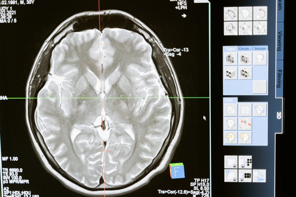
Scientists have concentrated on the grey matter of the cortex, composed of nerve cell bodies , while ignoring white matter, composed of axons, even though it makes up half the brain. Now, in the Proceedings of the National Academy of Sciences, Vanderbilt University researchers report strong signs of brain activity when performing certain tasks.

For several years, John Gore, PhD, director of the Vanderbilt University Institute of Imaging Science, and his colleagues have used functional magnetic resonance imaging (fMRI) to detect blood oxygenation-level dependent (BOLD) signals, a key marker of brain activity, in white matter.

In this latest paper, the researchers report that when people who are having their brains scanned by fMRI perform a task, like wiggling their fingers, BOLD signals increase in white matter throughout the brain.

“We don’t know what this means,” said the paper’s first author, Kurt Schilling, PhD, research assistant professor of Radiology and Radiological Sciences at VUMC. “We just know that something is happening. There truly is a powerful signal in the white matter.”

It is important to pursue this because disorders as diverse as epilepsy and multiple sclerosis disrupt the “connectivity” of the brain, Schilling said. This suggests that something is going on in white matter.

To find out, the researchers will continue to study changes in white matter signals they’ve previously detected in schizophrenia, Alzheimer’s disease and other brain disorders. Through animal studies and tissue analysis, they also hope to determine the biological basis for these changes.

In grey matter, BOLD signals reflect a rise in blood flow (and oxygen) in response to increased nerve cell activity.

Perhaps the axons, or the glial cells that maintain the protective myelin sheath around them, also use more oxygen when the brain is ‘working’. Or perhaps these signals are somehow related to what’s going on in the grey matter.

But even if nothing biological is going on in white matter, “there’s still something happening here,” Schilling said. “The signal is changing. It’s changing differently in different white matter pathways and it’s in all white matter pathways, which is a unique finding.”

One reason that white matter signals have been understudied is that they have lower energy than grey matter signals, and thus are more difficult to distinguish from the brain’s background “noise.”

The VUMC researchers boosted the signal-to-noise ratio by having the person whose brain was being scanned repeat a visual, verbal or motor task many times to establish a trend and by averaging the signal over many different white matter fibre pathways.

“For 25 or 30 years, we’ve neglected the other half of the brain,” Schilling said. Some researchers not only have ignored white matter signals but have removed them from their reports of brain function.

The Vanderbilt findings suggest that many fMRI studies thus “may not only underestimate the true extent of brain activation, but also … may miss crucial information from the MRI signal,” the researchers concluded.

Source: Vanderbilt University

A Hard-to-treat Subtype of Asthma in Older Men

Credit: Pixabay CC0

Scientists have uncovered a group of T cells that may drive severe asthma, which gather in the lungs and seem most harmful in men who develop asthma in later life. The new research, published in MED, suggests asthma patients with these cells in their lungs may be more likely to have hard-to-treat, and potentially fatal, asthma attacks. These cells do not respond to the usual general therapy for asthma patients.

The scientists, from the University of Southampton and La Jolla Institute for Immunology (LJI), in California, uncovered these T cells, called ‘cytotoxic CD4+ tissue-resident memory T cells’, thanks to volunteers enrolled in the NHS clinic-based WATCH study. It follows hundreds of asthma patients of different ages, sexes, and disease severities. By following patients over many years, and analysing their immune cell populations, researchers are making new connections between asthma symptoms and immune cell activity.

“If you are male and you develop asthma after age 40, there’s a high chance this T cell population is in your lungs,” says LJI Research Assistant Professor Gregory Seumois, who co-led the study with LJI Professor Pandurangan Vijayanand.

“Once you understand the role of cells like these T cells better, you can start to develop treatments that target those cells,” says WATCH study director Dr Ramesh Kurukulaaratchy, Associate Professor at the University of Southampton and researcher at the NIHR Southampton Biomedical Research Centre.

Scientists now hope to learn more about these cells and their role in asthma development in order to develop personalised therapies for asthma patients.

How harmful T cells drive asthma

The ‘memory’ T cells help protect the body from viruses and bacteria it has encountered before, but the same T cell memory is a big problem for asthma patients. Their misguided T cells see harmless molecules, such as pollen, and produce a dangerous inflammatory response.

Men who developed asthma later in life had an overwhelming number of these potentially harmful T cells. Their lungs should have been home to a diverse bunch of CD4+ T cell types but, in this group, more than 65% of their cells were cytotoxic CD4+ tissue-resident memory T cells.

Personalised asthma treatments

Single-cell RNA sequencing by LJI scientists provides a ‘biomarker’ to help detect cytotoxic CD4+ tissue-resident memory T cells in more patients going forward.

Finding this biomarker represents a “paradigm shift” in asthma research, says Dr Kurukulaaratchy. Before now, scientists and clinicians separated asthma patients into just two groups: ‘T2 high’ and T2 low’. In a study published earlier this year, the research team showed the importance of drilling down to identify many more asthma patient subgroups; their analysis reveals that 93% of WATCH subjects with severe asthma were in the T2 high category.

Study co-author Professor Hasan Arshad, Chair in Allergy and Clinical Immunology at the University of Southampton, researcher at the NIHR Southampton Biomedical Research Centre, and Director of The David Hide Asthma and Allergy Research Centre, Isle of Wight says: “We have to think of severe asthma as having different subtypes, and the treatment has to be tailored according to these subtypes because one size does not fit all.”

The researchers now want to use sequencing tools and other techniques to discover additional biomarkers and asthma patient subtypes.

Source: University of Southampton

Researchers Sum up Head and Neck Surgery Site Infection Risks and Treatment

Photo by cottonbro studio

In a new research perspective published in Oncoscience, researchers from Germany discuss the diagnosis and management of postoperative wound infections in the head and neck area. Key topics include patient risk factors, the importance of sterilisation, and the most common complications.

In everyday clinical practice at a department for oral and maxillofacial surgery, a large number of surgical procedures in the head and neck region take place under both outpatient and inpatient conditions. The basis of every surgical intervention is the patient’s consent to the respective procedure. Particular attention is drawn to the general and operation-specific risks. 

Particularly in the case of soft tissue procedures in the facial region, bleeding, secondary bleeding, scarring and infection of the surgical area are among the most common complications/risks, depending on the respective procedure. In their new perspective, researchers Filip Barbarewicz, Kai-Olaf Henkel and Florian Dudde from Army Hospital Hamburg in Germany discuss the diagnosis and management of postoperative infections in the head and neck region.

“In order to minimise the wound infections/surgical site infections, aseptic operating conditions with maximum sterility are required.”

Furthermore, depending on the extent of the surgical procedure and the patient‘s previous illnesses, peri- and/or postoperative antibiotics should be considered in order to avoid postoperative surgical site infection. Abscesses, cellulitis, phlegmone and (depending on the location of the procedure) empyema are among the most common postoperative infections in the respective surgical area. The main pathogens of these infections are staphylococci, although mixed (germ) patterns are also possible. 

“Risk factors for the development of a postoperative surgical site infection include, in particular, increased age, smoking, multiple comorbidities and/or systemic diseases (eg, diabetes mellitus type II) as well as congenital and/ or acquired immune deficiency.”