Day: October 24, 2023

Timed Cortisol Delivery Improves Adrenal Condition Symptoms

Photo by Karolina Grabowska on Pexels

A trial of a hormone replacement therapy that more closely replicates the natural circadian and ultradian rhythms of hormones has shown to improve symptoms in patients with adrenal conditions. Results from the University of Bristol-led clinical trial are published in the Journal of Internal Medicine.

Low cortisol levels typically result from conditions such as Addison’s and Congenital Adrenal Hyperplasia. The hormone regulates a range of vital processes, from cognitive processes such as memory formation, metabolism and immune responses, through to blood pressure and blood sugar levels. When low, it can trigger symptoms of debilitating fatigue, nausea, muscle weakness, dangerously low blood pressure and depression. Although rare, these adrenal conditions require lifelong daily hydrocortisone replacement therapy.

Although existing oral hormone replacement treatment can restore cortisol levels, it is still associated with an impaired quality of life for patients. Scientists believe this is because the current treatment does not mimic the body’s normal physiological timing, missing cortisol’s anticipatory rise and lacking its underlying ultradian and circadian rhythms.

The new ‘Pulsatility’ therapy, the culmination of ten years research by the Bristol team, is designed to deliver standard hydrocortisone replacement to patients via a pump which replicates more closely cortisol’s natural rhythmic secretion pattern. The pulsatile subcutaneous pump has now revealed promising results in its first clinical trial.

The double-blinded PULSES six-week trial recruited 20 participants aged 18 to 64 years with adrenal insufficiency conditions. They treated with usual dose hydrocortisone replacement therapy administered either via the pump or the standard three times daily oral treatment.

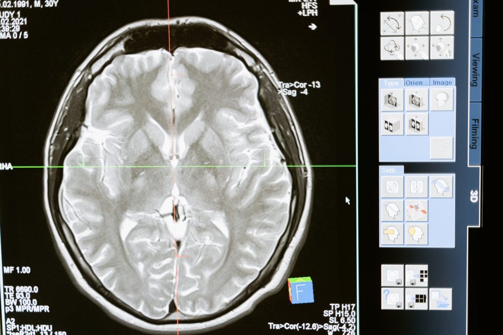
While only psychological and metabolic symptoms were assessed during the trial, results revealed the pump therapy decreased patient fatigue by approximately 10%, improved mood and increased energy levels by 30% first thing in the morning – when many patients struggle the most. MRI scans also revealed alteration in the way that the brain processes emotional information.

Dr Georgina Russell, Honorary Lecturer at the University’s Bristol Medical School, and the lead author, explained: “Patients on cortisol replacement therapy often have side effects which makes it difficult for them to lead normal lives. We hope this new therapy will offer greater hope for the thousands of people living with hormone insufficiency conditions.”

Stafford Lightman, a neuroendocrinology expert and Professor of Medicine at Bristol Medical School: Translational Health Sciences (THS), and the study’s joint lead author, added: “Besides reduction in dosage, cortisol replacement has remained unchanged for many decades. It is widely recognised that current replacement therapy is unphysiological due to its lack of pre-awakening surge, ultradian rhythmicity, and post dose supraphysiological peaks. The new therapy clearly shows that the timing of cortisol delivery, in line with the body’s own rhythmic pattern of cortisol secretion, is important for normal cognition and behaviour.

Source:

Physicians and Nurse Practitioners have Similar Prescribing Error Rates

Photo by Towfiqu Barbhuiya on Unsplash

A study of more than 73 000 primary care physicians (PCPs) and nurse practitioners (NPs) in the US suggest that  both are similarly likely to inappropriately prescribe medications to older patients. According to the authors, this study adds to growing evidence indicating that when prescriptive authority is expanded to include NPs, these new prescribers do not perform worse than physicians. The study is published in Annals of Internal Medicine.

NPs, registered nurses with advanced degrees and more experience, are helping to solve the ongoing PCP shortage problem in the United States. Currently, state laws determine what type of medical care NPs can provide, such as practicing independently and prescribing medications without physician supervision.

Researchers from University of California, Los Angeles, Yale Law School, and Stanford University calculated inappropriate prescribing rates for 23 669 NPs and 50 060 PCPs prescribing medications to patients aged 65 years and older across 29 states in the US where NPs are authorised to prescribe. Inappropriate prescribing was defined using the American Geriatrics Society’s Beers Criteria. The researchers found that both PCPs and NPs averaged approximately 1.7 inappropriate prescriptions for every 100 prescriptions written. However, NPs were overrepresented among clinicians with the highest and the lowest rates of inappropriate prescribing. According to the authors, these findings provide useful lessons for policymakers, lawmakers, and regulators. Use of clinician-level performance measures, coupled with efforts to improve prescribing at the organisational and individual levels, could help to address deficient performance among all clinicians who prescribe. The authors say that technologic interventions, such as prescription drug monitoring, have a role to play, as do initiatives aimed at ensuring better adherence to trusted guidelines like the Beers Criteria.

An accompanying editorial from authors at University of California, Los Angeles suggests that rates of inappropriate prescribing are too high among clinicians of all stripes. The authors note that NPs are providing a greater proportion of care to older adults outside of large metropolitan areas, many of whom would likely have no other source of primary care. They also emphasise that NPs will continue to serve critical roles in ensuring that older adults in areas with inadequate numbers of health care providers receive primary care. The goal of clinicians should be to reduce variation and improve prescribing quality among all clinicians who care for older adults.

Source: EurekAlert!

Scientists Record Powerful Signals in the Brain’s White Matter

Scientists have concentrated on the grey matter of the cortex, composed of nerve cell bodies , while ignoring white matter, composed of axons, even though it makes up half the brain. Now, in the Proceedings of the National Academy of Sciences, Vanderbilt University researchers report strong signs of brain activity when performing certain tasks.

For several years, John Gore, PhD, director of the Vanderbilt University Institute of Imaging Science, and his colleagues have used functional magnetic resonance imaging (fMRI) to detect blood oxygenation-level dependent (BOLD) signals, a key marker of brain activity, in white matter.

In this latest paper, the researchers report that when people who are having their brains scanned by fMRI perform a task, like wiggling their fingers, BOLD signals increase in white matter throughout the brain.

“We don’t know what this means,” said the paper’s first author, Kurt Schilling, PhD, research assistant professor of Radiology and Radiological Sciences at VUMC. “We just know that something is happening. There truly is a powerful signal in the white matter.”

It is important to pursue this because disorders as diverse as epilepsy and multiple sclerosis disrupt the “connectivity” of the brain, Schilling said. This suggests that something is going on in white matter.

To find out, the researchers will continue to study changes in white matter signals they’ve previously detected in schizophrenia, Alzheimer’s disease and other brain disorders. Through animal studies and tissue analysis, they also hope to determine the biological basis for these changes.

In grey matter, BOLD signals reflect a rise in blood flow (and oxygen) in response to increased nerve cell activity.

Perhaps the axons, or the glial cells that maintain the protective myelin sheath around them, also use more oxygen when the brain is ‘working’. Or perhaps these signals are somehow related to what’s going on in the grey matter.

But even if nothing biological is going on in white matter, “there’s still something happening here,” Schilling said. “The signal is changing. It’s changing differently in different white matter pathways and it’s in all white matter pathways, which is a unique finding.”

One reason that white matter signals have been understudied is that they have lower energy than grey matter signals, and thus are more difficult to distinguish from the brain’s background “noise.”

The VUMC researchers boosted the signal-to-noise ratio by having the person whose brain was being scanned repeat a visual, verbal or motor task many times to establish a trend and by averaging the signal over many different white matter fibre pathways.

“For 25 or 30 years, we’ve neglected the other half of the brain,” Schilling said. Some researchers not only have ignored white matter signals but have removed them from their reports of brain function.

The Vanderbilt findings suggest that many fMRI studies thus “may not only underestimate the true extent of brain activation, but also … may miss crucial information from the MRI signal,” the researchers concluded.

Source: Vanderbilt University

A Hard-to-treat Subtype of Asthma in Older Men

Credit: Pixabay CC0

Scientists have uncovered a group of T cells that may drive severe asthma, which gather in the lungs and seem most harmful in men who develop asthma in later life. The new research, published in MED, suggests asthma patients with these cells in their lungs may be more likely to have hard-to-treat, and potentially fatal, asthma attacks. These cells do not respond to the usual general therapy for asthma patients.

The scientists, from the University of Southampton and La Jolla Institute for Immunology (LJI), in California, uncovered these T cells, called ‘cytotoxic CD4+ tissue-resident memory T cells’, thanks to volunteers enrolled in the NHS clinic-based WATCH study. It follows hundreds of asthma patients of different ages, sexes, and disease severities. By following patients over many years, and analysing their immune cell populations, researchers are making new connections between asthma symptoms and immune cell activity.

“If you are male and you develop asthma after age 40, there’s a high chance this T cell population is in your lungs,” says LJI Research Assistant Professor Gregory Seumois, who co-led the study with LJI Professor Pandurangan Vijayanand.

“Once you understand the role of cells like these T cells better, you can start to develop treatments that target those cells,” says WATCH study director Dr Ramesh Kurukulaaratchy, Associate Professor at the University of Southampton and researcher at the NIHR Southampton Biomedical Research Centre.

Scientists now hope to learn more about these cells and their role in asthma development in order to develop personalised therapies for asthma patients.

How harmful T cells drive asthma

The ‘memory’ T cells help protect the body from viruses and bacteria it has encountered before, but the same T cell memory is a big problem for asthma patients. Their misguided T cells see harmless molecules, such as pollen, and produce a dangerous inflammatory response.

Men who developed asthma later in life had an overwhelming number of these potentially harmful T cells. Their lungs should have been home to a diverse bunch of CD4+ T cell types but, in this group, more than 65% of their cells were cytotoxic CD4+ tissue-resident memory T cells.

Personalised asthma treatments

Single-cell RNA sequencing by LJI scientists provides a ‘biomarker’ to help detect cytotoxic CD4+ tissue-resident memory T cells in more patients going forward.

Finding this biomarker represents a “paradigm shift” in asthma research, says Dr Kurukulaaratchy. Before now, scientists and clinicians separated asthma patients into just two groups: ‘T2 high’ and T2 low’. In a study published earlier this year, the research team showed the importance of drilling down to identify many more asthma patient subgroups; their analysis reveals that 93% of WATCH subjects with severe asthma were in the T2 high category.

Study co-author Professor Hasan Arshad, Chair in Allergy and Clinical Immunology at the University of Southampton, researcher at the NIHR Southampton Biomedical Research Centre, and Director of The David Hide Asthma and Allergy Research Centre, Isle of Wight says: “We have to think of severe asthma as having different subtypes, and the treatment has to be tailored according to these subtypes because one size does not fit all.”

The researchers now want to use sequencing tools and other techniques to discover additional biomarkers and asthma patient subtypes.

Source: University of Southampton

Researchers Sum up Head and Neck Surgery Site Infection Risks and Treatment

Photo by cottonbro studio

In a new research perspective published in Oncoscience, researchers from Germany discuss the diagnosis and management of postoperative wound infections in the head and neck area. Key topics include patient risk factors, the importance of sterilisation, and the most common complications.

In everyday clinical practice at a department for oral and maxillofacial surgery, a large number of surgical procedures in the head and neck region take place under both outpatient and inpatient conditions. The basis of every surgical intervention is the patient’s consent to the respective procedure. Particular attention is drawn to the general and operation-specific risks. 

Particularly in the case of soft tissue procedures in the facial region, bleeding, secondary bleeding, scarring and infection of the surgical area are among the most common complications/risks, depending on the respective procedure. In their new perspective, researchers Filip Barbarewicz, Kai-Olaf Henkel and Florian Dudde from Army Hospital Hamburg in Germany discuss the diagnosis and management of postoperative infections in the head and neck region.

“In order to minimise the wound infections/surgical site infections, aseptic operating conditions with maximum sterility are required.”

Furthermore, depending on the extent of the surgical procedure and the patient‘s previous illnesses, peri- and/or postoperative antibiotics should be considered in order to avoid postoperative surgical site infection. Abscesses, cellulitis, phlegmone and (depending on the location of the procedure) empyema are among the most common postoperative infections in the respective surgical area. The main pathogens of these infections are staphylococci, although mixed (germ) patterns are also possible. 

“Risk factors for the development of a postoperative surgical site infection include, in particular, increased age, smoking, multiple comorbidities and/or systemic diseases (eg, diabetes mellitus type II) as well as congenital and/ or acquired immune deficiency.”