Month: July 2023

Novel Metal Complex Treatment Kills Antibiotic-resistant Bacteria

Photo by CDC on Unsplash

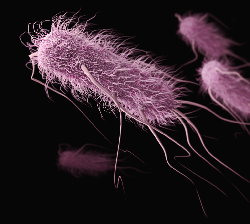
An innovative treatment paves the way for reducing antimicrobial resistance in the treatment of a deadly infection in chickens, according to a new study in Veterinary Microbiology. The ground-breaking study investigated the effectiveness of a novel metal-derived complex in treating Avian Pathogenic Escherichia coli (APEC), a serious respiratory infection of chickens which has become increasingly more resistant to antibiotics. A growing body of evidence indicates that the APEC could potentially spread to humans.

University of Surrey’s Professor Roberto La Ragione said: “Antimicrobial resistance is one of the biggest threats to human and animal health. Not being able to use antibiotics to treat an infection not only prolongs an illness and associated welfare issues, but also increases the likelihood of it spreading.

“Coronavirus demonstrated how easily a pandemic can happen, and the threat of another is looking more likely as antibiotics to treat simple bacterial infections are no longer working.”

To test the effectiveness of the metal complex, manganese carbonyl, researchers worked with the Greater Wax Moth larvae and APEC. Split into two groups, the first received manganese carbonyl, whilst the second, the controls, received either a phosphate-buffered saline (PBS) or dimethyl sulfoxide (DMSO). After four days, the survival rate for the larvae which received manganese carbonyl was between 56–75%, whereas in the control group, the survival rate was between 25–45% (PBS) and 19-45 per cent (DMSO), demonstrating the protective effect of the complex.

The test was repeated in chickens infected with APEC, who again received either manganese carbonyl or PBS. Bacterial shedding identified in the faeces of the chickens was significantly lower 24 hours post-treatment in those who received manganese carbonyl compared to the PBS control group, indicating bacterial killing induced by the compound. This is supported by caecal samples taken three days post-treatment which again found significantly fewer bacteria in those that received manganese carbonyl. Examination of tissue samples from the livers of the birds indicated no toxic effects from the metal compound, which was observed in the larvae.

Dr Jonathan Betts, a Research Fellow at the University of Surrey School of Veterinary Medicine, said:

“The development of alternatives to antibiotics is vital to safeguard our future health. Metal complexes such as manganese carbonyl could do this, as we have shown not only are they effective, but they are much cheaper to produce than traditional antibiotics.

“Discovering the effectiveness of manganese carbonyl in treating APEC is a monumental step forward in tackling antimicrobial resistance as it shows we don’t necessarily need more antibiotics; we just need to think more innovatively in developing treatments.”

The international research team also included the University of Surrey, the Animal and Plant Health Agency, the University of Connecticut, the University of Sheffield and Institut für Anorganische Chemie, Julius-Maximilians-Universität Würzburg.

This study was made possible by a BBSRC grant to Professor La Ragione and Professor Poole.

Source: University of Surrey

Black Adults Experience Less Cognitive Decline after Retirement

Photo by Barbara Olsen on Pexels

A study published in the Journal of the American Geriatrics Society found that immediately after retirement, white adults tended to experience a significant decline in cognitive function, whereas Black adults experienced minimal cognitive decline. White men showed the steepest post-retirement cognitive decline across sex/race combinations, whereas Black women showed the least decline.

White women performed better cognitively at retirement than other race/sex subgroups, and after retirement, their cognitive functioning declined at a rate that was slightly less than the average for this study. Results were adjusted for sociodemographics and physical and mental health indicators.

The study, which included 2226 US participants followed for up to 10 years, revealed greater post-retirement cognitive decline among individuals who attended college compared with those who did not.

“The results seem to point to the possibility that better job opportunities could lead to greater cognitive losses after retirement whereas exposure to lifelong structural inequalities may actually ease transition to retirement with respect to cognitive aging,” said lead author Ross Andel, PhD, of Arizona State University’s Edson College of Nursing and Health Innovation.

Source: Wiley

Microwaving Plastic Baby Food Container Releases Billions of Plastic Nanoparticles

Photo by Burst on Pexels

Experiments have shown that microwaving plastic baby food containers available on the shelves of US stores can release huge numbers of micrometre or smaller-sized plastic particles – in some cases, more than 2 billion nanoplastics and 4 million microplastics for every square centimetre of container.

Though the health effects of consuming micro- and nanoplastics remain unclear, the University of Nebraska-Lincoln researchers further found that three-quarters of cultured embryonic kidney cells had died after two days of being introduced to those same particles. A 2022 report from the World Health Organization recommended limiting exposure to such particles.

“It is really important to know how many micro- and nanoplastics we are taking in,” said Kazi Albab Hussain, the study’s lead author and a doctoral student in civil and environmental engineering at the University of Nebraska-Lincoln. “When we eat specific foods, we are generally informed or have an idea about their caloric content, sugar levels, other nutrients. I believe it’s equally important that we are aware of the number of plastic particles present in our food.

“Just as we understand the impact of calories and nutrients on our health, knowing the extent of plastic particle ingestion is crucial in understanding the potential harm they may cause. Many studies, including ours, are demonstrating that the toxicity of micro- and nanoplastics is highly linked to the level of exposure.”

The team embarked on its study in 2021, the same year that Hussain became a father. While prior research had investigated the release of plastic particles from baby bottles, the team realised that no studies had examined the sorts of plastic containers and pouches that Hussain found himself shopping for, and that millions of other parents regularly do, too.

Hussain and his colleagues decided to conduct experiments with two baby food containers made from polypropylene and a reusable pouch made of polyethylene, both FDA-approved plastics. In one experiment, the researchers filled the containers with either deionised water or 3% acetic acid (the latter intended to simulate dairy products, fruits, vegetables and other relatively acidic consumables) then heated them at full power for three minutes in a 1000-watt microwave. Afterward, they analysed the liquids for evidence of micro- and nanoplastics: the micro- being particles at least a micrometre in diameter, the nano- any particles smaller.

The actual number of each particle released by the microwaving depended on multiple factors, including the plastic container and the liquid within it. But based on a model that factored in particle release, body weight, and per-capita ingestion of various food and drink, the team estimated that infants drinking products with microwaved water and toddlers consuming microwaved dairy products are taking in the greatest relative concentrations of plastic. Experiments designed to simulate the refrigeration and room-temperature storage of food or drink over a six-month span also suggested that both could lead to the release of micro- and nanoplastics.

“For my baby, I was unable to completely avoid the use of plastic,” Hussain said. “But I was able to avoid those (scenarios) which were causing more of the release of micro- and nanoplastics. People also deserve to know those, and they should choose wisely.”

With the help of Svetlana Romanova from the University of Nebraska Medical Center, the team then cultured and exposed embryonic kidney cells to the actual plastic particles released from the containers – a first, as far as Hussain can tell. Rather than introduce just the number of particles released by one container, the researchers instead exposed the cells to particle concentrations that infants and toddlers might accumulate over days or from multiple sources.

After two days, just 23% of kidney cells exposed to the highest concentrations had managed to survive – a much higher mortality rate than that observed in earlier studies of micro- and nanoplastic toxicity. The team suspects that kidney cells might be more susceptible to the particles than are other cell types examined in prior research. But those earlier studies also tended to examine the effects of larger polypropylene particles, some of them potentially too large to penetrate cells. If so, the Hussain-led study could prove especially sobering: Regardless of its experimental conditions, the Husker team found that polypropylene containers and polyethylene pouches generally release about 1000 times more nanoplastics than microplastics.

The question of cell infiltration is just one among many that will require answers, Hussain said, before determining the true risks of consuming micro- and nanoplastics. But to the extent that they do pose a health threat – and that plastics remain a go-to for baby food storage – parents would have a vested interest in seeing that the companies manufacturing plastic containers seek out viable alternatives, he said.

“We need to find the polymers which release fewer (particles),” Hussain said. “Probably, researchers will be able to develop plastics that do not release any micro- or nanoplastics – or, if they do, the release would be negligible.

“I am hopeful that a day will come when these products display labels that read ‘microplastics-free’ or ‘nanoplastics-free.'”

Source: University of Nebraska-Lincoln

Going Viral: Dr Chivaugn Gordon on Medical School with a Difference

Dr Chivaugn Gordon, head of undergraduate education at UCT’s Department of Obstetrics and Gynaecology, reflects on her love of teaching future doctors about women’s health issues. PHOTO: Nasief Manie/Spotlight

By Biénne Huisman for Spotlight

With humour and wearing an occasional wig, Dr Chivaugn Gordon teaches medical students about serious women’s health issues. During hard lockdown she delighted students at the University of Cape Town (UCT) with educational videos using household items as props. For example, she created an endometrium (the inner lining of the uterus) from hair gel and red glitter, performed a biopsy on a potato, and showed a chicken hand puppet go into labour.

One video features a patient named Zoya Lockdownikoff – who is a spy – consulting with her doctor about abnormal menstrual bleeding. Gordon, in a blonde wig with round sunglasses, plays Lockdownikoff; and Gordon’s husband, Dr Adalbert Ernst, plays her doctor.

Lockdownikoff explains that the bleeding started when she “did a very complicated backflip to escape a very compromising situation” and that it’s ruining her expensive super-spy coats.

Gordon is head of undergraduate education at UCT’s Department of Obstetrics and Gynaecology, while Ernst is with the university’s Department of Anaesthesia and Perioperative Medicine.

Speaking from her yellow-walled lounge in Cape Town’s Bergvliet, Gordon says: “I became a doctor because I love working with patients. And then I realised, oh cool, I love teaching too. And now I can do these two things together.”

Interest in IPV

For Gordon a driving interest has been intimate partner violence (IPV) which she introduced into her undergraduate curriculum in 2015.

“The aim is to have graduating doctors who are able to recognise intimate partner violence. Everybody thinks that you can’t possibly be abused unless you have a black eye or a fractured arm. But actually, IPV is often more psychological. It’s often psychological abuse. So the challenge is to teach young doctors what are the red flags in someone’s behaviour, or in their clinical presentation, that might indicate IPV.”

Published online in April, Gordon delivered a talk for TEDxUCT called “Tackling IPV, one awkward dad conversation at a time”, in which she notes IPV is “a global pandemic that has been ongoing since time began”. The title refers to Gordon’s father who raised her.

According to a paper published in the journal Lancet Psychiatry last year, IPV is the most common form of violence worldwide; it is most prevalent in unequal societies, and its victims are mostly women and girls. The paper states that worldwide 27% of women and girls aged 15 and older have experienced physical or sexual IPV, but in South Africa the figure is estimated to be much higher, between 33 and 50%.

Gordon contributed to South Africa’s revised Domestic Violence Amendment Act of 2021, through UCT’s Gender Health and Justice Research Unit.

The new legislation broadens the definition of domestic violence to include (above and beyond physical and sexual abuse) emotional, verbal or psychological abuse, which is described as “a pattern of degrading, manipulating, threatening, offensive, intimidating or humiliating conduct towards a complainant that causes mental or psychological harm…including (repeated) insults, ridicule or name calling; (repeated) threats to cause emotional pain; the (repeated) exhibition of obsessive possessiveness or jealousy…”

Gordon highlights the term coercive control. “Because that underpins most serious intimate partner violence. So, somebody who is extremely controlling; they want their partner to do what they want, when they want, and how they want immediately. They normally start isolating you from friends and family so they can spin a narrative of your reality that can’t be contested by anyone else. And it also makes it more difficult to leave.”

Red flags

Gordon highlights some of the IPV red flags that doctors should look for in their patients.

“Depression, anxiety, PTSD, insomnia, [and] things like self-medicating with substances,” she says. “Because when you are living in absolute, abject terror every day of your life, it’s going to manifest in some kind of psychological manner. So, when people have been broken down and worn down and their self-esteem has been eroded it also affects the way they might interact with the healthcare professional.

“Big red flags come out in body language. Usually when someone goes to a doctor, they tell you everything about all their symptoms, because they want you to make them better. So, if you’ve got a patient who is closed off, they’re not making eye contact, they’re avoiding answering your questions, they’re just very reticent and you can’t get anything out of them…then you’ve got to think.”

Gordon stresses that IPV happens across economic strata and in all walks of life. “Every time I run this workshop, a medical student who comes from a very privileged background, from a very financially stable, loving home, comes to me, saying this is happening to her. It happens everywhere. I’ve got medical colleagues, several, who have experienced intimate partner violence. It doesn’t discriminate.”

Republished from Spotlight under a Creative Commons Licence.

Source: Spotlight

Single Injection of New Anthypertensive Drug Could Reduce BP for Six Months

Pexels Photo by Thirdman

A new drug which interferes with the production of angiotensin could help lower persistent hypertension for up to six months following a single injection, a study suggests. The results were published in the New England Journal of Medicine. If successful, the drug, called zilebesiran, would be more convenient for hypertension management than daily oral antihypertensive drugs.

More than half of patients with hypertension fail to take all of their prescribed medicine, leading to inconsistent blood pressure control. Better management of the condition could reduce the risk of stroke, heart attack and premature death, experts say.

Sustained drop

The international team, including experts from the University of Edinburgh’s Centre for Cardiovascular Science, ran the early stage clinical trials across four UK sites.

Patients who received zilebesiran experienced a substantial reduction in systolic blood pressure which lasted up to six months.

On average, systolic blood pressure lowered by over 10mmHg at a 200mg dose or more of the drug, and more than 20mmHg at the highest dose of 800mg. A drop of this size can take someone with hypertension to within a much safer range.

Blood pressure naturally goes up and down throughout the course of the day, making it difficult to treat – but the study found that the drop in blood pressure seen in patients who were treated with zilebesiran was consistent over 24 hours.

Novel approach

Developed by US-based company Alnylam, zilebesiran works by preventing the production of angiotensin, a hormone in the body that narrows blood vessels, leading to raised blood pressure. A number of existing antihypertensives also target angiotensin.

Zilebesiran uses a novel approach to interfere with the machinery in the liver that makes the protein angiotensinogen, the source of all forms of angiotensin.

Known as small interfering RNA (siRNA), zilebesiran turns off the gene responsible for producing angiotensinogen, preventing it from being made. 

The siRNA approach has already been used to develop treatments for a number of other conditions, with the ability to silence specific genes with high accuracy and effects lasting many months.

Safety data

107 patients with hypertension took part in the trial – 80 received a single injection of zilebesiran under the skin, while 32 received a placebo containing no active ingredients. Five patients who initially received the placebo were later moved to zilebesiran.

Experts caution that further studies involving a larger number of patients are needed to robustly investigate the safety of the drug and provide further insights into its potential to improve clinical outcomes in people with hypertension.

This is a potentially major development in hypertension. There has not been a new class of drug licensed for the treatment of high blood pressure in the last 17 years. This novel approach leads to a substantial reduction in blood pressure, both by day and night, that lasts for around six months after a single injection. This is attractive because it helps avoid the difficulty with adherence to treatment seen with current medicines. The next stage of clinical trials will focus on developing robust safety data, and broader evidence of efficacy, before zilebesiran can be licensed for use.

Professor David WebbChristison Chair of Therapeutics and Clinical Pharmacology at the University of Edinburgh, who led the Edinburgh study site

Source: University of Edinburgh

T Cell Monitoring may Help Prevent Type 1 Diabetes

A 3D map of the islet density routes throughout the healthy human pancreas. Source: Wikimedia CC0

Scripps Research scientists have shown that people at risk of developing type 1 diabetes could be identified by analysis of the T cells which drive the disease. The new approach, if validated in further studies, could be used to select suitable patients for a newly FDA-approved treatment that stops the autoimmune process, thereby making type 1 diabetes a preventable condition.

In the study, which appears in Science Translational Medicine, the researchers isolated T cells from mouse and human blood samples. By analysing the T cells that can cause type 1 diabetes, they were able to distinguish the at-risk patients who had active autoimmunity from those who had no significant autoimmunity – with 100% accuracy in a small sample.

“These findings represent a big step forward because they offer the possibility of catching this autoimmune process while there is still time to prevent or greatly delay diabetes,” says study senior author Luc Teyton, MD, PhD, professor in the Department of Immunology and Microbiology at Scripps Research.

The study’s first authors were graduate student Siddhartha Sharma and research assistants Josh Boyer and Xuqian Tan, all of the Teyton lab at the time of the study.

Type 1 diabetes usually occurs in childhood or early adulthood, in an autoimmune process that destroys the pancreas’s insulin-producing islet cells. The process can last years, with multiple starts and stops. Exactly how the process begins is not well understood, though it is known to involve genetic factors and may be triggered by routine viral infections.

In 2022, the US Food & Drug Administration approved an immune-suppressing therapy that can protect islet cells and at least delay diabetes onset by months to years if given in the early stages of autoimmunity. However, doctors have not had a good method for identifying people who could benefit from such treatment. They have traditionally examined levels of anti-islet antibodies in patient blood samples, but this antibody response has not been a very accurate measure of autoimmune progression.

“Anti-islet antibody levels are poorly predictive at the individual level, and type 1 diabetes is fundamentally a T cell-driven disease,” Teyton says.

In the study, Teyton and his team constructed protein complexes to mimic the mix of immune proteins and insulin fragments that CD4 T cells normally would recognise to initiate the autoimmune reaction. They used these constructs as bait to capture anti-insulin CD4 T cells in blood samples. They then analysed the gene activity within the captured T cells, and expression of proteins on the cells, to gauge their state of activation.

In this way, they were able to develop a classification algorithm that correctly identified which at-risk patients, in a set of nine, had ongoing anti-islet autoimmunity.

Teyton now hopes to validate the CD4 T cell-based approach with a long-term study in a larger cohort of participants, comparing this approach to the traditional approach of quantifying anti-islet antibodies.

Teyton and his colleagues also are working to make the process of isolating and analysing anti-islet T cells in blood samples more affordable and convenient, so that it can be used more easily in a clinical setting.

“If we can develop this into a useful method for identifying at-risk patients and tracking their autoimmunity status, we not only would have a way of getting the right people into treatment, but also would be able to monitor their disease progress and evaluate potential new preventive therapies,” Teyton says.

Source: Scripps Research Institute

Review Links Private Equity Takeovers of Healthcare Services to Patient Harm

Photo by Hush Naidoo Jade Photography on Unsplash

Private equity ownership of healthcare services such as nursing homes and hospitals is associated with harmful impacts on costs and quality of care, suggests a review of the latest evidence published by The BMJ. No consistently beneficial impacts of private equity ownership were identified, and the researchers say these results confirm the need for more research on private equity ownership in healthcare and possibly increased regulation.

Private equity firms use capital from wealthy individuals and large institutional investors to buy companies, and, after a relatively brief period of ownership, sell them for substantial returns. Over the past decade, private equity firms have increasingly invested in, acquired, and consolidated healthcare facilities, with global healthcare buyouts exceeding $200bn since 2021 alone. But despite much speculation, it’s still not clear what impact private equity ownership of healthcare operators has on costs, quality of care, and health outcomes.

To address this uncertainty, researchers analysed the results of 55 studies (47 focused exclusively on the US) published in peer reviewed journals in the past two decades.

Nursing homes were the most commonly studied settings, followed by hospitals and dermatology facilities. The studies were designed differently, and were of varying quality, but the researchers were able to allow for that in their analysis.

Nine of 12 studies showed higher costs to patients or payers at health facilities owned by private equity firms (harmful impact), three found no differences, and none showed lower costs (beneficial impact).

Private equity ownership was also associated with mixed to harmful impacts on quality. Of 27 studies that assessed healthcare quality, 12 found harmful impacts, three found beneficial impacts, nine found mixed impacts (some quality measures declined, some improved), and in three the results were neutral.

Health outcomes showed both beneficial and harmful results, as did costs to operators, but the volume of studies for these outcomes was too low for any definitive conclusions to be drawn.

When nursing homes were analysed separately, private equity ownership often had mixed impacts on quality, but the researchers point out that more evidence suggests a degradation rather than an improvement in quality, such as a decrease in nurse staffing or a shift to lower nursing skill mix.

The researchers acknowledge that they did not differentiate between different types of private equity investment and ownership, and were unable to assess larger possible impacts of private equity on access to care. And because most of the included studies occurred in the US, the impacts identified may not apply to all global settings.

Nevertheless, they say this study fills a gap in the current literature on private equity ownership in healthcare, and presents emergent patterns related to private equity ownership that other studies have been unable to synthesise.

As such, they say: “The results of this study confirm the need for increased rigorous research on private equity ownership in healthcare, particularly its impacts on health outcomes and system costs and in other non-US settings, such as Europe.”

“This said, the current body of evidence is robust enough to confirm that private equity ownership is a consequential and increasingly prominent element in healthcare, warranting surveillance, reporting, and possibly increased regulation.”

Source: EurekAlert!

Scientists Unravel The Reason Why NSAIDs Exacerbate C. Diff Infections

Clostridioides difficile. Credit: CDC

Nonsteroidal anti-inflammatory drugs (NSAIDs) exacerbate gastrointestinal infections by Clostridioides difficile, the leading cause of antibiotic-associated diarrhoea worldwide – but the reason why has long eluded medical science. In a new paper published in Science Advances, researchers have begun to answer that question, showing that NSAIDs disrupt the mitochondria of cells lining the colon, sensitising them to damage by pathogenic toxins.

C. difficile is a bacterium that leads to a wide range of symptoms, from mild diarrhoea to complex infection and death. The factors that influence this wide spectrum of clinical outcomes remain largely unclear, but emerging evidence suggests that factors like diet and pharmaceutical drugs influence both susceptibility to infection and disease progression. However, little is known about how these factors impact the course of C. difficile infection.

Prior studies have shown that NSAIDs like indomethacin, aspirin, and naproxen negatively affect the gut, both in patients with C. difficile infection and other conditions like inflammatory bowel disease (IBD). Long-term NSAID use can lead to stomach ulcers and intestinal injuries. One hypothesis that this is due to the effects of NSAIDs on cyclooxygenase (COX) enzymes; a process that helps reduce inflammation and pain but also impairs mucosal function in the upper gastrointestinal tract. NSAIDs also have off-target effects and have been shown to affect cellular mitochondria by uncoupling cellular mitochondrial functions, but these had not been studied in C. difficile infections.

To define these effects, Children’s Hospital of Philadelphia (CHOP) researchers, led by graduate student Joshua Soto Ocaña, used in vitro and mouse models of C. difficile infection to test how permeable colonic epithelial cells are in the presence of the NSAID indomethacin. The researchers observed that both indomethacin and C. difficile toxins increased epithelial cell barrier permeability and inflammatory cell death. They also found that the effect was additive: the combined effect on cell permeability of both toxins and indomethacin was increased compared to each independently, suggesting a synergistic effect of NSAIDs and C. difficile in increasing this pathogen’s virulence.

Surprisingly, the researchers found that NSAIDs exacerbate C. difficile infection independent of COX inhibition and instead through off-target effects on mitochondria. They did so by treating colonic epithelial cells with a precursor molecule that is similar in structure to indomethacin but lacks the ability to inhibit the COX enzyme. Not only did they find that this NSAID-like molecule induced cell death, but they also found that adding selective COX inhibitors did not increase cell death, demonstrating that COX enzyme inhibition is not required to induce epithelial cell damage during C. difficile infection and that, instead, this damage occurs through off-target effects of NSAIDs.

To test the role of off-target effects during C. difficile infection, the researchers used mice pretreated either with indomethacin or the NSAID-precursor molecule. When exposed to C. difficile, both groups of mice showed equal enhancement in disease severity and mortality compared to untreated control mice infected with C. difficile only. The researchers also observed a similar effect in mice who were pretreated with the NSAID aspirin. To further define the specific mechanisms driving these off-target effects of NSAIDs, researchers looked at mitochondrial functions in colonic epithelial cells in vitro and in mice. They observed that the combination of NSAIDs and C. difficile toxins increased damage to colonic epithelial cell mitochondria and disrupted several important mitochondrial functions.

“Our work further demonstrates the clinical importance of NSAIDs in patients with C. diff infection and sheds light on why the combination of these two may be so detrimental,” said senior author Joseph P. Zackular, PhD, Investigator and Assistant Professor of Pathology and Laboratory Medicine at CHOP. “Our mechanistic findings are a starting point for further research that aims to understand the impact of mitochondrial functions during C. diff infection. These data could also inform how NSAID-mediated mitochondrial uncoupling affects other diseases, such as small intestinal injury, IBD, and colorectal cancer.”

Source: Children’s Hospital of Philadelphia

Scientists Discover That a Key Protein Boosts Cell Repair and Healthy Ageing

Photo by Ravi Patel on Unsplash

Researchers have found an anti-ageing function in a protein deep within human cells. They discovered that a protein called ATSF-1 controls a fine balance between the creation of new mitochondria and the repair of damaged mitochondria. Their findings were published in Nature Cell Biology.

Mitochondria create toxic by-products during their energy production process, which contributes to the rate at which the cell ages.

Associate Professor Steven Zuryn and Dr Michael Dai at the Queensland Brain Institute made the discovery of a key repair protein. “In conditions of stress, when mitochondrial DNA has been damaged, the ATSF-1 protein prioritises repair which promotes cellular health and longevity,” Dr Zuryn said.

As an analogy, Dr Zuryn likened the relationship to a race car needing a pitstop.

“ATSF-1 makes the call that a pitstop is needed for the cell when mitochondria need repairs,” he said.

“We studied ATFS-1 in C. elegans, or round worms and saw that enhancing its function promoted cellular health, meaning the worms became more agile for longer. They didn’t live longer, but they were healthier as they aged.”

“Mitochondrial dysfunction lies at the core of many human diseases, including common age-related diseases such as dementias and Parkinson’s. Our finding could have exciting implications for healthy ageing and for people with inherited mitochondrial diseases.”

Understanding how cells promote repair is an important step towards identifying possible interventions to prevent mitochondrial damage.

“Our goal is to prolong the tissue and organ functions that typically decline during ageing by understanding how deteriorating mitochondria contribute to this process,” Dr Dai said. “We may ultimately design interventions that keep mitochondrial DNA healthier for longer, improving our quality of life.”

Source: University of Queensland

Green Tea Extract may Reduce Uterine Fibroids

Photo by Andrea Piacquadio on Pexels

In a pre-clinical, proof-of-concept study from Johns Hopkins Medicine, researchers found that epigallocatechin gallate (EGCG), a green tea compound with powerful antioxidant properties, could be promising for both treating and preventing uterine fibroids. Results of the study, appearing in Scientific Reports, add to growing evidence that EGCG may reduce fibroid cell growth, though the study is still at an early stage. The study was specifically designed to identify the biochemical mechanisms responsible for EGCG action in fibroid cells.

The investigators emphasise that their study involves in vitro human fibroid cells treated with EGCG extract to explore the possibility of oral EGCG supplementation as a therapy, rather than just drinking cups of green tea as a preventative measure for uterine fibroids.

“The purpose of this study was to examine how EGCG works to treat and prevent uterine fibroids,” says James Segars Jr., MD, professor of gynaecology and obstetrics. “There is no standard protocol for uterine fibroid disease management or prevention, no tools to prevent their growth, so finding a safe nonsurgical therapy is important.”

Uterine fibroids are the most common benign tumours of the uterus. Made up of smooth muscle cells and a large matrix of connective tissue, the fibroids range in size from nearly microscopic to bulky masses that can enlarge and distort the uterus.

An estimated 77% of women will develop fibroids in their lifetime, most of them by age 50. Black and Hispanic women develop them at 1.5 to two times the rate of white women.

While many people with uterine fibroids are symptom-free, about 25% experience significant symptoms including heavy uterine bleeding, pelvic pain and infertility. In addition to complete removal of the uterus, surgical treatment may include various means of removing fibroid tumours from the uterine wall.

For the new study, researchers used laboratory cultures of uterine fibroids collected from living patients. Because uterine fibroid cells have a large extracellular matrix compared to normal cells, researchers designed their experiments to see if treatment of cells with EGCG affects protein expression associated with this matrix. Specifically, they studied fibronectin, a matrix protein; cyclin D1, a protein involved with cell division; and connective tissue growth factor (CTGF) protein.

Cells were dosed with 100mmol/L of EGCG in growth media for 24 hours, and then a Western blot was performed. In this study, researchers looked for levels of cyclin D1 and CTGF proteins in EGCG-treated fibroid cells compared to untreated cell.

They found that EGCG reduced protein levels of fibronectin by 46% to 52%, compared with an untreated controls. They also found that EGCG disrupted pathways involved in fibroid tumour cell growth, movement, signalling and metabolism, and they saw up to an 86% decrease in CTGF proteins compared with the control group.

“The results from this study show that EGCG targets many signalling pathways involved in fibroid growth, particularly the extracellular matrix,” says study lead author Md Soriful Islam, PhD, MSc. “EGCG supplements could be an easily accessible and natural way to relieve symptoms and slow fibroid growth.”

These results lend support to the FRIEND study, an ongoing clinical trial of EGCG in women with fibroids who are seeking pregnancy. While results from this study show promise, researchers caution that more studies need to be done, and consumers should not try to self-dose with green tea supplements. Future research on EGCG will include clinical trials with large and diverse patient groups to determine optimal doses as well as possible side effects of EGCG supplementation.

Source: John Hopkins Medicine