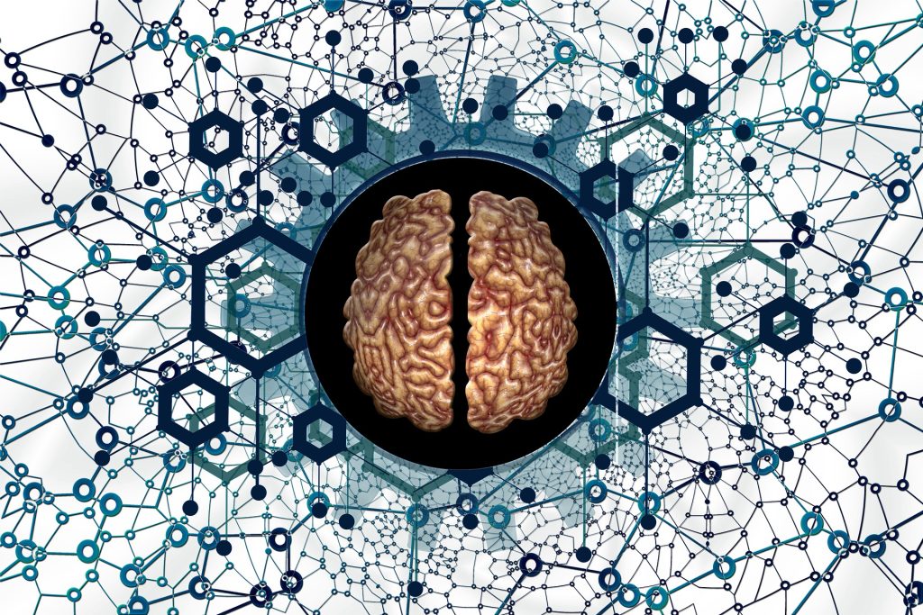
Elon Musk’s company Neuralink had finally received approval for human testing of its brain-computer interface (BCI). After initially rejecting the application, the US Food and Drug Administration finally gave the company the go-ahead on Thursday.
Neuralink, which aims to develop an implant that would allow humans to interface directly with computers as well as enabling medical applications such as controlling prostheses. Last year, the company showed off a monkey that was able to play the simple video game Pong on a monitor using its mind.
Neuralink is by no means the first company to try to achieve these goals. Many other institutions have made advances over the past decades, but the field is a difficult one and progress is slow. In its previous rejection, the FDA cited concerns such as the devices using lithium for their batteries, migration of the wires inside the brain and the difficulty of extracting the devices without harming brain tissue.
The company’s use of animals to develop the technology has infuriated activists, but this is a standard practice in development of BCI technology. Last year, whistleblowers accused the company of killing 1500 animals since its inception.
In a guidance document, the FDA says that, “The field of implanted BCI devices is progressing rapidly from fundamental neuroscience discoveries to translational applications and market access. Implanted BCI devices have the potential to bring benefit to people with severe disabilities by increasing their ability to interact with their environment, and consequently, providing new independence in daily life.”
China is also aggressively pursuing the development of BCIs as part of their ‘China Brain Project’, as discussed in the journal Neuron. It has a significant advantage as it has a large population of macaques to draw on, along with fewer ethical concerns and policies expediting biotech research.
Neuroscientists published in the Journal of Neurochemistry, shows that maternal levels of vitamin D are key in the development of dopaminergic neurons, which are thought to be involved in schizophrenia.
Professor Darryl Eyles has built on past research out of his laboratory at the Queensland Brain Institute linking maternal vitamin D deficiency and brain development disorders, such as schizophrenia, to understand the functional changes taking place in the brain.
Schizophrenia is associated with many developmental risk factors, both genetic and environmental. While the precise neurological causes of the disorder are unknown, what is known is that schizophrenia is associated with a pronounced change in the way the brain uses dopamine, the neurotransmitter often referred to as the brain’s ‘reward molecule’.
Professor Eyles has followed the mechanisms that might relate to abnormal dopamine release and discovered that maternal vitamin D deficiency affects the early development and later differentiation of dopaminergic neurons.
The team at the Queensland Brain Institute developed dopamine-like cells to replicate the process of differentiation into early dopaminergic neurons that usually takes place during embryonic development.
They cultured the neurons both in the presence and absence of the active vitamin D hormone. In three different model systems they showed dopamine neurite outgrowth was markedly increased. They then showed alterations in the distribution of presynaptic proteins responsible for dopamine release within these neurites.
“What we found was the altered differentiation process in the presence of vitamin D not only makes the cells grow differently, but recruits machinery to release dopamine differently,” Professor Eyles said.
Using a new visualisation tool known as false fluorescent neurotransmitters, the team could then analyse the functional changes in presynaptic dopamine uptake and release in the presence and absence of vitamin D.
They showed that dopamine release was enhanced in cells grown in the presence of the hormone compared to a control.
“This is conclusive evidence that vitamin D affects the structural differentiation of dopaminergic neurons.”
Leveraging advances in targeting and visualising single molecules within presynaptic nerve terminals has enabled Professor Eyles and his team to further explore their long-standing belief that maternal vitamin D deficiency changes how early dopaminergic circuits are formed.
The team is now exploring whether other environmental risk factors for schizophrenia such as maternal hypoxia or infection similarly alter the trajectory of dopamine neuron differentiation.
Eyles and his team believe such early alterations to dopamine neuron differentiation and function may be the neurodevelopmental origin of dopamine dysfunction later in adults who develop schizophrenia.
Regular physical activity and exercise may reduce bleeding in individuals with intracerebral haemorrhage, a University of Gothenburg study shows. The study, published in the journal Stroke and Vascular Neurology, analysed data on 686 people treated for intracerebral haemorrhage at Sahlgrenska University Hospital in Gothenburg during the years 2014 to 2019.
The study was a retrospective analysis and could not determine causation. Nonetheless, it was clear that those who reported regular physical activity had smaller haemorrhages than those who reported being inactive.
Physically active was defined as engaging in at least light physical activity, such as walking, cycling, swimming, gardening, or dancing, for at least four hours weekly.
50 percent less bleeding volume
The main author of the study is Adam Viktorisson, a PhD student in clinical neuroscience at Sahlgrenska Academy, University of Gothenburg, and doctor in general practice at Sahlgrenska University Hospital.
“We found that individuals who engage in regular physical activity had, on average, bleeding volumes that were 50 percent smaller upon arriving to the hospital. A similar connection has previously been seen in animal studies, but no prior study has demonstrated this in humans.”
Everyone who comes to the hospital with a suspected intracerebral haemorrhage undergoes a computerized tomography (CT) scan of the brain. Depending on the severity of the haemorrhage, neurosurgery may be required. However, in most cases, non-surgical methods and medications are used to manage symptoms and promote patient recovery.
Intracerebral haemorrhage is the most dangerous type of stroke and can lead to life-threatening conditions. The risk of severe consequences from the haemorrhage increases with the extent of the bleeding.
“In cases of major intracerebral haemorrhages, there is a risk of increased pressure within the skull that can potentially lead to fatal outcomes” says Thomas Skoglund, associate professor of neurosurgery at the University of Gothenburg, neurosurgeon at the University Hospital, and one of the study’s co-authors.
Better understanding of intracerebral hemorrhages
The findings were significant regardless of the location within the cerebrum. Physically active individuals exhibited reduced bleeding in both the deep regions of the brain, which are often associated with high blood pressure, and the surface regions, which are linked to age-related conditions like dementia.
The study creates scope for further research on intracerebral haemorrhages and physical activity. Katharina Stibrant Sunnerhagen, professor of rehabilitation medicine at the University of Gothenburg and senior consultant physician at Sahlgrenska University Hospital, oversees the study.
“We hope that our findings contribute to a deeper understanding of intracerebral haemorrhages and aid in the development of more effective preventive measures” she concludes.
New research from Oregon Health & Science University is helping explain why at least five people have become HIV-free after receiving a stem cell transplant. The study’s insights may bring scientists closer to developing what they hope will become a widespread cure for HIV, hopefully without the need for costly techniques like stem cell therapy.
Published today in the journal Immunity, the OHSU-led study describes how two nonhuman primates were cured of the monkey form of HIV after receiving a stem cell transplant. It also reveals that two circumstances must co-exist for a cure to occur and documents the order in which HIV is cleared from the body – details that can inform efforts to make this cure applicable to more people.
“Five patients have already demonstrated that HIV can be cured,” said the study’s lead researcher, Jonah Sacha, PhD, OHSU professor.
“This study is helping us home in on the mechanisms involved in making that cure happen,” Sacha continued. “We hope our discoveries will help to make this cure work for anyone, and ideally through a single injection instead of a stem cell transplant.”
The first known case of HIV being cured through a stem cell transplant was reported in 2009. A man who was living with HIV was also diagnosed with acute myeloid leukemia, a type of cancer, and underwent a stem cell transplant in Berlin, Germany. Stem cell transplants, which are also called bone marrow transplants, are used to treat some forms of cancer. Known as the Berlin patient, he received donated stem cells from someone with a mutated CCR5 gene, which normally codes for a receptor on the surface of white blood cells that HIV uses to infect new cells. A CCR5 mutation makes it difficult for the virus to infect cells, and can make people resistant to HIV. Since the Berlin patient, four more people have been similarly cured.
This study was conducted with a species of nonhuman primate known as Mauritian cynomolgus macaques, which the research team previously demonstrated can successfully receive stem cell transplants. While all of the study’s eight subjects had HIV, four of them underwent a transplant with stem cells from HIV-negative donors, and the other half served as the study’s controls and went without transplants.
Of the four that received transplants, two were cured of HIV after successfully being treated for graft-versus-host disease, which is commonly associated with stem cell transplants.
Other researchers have tried to cure nonhuman primates of HIV using similar methods, but this study marks the first time that HIV-cured research animals have survived long term. Both remain alive and HIV-free today, about four years after transplantation. Sacha attributes their survival to exceptional care from Oregon National Primate Research Center veterinarians and the support of two study coauthors, OHSU clinicians who care for people who undergo stem cell transplants: Richard T. Maziarz, M.D., and Gabrielle Meyers, M.D.
“These results highlight the power of linking human clinical studies with pre-clinical macaque experiments to answer questions that would be almost impossible to do otherwise, as well as demonstrate a path forward to curing human disease,” said Maziarz, a professor of medicine in the OHSU School of Medicine and medical director of the adult blood and marrow stem cell transplant and cellular therapy programs in the OHSU Knight Cancer Institute.
The how behind the cure
Although Sacha said it was gratifying to confirm stem cell transplantation cured the nonhuman primates, he and his fellow scientists also wanted to understand how it worked. While evaluating samples from the subjects, the scientists determined there were two different, but equally important, ways they beat HIV.
First, the transplanted donor stem cells helped kill the recipients’ HIV-infected cells by recognizing them as foreign invaders and attacking them, similar to the process of graft-versus-leukaemia that can cure people of cancer.
Second, in the two subjects that were not cured, the virus managed to jump into the transplanted donor cells. A subsequent experiment verified that HIV was able to infect the donor cells while they were attacking HIV. This led the researchers to determine that stopping HIV from using the CCR5 receptor to infect donor cells is also needed for a cure to occur.
The researchers also discovered that HIV was cleared from the subjects’ bodies in a series of steps. First, the scientists saw that HIV was no longer detectable in blood circulating in their arms and legs. Next, they couldn’t find HIV in lymph nodes, or lumps of immune tissue that contain white blood cells and fight infection. Lymph nodes in the limbs were the first to be HIV-free, followed by lymph nodes in the abdomen.
The step-wise fashion by which the scientists observed HIV being cleared could help physicians as they evaluate the effectiveness of potential HIV cures. For example, clinicians could focus on analysing blood collected from both peripheral veins and lymph nodes. This knowledge may also help explain why some patients who have received transplants initially have appeared to be cured, but HIV was later detected. Sacha hypothesises that those patients may have had a small reservoir of HIV in their abdominal lymph nodes that enabled the virus to persist and spread again throughout the body.
Sacha and colleagues continue to study the two nonhuman primates cured of HIV. Next, they plan to dig deeper into their immune responses, including identifying all of the specific immune cells involved and which specific cells or molecules were targeted by the immune system.
Almost 100 former nurses at Jubilee District Hospital in Hammanskraal are calling on the Gauteng Department of Health to employ them permanently. Originally contracted in July 2020 to deal with the Covid pandemic, their employment contracts were periodically renewed but terminated at the end of March 2023.
The nurses have been sitting outside the hospital since Monday.
“They want us to work under agencies, and we don’t want that,” said a nurse.
“This hospital is very understaffed, but they are being stubborn. Inside the wards there are only two nurses working, and they are overstretched. They are struggling but the department doesn’t want to employ more nursing staff,” she said.
“It’s heartbreaking to see our people in distress, and I know I am a qualified person and could help. We are told there is no budget for us,” she said.
In a statement on Monday, the Democratic Nursing Organisation of South Africa (DENOSA), said as a result of the cholera outbreak “Jubilee Hospital is now experiencing an influx of patients, which is stretching the facility to breaking point.”
“Nurses in the facilities in the area will also be made to perform duties that are outside their scope of practice where they may be expected to carry water buckets to the water tankers. DENOSA does not encourage that nurses perform duties that are outside their scope.”
DENOSA Gauteng provincial secretary Bongani Mazibuko said there was a shortage of nurses and that it had been agreed at the provincial level to extend the contracts of Covid contract nurses.
Mazibuko said the contracts were due to end on the 31 March 2023 and the Gauteng health department was supposed to have given the nurses new contracts for 1 April 2023 to March 2024.
He said nurses whose contracts had been terminated should contact the union.
GroundUp made several attempts, all in vain, to get comment from the Gauteng health department.