Month: April 2023

Improper ‘Pruning’ of Brain Connections may Cause Teen Mental Health Disorders

Photo by Andrew Neel on Unsplash

Problems with the brain’s ability to ‘prune’ itself of unnecessary connections may underlie a wide range of mental health disorders that begin during adolescence, according to research published in Nature Medicine.

The findings, from an international collaboration, led by researchers in the UK, China and Germany, may help explain why people are often affected by more than one mental health disorder, and may in future help identify those at greatest risk.

One in seven adolescents (aged 10-19 years old) worldwide experiences mental health disorders, according to the World Health Organization (WHO). Depression, anxiety and behavioural disorders, such as attention deficit hyperactivity disorder (ADHD), are among the leading causes of illness and disability among young people, and adolescents will commonly have more than one mental health disorder.

Many mental health problems emerge during adolescence, such as depression and anxiety, which manifest as ‘internalising’ symptoms, including low mood and worrying. Other conditions such as attention deficit hyperactivity disorder (ADHD) manifest as ‘externalising’ symptoms, such as impulsive behaviour.

Professor Barbara Sahakian from the Department of Psychiatry at the University of Cambridge said: “Young people often experience multiple mental health disorders, beginning in adolescence and continuing – and often transforming – into adult life. This suggests that there’s a common brain mechanism that could explain the onset of these mental health disorders during this critical time of brain development.”

In the study, the researchers say they have identified a characteristic pattern of brain activity among these adolescents, which they have termed the ‘neuropsychopathological factor’, or NP factor for short.

The team examined data from 1,750 adolescents, aged 14 years, from the IMAGEN cohort, a European research project examining how biological, psychological, and environmental factors during adolescence may influence brain development and mental health. In particular, they examined imaging data from brain scans taken while participants took part in cognitive tasks, looking for patterns of brain connectivity – in other words, how different regions of the brain communicate with each other.

Adolescents who experienced mental health problems – regardless of whether their disorder was one of internalising or externalising symptoms, or whether they experienced multiple disorders – showed similar patterns of brain activity. These patterns – the NP factor – were largely apparent in the frontal lobes, the area at the front of the brain responsible for executive function which, among other functions, controls flexible thinking, self-control and emotional behaviour.

The researchers confirmed their findings by replicating them in 1799 participants from the ABCD Study in the USA, a long-term study of brain development and child health, and by studying patients who had received psychiatric diagnoses.

When the team looked at genetic data from the IMAGEN cohort, they found that the NP factor was strongest in individuals who carried a particular variant of the gene IGSF11 that has been previously associated with multiple mental health disorders. This gene is known to play an important role in synaptic pruning, a process whereby unnecessary brain connections – synapses – are discarded. Problems with pruning may particularly affect the frontal lobes, since these regions are the last brain areas to complete development in adolescents and young adults.

Dr Tianye Jia from the Institute of Science and Technology for Brain-Inspired Intelligence, Fudan University, Shanghai, China and the Institute of Psychiatry, Psychology & Neuroscience, King’s College London, London, UK said: “As we grow up, our brains make more and more connections. This is a normal part of our development. But too many connections risk making the brain inefficient. Synaptic pruning helps ensure that brain activity doesn’t get drowned out in ‘white noise’.

“Our research suggests that when this important pruning process is disrupted, it affects how brain regions talk to each other. As this impact is seen most in the frontal lobes, this then has implications for mental health.”

The researchers say that the discovery of the NP factor could help identify those young people at greatest risk of compounding mental health problems.

Professor Jianfeng Feng from Fudan University in Shanghai, China, and the University of Warwick, UK, said: “We know that many mental health disorders begin in adolescence and that individuals who develop one disorder are at increased risk of developing other disorders, too. By examining brain activity and looking for this NP factor, we might be able to detect those at greatest risk sooner, offering us more opportunity to intervene and reduce this risk.”

Source: University of Cambridge

Chinese Traditional Medicine in Pregnancy Linked to Increased Congenital Malformations

A new study published in the journal Acta Obstetricia et Gynecologica Scandinavica has uncovered an association between the use of traditional Chinese medicine during pregnancy and congenital malformations, including heart defects, in children.

In the prospective study, 16 751 women who received obstetrics care from hospitals in China completed a survey on their use of traditional Chinese medicine before and during pregnancy. Among foetuses, there were 273 congenital malformations.

Foetuses exposed to traditional Chinese medicine had 2.1-times higher odds of developing congenital malformations compared with those without exposure. There were significant associations with congenital malformations in women with early pregnant use of traditional Chinese medicine and for those who used two or more types of these products.

“To improve traditional Chinese medicine, we should pay more attention to its hazards – especially the identification of teratogenic ingredients – while also evaluating its therapeutic effects,” said corresponding author Jiang-Nan Wu, of Obstetrics and Gynecology Hospital of Fudan University, in China.

Source: Wiley

Suicide Attempts Show Increasing Exposures to Cannabis

Photo by Kindel Media on Unsplash

Analysis of US poison centre data has shown that suspected suicidal cannabis exposures have increased 17% annually, over a period of 12 years. According to the study published in the journal JAMA Network Open, more than 92%, involved other substances in addition to cannabis, and the data cannot show a direct causal link between cannabis and suicide attempts.

Still, the findings are cause for concern, the researchers said, especially since the increase was more pronounced among children and women during and after the pandemic.

“This study adds to already ample evidence that cannabis use, particularly by younger people, has significant implications for mental health,” said study co-author Tracy Klein, a WSU associate professor of nursing. “We don’t have evidence that cannabis alone was the primary driver of a suicide attempt, but we do know that cannabis can worsen certain mental health conditions and increase impulsivity.”

The researchers found 18,698 cases of intentional, suspected suicide cannabis exposures reported to U.S. poison centers from 2009 to 2021. Of these cases, 9.6% resulted in death or major outcomes such as permeant disability. The researchers noted that while more of these exposures involved younger people, severe consequences occurred more often among people 65 and older.  

U.S. poison centers take calls 24-hours a day from households and healthcare facilities to provide toxicology expertise in suspected poisoning cases. They also investigate the causes, often following up with patients and doctors to determine if patients took substances intentionally or not.

It is well known that accidental cannabis poisonings have been increasing since many states legalized cannabis. Some policies can help prevent these unintentional cases, Klein said, such as packaging guidelines so edible cannabis products are not mistaken for candy.

Intentional cannabis poisonings, on the other hand, have not been well studied, which is one of the reasons the researchers undertook this analysis, and their findings point to the need for more mental health services.

“We have a significant shortage of mental health and primary care providers in the United States,” Klein said. “We know that mental health needs not only changed but became even more acute during the COVID-19 emergency. Cannabis is one part of that.”

Other research has shown that cannabis use is associated with depression and anxiety in youth and that it may interfere with brain development as well. Recent studies have also suggested a link between suicidal ideation and cannabis use in young people. Given this evidence, it is especially important to limit youth access to cannabis, said Janessa Graves, first author and a WSU nursing associate professor.  

“Children and adolescents shouldn’t be able to purchase or access cannabis,” Graves said. “We also need to educate kids and parents around the risks of cannabis. I think many people just aren’t aware the impacts cannabis can have on brain development, and on behavioural and mental health, especially in adolescents and young adults.”

Source: Washington State University

Experimental Biologic Drug Defeats MRSA in Early Lab Tests

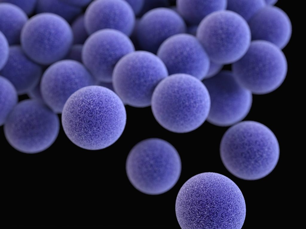
Methicillin-resistant Staphylococcus aureus (MRSA). Image by CDC on Unsplash

Researchers reported in Cell Host & Microbe that early tests of a bioengineered drug candidate were successful in countering Staphylococcus aureus, a bacteria particularly dangerous to hospitalised patients. 

Experiments demonstrated that SM1B74, an antibacterial biologic agent, was superior to a standard antibiotic drug at treating mice infected with S. aureus, including its treatment-resistant form known as MRSA.

The researchers tested mAbtyrins, a combination molecule based on an engineered version of a human monoclonal antibody (mAb), a protein that clings to and marks S. aureus for uptake and destruction by immune cells. Attached to the mAb are centyrins, small proteins that prevent these bacteria from boring holes into the human immune cells in which they hide. As the invaders multiply, these cells die and burst, eliminating their threat to the bacteria.

Together, the experimental treatment targets ten disease-causing mechanisms employed by S. aureus, but without killing it, say the study authors. This approach promises to address antibiotic resistance, say the researchers, where antibiotics kill vulnerable strains first, only to make more space for others that happen to be less vulnerable until the drugs no longer work.

“To our knowledge, this is the first report showing that mAbtyrins can drastically reduce the populations of this pathogen in cell studies, and in live mice infected with drug-resistant strains so common in hospitals,” said lead study author Victor Torres, PhD, the C.V. Starr Professor of Microbiology and director of the NYU Langone Health Antimicrobial-Resistant Pathogen Program.”Our goal was to design a biologic that works against S. aureus inside and outside of cells, while also taking away the weapons it uses to evade the immune system.”

Inside Out

The new study is the culmination of a five-year research partnership between scientists at NYU Grossman School of Medicine and Janssen to address the unique nature of S. aureus.

The NYU Langone team together with Janssen researchers, published in 2019 a study that found that centyrins interfere with the action of potent toxins used by S. aureus to bore into immune cells. They used a molecular biology technique to make changes in a single parental centyrin, instantly creating a trillion slightly different versions of it via automation. Out of this “library,” careful screening revealed a small set of centyrins that cling more tightly to the toxins blocking their function.

Building on this work, the team fused the centyrins to a mAb originally taken from a patient recovering from S. aureus infection. Already primed by its encounter with the bacteria, the mAb could label the bacterial cells such that they are pulled into bacteria-destroying pockets inside of roving immune cells called phagocytes. That is unless the same toxins that enable S. aureus to drill into immune cells from the outside let it drill out of the pockets to invade from the inside.

In a “marvel of bioengineering,” part of the team’s mAbtyrin serves as the passport recognised by immune cells, which then engulf the entire, attached mAbtyrin, along with its centyrins, and fold it into the pockets along with bacteria. Once inside, the centyrins block the bacterial toxins there. This, say the authors, sets their effort apart from antibody combinations that target the toxins only outside of cells.

The team made several additional changes to their mAbtyrin that defeat S. aureus by, for instance, activating chain reactions that amplify the immune response, as well by preventing certain bacterial enzymes from cutting up antibodies and others from gumming up their action.

The researchers tracked the growth of S. aureus strains commonly occurring in US communities in the presence of primary human immune cells (phagocytes). Bacterial populations grew almost normally in the presence of the parental antibody, slightly less well in the presence of the team’s engineered mAb, and half as fast when the mAbtyrin was used.

In another test, 98% of mice treated with a control mAb (no centyrins) developed bacteria-filled sores on their kidneys when infected with a deadly strain of S. aureus, while only 38% of mice did so when treated with the mAbtyrin. Further, when these tissues were removed and colonies of bacteria in them counted, the mice treated with the mAbtyrin had one hundred times (two logs) fewer bacterial cells than those treated with a control mAb.

Finally, the combination of small doses of the antibiotic vancomycin with the mAbtyrin in mice significantly improved the efficacy of the mAbtyrin, resulting in maximum reduction of bacterial loads in the kidneys and greater than 70% protection from kidney lesions.

“It is incredibly important,” said Torres, “that we find new ways to boost the action of vancomycin, a last line of defence against MRSA.”

Source: NYU Langone Health / NYU Grossman School of Medicine

Researchers Quantify Risk Factors for Oral Diclofenac

Safety concerns related to the widely used analgesic diclofenac may be tied to a little-studied drug-metabolising enzyme which can vary as much as 3000 times across individuals, according to new research published in the journal Clinical Pharmacology & Therapeutics.

The findings could be used to develop ways to identify those at risk of serious side effects from the drug. They may help determine safer dosing standards for specific populations, including women, young children and people of certain ethnicities.

Used to treat arthritis-associated pain and inflammation, diclofenac was available over-the-counter in the US until 2013, when the Food and Drug Administration restricted it to prescription-only use following reports of the drug causing heart damage. More than 10 million prescriptions per year are written for it in the US. It is also one of the most widely used non-steroidal anti-inflammatory drugs worldwide, and is still over-the-counter in many countries. 

“Most patients who are using diclofenac have arthritis, and many of them are at risk of heart disease,” senior author Bhagwat Prasad, an associate professor at Washington State University. “So there is a concern that taking diclofenac may be putting them at even greater risk of cardiovascular events such as heart attack and stroke.”

Previous findings by the WSU team had found a high degree of variability in the expression of UGT2B17, an enzyme that is a known player in diclofenac metabolism. They found that the enzyme is present at much lower levels in women than in men, which could explain the increased risk of heart damage seen in women taking diclofenac. The enzyme was also mostly absent in children under age 9 and discovered large ethnicity-based differences in the number of people who lack the gene for the enzyme altogether, which ranges from around 20% of Caucasians up to around 90% of Japanese people.

In this new study, the WSU researchers used human liver and intestinal samples along with computer modelling to quantify the degree to which this enzyme contributes to diclofenac metabolism relative to other related enzymes. They found it to be a major player, supporting the idea that low levels of the UGT2B17 enzyme may be the cause of heart damage tied to diclofenac use.

“No one knew why this heart toxicity is happening in some individuals,” said first author Deepak Ahire, a graduate student in the WSU College of Pharmacy and Pharmaceutical Sciences. “Our study showed, for the first time, that UGT2B17 is important in diclofenac metabolism and suggests that differences in UGT2B17 expression are what makes people’s response to diclofenac so variable, leading to toxicity in some whereas for others the drug simply does not work.”

Ahire said that their study found that this enzyme metabolises diclofenac mainly in the intestine, unlike other related enzymes that are active mostly in the liver. Thus the observed effects are limited to diclofenac tablets taken by mouth, which provides for the quickest absorption and pain relief.

The findings suggest genetic testing could help healthcare providers evaluate safety risks before prescribing diclofenac. Prasad also noted that drug regulatory authorities in countries where diclofenac is still available over the counter should consider doing efficacy testing to determine the optimal dose of the drug for their local market.

The WSU researchers are currently in the process of confirming their findings in a pilot clinical trial. Their next step would be to pursue collaborations with large hospitals to study the connection between diclofenac and heart damage in patients’ electronic medical records.

Source: Washington State University

Grey Hairs Result from Stem Cells Getting ‘Stuck’

Photo by Natasha Brazil on Unsplash

Certain stem cells have a unique ability to move between growth compartments in hair follicles, but get stuck as people age and so lose their ability to mature and maintain hair colour, a new Nature study shows.

Led by researchers from NYU Grossman School of Medicine, the new work focused on cells in the skin of mice and also found in humans called melanocyte stem cells, or McSCs. Hair colour is controlled by whether nonfunctional but continually multiplying pools of McSCs within hair follicles get the signal to become mature cells that make the protein pigments responsible for color.

The new study showed that McSCs are remarkably plastic. This means that during normal hair growth, such cells continually move back and forth on the maturity axis as they transit between compartments of the developing hair follicle. It is inside these compartments where McSCs are exposed to different levels of maturity-influencing protein signals.

Specifically, the research team found that McSCs transform between their most primitive stem cell state and the next stage of their maturation, the transit-amplifying state, and depending on their location.

The researchers found that as hair ages, sheds, and then repeatedly grows back, increasing numbers of McSCs get stuck in the stem cell compartment called the hair follicle bulge. There, they remain, do not mature into the transit-amplifying state, and do not travel back to their original location in the germ compartment, where WNT proteins would have prodded them to regenerate into pigment cells.

“Our study adds to our basic understanding of how melanocyte stem cells work to colour hair,” said study lead investigator Qi Sun, PhD, a postdoctoral fellow at NYU Langone Health. “The newfound mechanisms raise the possibility that the same fixed-positioning of melanocyte stem cells may exist in humans. If so, it presents a potential pathway for reversing or preventing the graying of human hair by helping jammed cells to move again between developing hair follicle compartments.”

Researchers say McSC plasticity is not present in other self-regenerating stem cells, such as those making up the hair follicle itself, which are known to move in only one direction along an established timeline as they mature. For example, transit-amplifying hair follicle cells never revert to their original stem cell state. This helps explain in part why hair can keep growing even while its pigmentation fails, says Sun.

Earlier work by the same research team at NYU showed that WNT signalling was needed to stimulate the McSCs to mature and produce pigment. That study had also shown that McSCs were many trillions of times less exposed to WNT signalling in the hair follicle bulge than in the hair germ compartment, which is situated directly below the bulge.

In the latest experiments in mice whose hair was physically aged by plucking and forced regrowth, the number of hair follicles with McSCs lodged in the follicle bulge increased from 15% before plucking to nearly half after forced aging. These cells remained incapable of regenerating or maturing into pigment-producing melanocytes.

The stuck McSCs, the researchers found, ceased their regenerative behaviour as they were no longer exposed to much WNT signalling and hence their ability to produce pigment in new hair follicles, which continued to grow.

By contrast, other McSCs that continued to move back and forth between the follicle bulge and hair germ retained their ability to regenerate as McSCs, mature into melanocytes, and produce pigment over the entire study period of two years.

“It is the loss of chameleon-like function in melanocyte stem cells that may be responsible for greying and loss of hair colour,” said study senior investigator Mayumi Ito, PhD, a professor in the Ronald O. Perelman Department of Dermatology and the Department of Cell Biology at NYU Langone Health.

“These findings suggest that melanocyte stem cell motility and reversible differentiation are key to keeping hair healthy and coloured,” said Ito, who is also a professor in the Department of Cell Biology at NYU Langone.

Ito says the team has plans to investigate means of restoring motility of McSCs or of physically moving them back to their germ compartment, where they can produce pigment.

Source: NYU Langone Health / NYU Grossman School of Medicine

A Sleeping Pill Lowers Alzheimer’s Protein Levels

An early sign of Alzheimer’s disease is sleep disturbance – many people eventually diagnosed with Alzheimer’s start experiencing difficulty falling and staying asleep years before the emergence of cognitive problems such as memory loss and confusion. In a vicious circle, Alzheimer’s disease disrupts sleep, and poor sleep accelerates harmful changes to the brain.

Now, researchers at Washington University School of Medicine in St. Louis have identified a possible way to help break that cycle. Published in Annals of Neurology, a small, two-night study has shown that people who took a sleeping pill before bed experienced a drop in the levels of key Alzheimer’s proteins – a good sign, since higher levels of such proteins tracks with worsening disease. The study, which involved a sleeping aid known as suvorexant that is already approved by the Food and Drug Administration (FDA) for insomnia, hints at the potential of sleep medications to slow or stop the progression of Alzheimer’s disease, although much more work is needed to confirm the viability of such an approach.

“This is a small, proof-of-concept study. It would be premature for people who are worried about developing Alzheimer’s to interpret it as a reason to start taking suvorexant every night,” said senior author Brendan Lucey, MD, an associate professor of neurology and director of Washington University’s Sleep Medicine Center. “We don’t yet know whether long-term use is effective in staving off cognitive decline, and if it is, at what dose and for whom. Still, these results are very encouraging. This drug is already available and proven safe, and now we have evidence that it affects the levels of proteins that are critical for driving Alzheimer’s disease.”

Suvorexant belongs to a class of insomnia medications known as dual orexin receptor antagonists. Orexin is a natural biomolecule that promotes wakefulness. When orexin is blocked, people fall asleep. Three orexin inhibitors have been approved by the FDA, and more are in the pipeline.

Alzheimer’s disease begins when plaques of the protein amyloid beta start building up in the brain. After years of amyloid accumulation, a second brain protein, tau, begins to form tangles that are toxic to neurons. People with Alzheimer’s disease start experiencing cognitive symptoms such as memory loss around the time tau tangles become detectable.

Lucey and colleagues were among the first to show in people that poor sleep is linked to higher levels of both amyloid and tau in the brain. The question remains as to whether good sleep has the opposite effect – a reduction in amyloid and tau levels, and a halt in or reversal of the progress of Alzheimer’s disease – but mouse studies with orexin inhibitors have been promising.

As a first step to assess the effect of orexin inhibitors on people, Lucey and colleagues recruited 38 participants ages 45 to 65 and with no cognitive impairments to undergo a two-night sleep study. The participants were given a lower dose (10 mg) of suvorexant (13 people), a higher dose (20 mg) of suvorexant (12 people) or a placebo (13 people) at 9 p.m. and then went to sleep in a clinical research unit at Washington University. Researchers withdrew a small amount of cerebrospinal fluid via spinal tap every two hours for 36 hours, starting one hour before the sleeping aid or placebo was administered, to measure how amyloid and tau levels changed over the next day and a half.

Amyloid levels dropped 10% to 20% in the cerebrospinal fluid of people who had received the high dose of suvorexant compared to people who had received placebo, and the levels of a key form of tau known as hyperphosphorylated tau dropped 10% to 15%, compared to people who had received placebo. Both differences are statistically significant. There was not a significant difference between the people who received a low dose of suvorexant and those who received the placebo.

By 24 hours after the first dose, hyperphosphorylated tau levels in the high-dose group had risen, while amyloid levels remained low compared to the placebo group. A second dose of suvorexant, administered on the second night, sent the levels of both proteins down again for people in the high-dose group.

“If we can lower amyloid every day, we think the accumulation of amyloid plaques in the brain will decrease over time,” Lucey said. “And hyperphosphorylated tau is very important in the development of Alzheimer’s disease, because it’s associated with forming tau tangles that kill neurons. If you can reduce tau phosphorylation, potentially there would be less tangle formation and less neuronal death.”

The study is preliminary, since it only looked at the effect of two doses of the drug in a small group of participants. Lucey has studies underway to assess the longer-term effects of orexin inhibitors in people at higher risk of dementia.

“Future studies need to have people taking these drugs for months, at least, and measuring the effect on amyloid and tau over time,” Lucey said. “We’re also going to be studying participants who are older and may still be cognitively healthy, but who already have some amyloid plaques in their brains. This study involved healthy middle-aged participants; the results may be different in an older population.

“I’m hopeful that we will eventually develop drugs that take advantage of the link between sleep and Alzheimer’s to prevent cognitive decline,” he continued. “We’re not quite there yet. At this point, the best advice I can give is to get a good night’s sleep if you can, and if you can’t, to see a sleep specialist and get your sleep problems treated.”

Source: Washington University School of Medicine

Scientists Probe Links Between Chronic Alcohol Use and Pain Sensitivity

The links between alcohol and pain are complex. Research published in the British Journal of Pharmacology suggests that chronic alcohol consumption may increase drinkers’ pain sensitivity through two different molecular mechanisms – the first driven by alcohol intake and the second by alcohol withdrawal.

The research also suggests potential new drug targets for treating alcohol-associated chronic pain and hypersensitivity.

“There is an urgent need to better understand the two-way street between chronic pain and alcohol dependence,” says senior author Marisa Roberto, PhD, the Schimmel Family Chair of Molecular Medicine, and a professor of neuroscience at Scripps Research. “Pain is both a widespread symptom in patients suffering from alcohol dependence, as well as a reason why people are driven to drink again.”

Over time, alcohol use disorder (AUD) can trigger the development of numerous chronic diseases, including heart disease, stroke, liver disease and some cancers. Among the many impacts of long-term alcohol consumption is pain: more than half of people with AUD experience persistent pain of some type. This includes alcoholic neuropathy, which is nerve damage that causes chronic pain and other symptoms.

Studies have also found that AUD is associated with changes in how the brain processes pain signals, as well as changes to how immune system activation occurs. In turn, this pain can lead to increased alcohol consumption. Moreover, during withdrawal, people with AUD can experience allodynia, in which a harmless stimulus is perceived as painful.

Roberto and her colleagues were interested in learning the underlying causes of these different types of alcohol-related pain. In the new study, they compared three groups of adult mice: animals that were dependent on alcohol (excessive drinkers), animals that had limited access to alcohol and were not considered dependent (moderate drinkers), and those that had never been given alcohol.

In dependent mice, allodynia developed during alcohol withdrawal, and subsequent alcohol access significantly decreased pain sensitivity. Separately, about half of the mice that were not dependent on alcohol also showed signs of increased pain sensitivity during alcohol withdrawal but, unlike the dependent mice, this neuropathy was not reversed by re-exposure to alcohol.

When Roberto’s group then measured levels of inflammatory proteins in the animals, they discovered that while inflammation pathways were elevated in both dependent and non-dependent animals, specific molecules were only increased in dependent mice. This indicates that different molecular mechanisms may drive the two types of pain. It also suggests which inflammatory proteins may be useful as drug targets to combat alcohol-related pain.

“These two types of pain vary greatly, which is why it is important to be able to distinguish between them and develop different ways to treat each type,” says first author Vittoria Borgonetti, PhD, a postdoctoral associate at Scripps Research.

Roberto’s group is continuing studies on how these molecules might be used to diagnose or treat alcohol-related chronic pain conditions.

“Our goal is to unveil new potential molecular targets that can be used to distinguish these types of pain and potentially be used in the future for the development of therapies,” says co-senior author Nicoletta Galeotti, PhD, associate professor of preclinical pharmacology at the University of Florence.

Source: Scripps Research Institute

Cranberry Products for UTI Prevention Really Do Work

A global study looking at the benefits of cranberry products has determined that cranberry juice, and its supplements, reduce the risk of repeat symptomatic UTIs in women by more than a quarter. The study researchers, from Flinders University and The Children’s Hospital at Westmead, also found that was reduced in children by more than half, and in people susceptible to UTI following medical interventions by about 53%.

Cranberry juice and healthcare supplements that commonly include the fruit, such as capsules and tablets, have long been promoted as a readily available solution to ward off the infection but the most recent review in 2012, with evidence from 24 trials, showed no benefit from the products.

The medical scientists behind this updated review published in Cochrane Reviews aimed to update these findings, as by looking at 50 more recent trials that included almost 9000 participants.

“This incredible result didn’t really surprise us, as we’re taught that when there’s more and better evidence, the truth will ultimately come out. UTIs are horrible and very common; about a third of women will experience one, as will many elderly people and also people with bladder issues from spinal cord injury or other conditions,” says the study lead author Dr Gabrielle Williams.

“Even back in 1973, my mum was told to try cranberry juice to prevent her horrible and frequent UTIs, and for her it’s been a saviour. Despite me niggling in her ear about evidence, she’s continued to take it daily, first as the nasty sour juice and in recent years, the easy to swallow capsules. As soon as she stops, wham the symptoms are back. As usual, it turns out that mum was right! Cranberry products can help some women prevent UTIs.”

Flinders University epidemiologist Dr Jacqueline Stephens, a co-author of the study, says if the UTI persists untreated it can move to the kidneys and cause pain and more complications, including sepsis in very severe cases, so prevention is the most effective way to reduce risks.

“Most UTIs are effectively, and pretty quickly, treated with antibiotics, sometimes as little as one dose can cure the problem. Unfortunately, in some people UTIs keep coming back. Without being sure if or how it works, some healthcare providers began suggesting it to their patients. It was a harmless, easy option at the time. Even centuries ago, Native Americans reportedly ate cranberries for bladder problems, leading somewhat more recently, to laboratory scientists exploring what it was in cranberries that helped and how it might work.”

“The studies we looked at included a range of methods to determine the benefits of cranberry products. The vast majority compared cranberry products with a placebo or no treatment for UTI and determined drinking cranberries as a juice or taking capsules reduced the number of UTIs in women with recurrent cases, in children and in people susceptible to UTi’s following medical interventions such as bladder radiotherapy.”

“It’s also important to consider that few people reported any side effects with the most common being tummy pain based on the results. We also did not find enough information to determine if cranberry products are more or less effective compared with antibiotics or probiotics in preventing further UTIs.”

The data also doesn’t show any benefit for elderly people, pregnant women or in people with bladder emptying problems.

The study’s senior author, Professor Jonathan Craig, says the real benefits of cranberry products became clear when the researchers expanded the scope of the review to include the most recently available clinical data.

“This is a review of the totality of the evidence and as new evidence emerges, new findings might occur. In this case, the new evidence shows a very positive finding that cranberry juice can prevent UTI in susceptible people,” says Professor Craig.

“We have shown the efficacy of cranberry products for the treatment of UTIs using all the evidence published on this topic since the mid-nineties. The earlier versions of this review didn’t have enough evidence to determine efficacy and subsequent clinical trials showed varied results, but in this updated review the volume of data has shown this new finding.”

The study authors conclude that while cranberry products do help prevent UTIs in women with frequent recurrence, more studies are needed to further clarify who with UTI would benefit most from cranberry products.

Source: Flinders University

More Physical Activity a Health Benefit of a Three-day Weekend

Photo by Julian Jagtenberg on Pexels

With a three-day weekend being trialled in countries across the globe, new empirical research by Australian researchers shows that the extra time off increases active healthy behaviours, lending extra support to a shorter but still productive work week.

The researchers assessed study participants’ changes in daily movements before, during and after holidays, finding that physical activity increased when they were on holiday, even with just a three-day break. Their findings were published in the International Journal of Behavioral Nutrition and Physical Activity.

Across the 13-month study period, participants generally took two to three holidays on average, each being around 12 days. The most common holiday type was ‘outdoor recreation’ (35%), followed by ‘family/social events’ (31%), ‘rest and relaxation’ (17%) and ‘non-leisure pursuits’ such as caring for others or home renovations (17%).

Specifically, it showed that on holiday, participants:

  • engaged in 13% more moderate-to-vigorous physical activity (MVPA) each day (or five min/day more)
  • were 5% less sedentary each day (or 29 min/day less)
  • slept 4% more each day (or 21 min/day more).

University of South Australia researcher Dr Ty Ferguson says that the research indicates that people display healthier behaviours when they are on holiday.

“When people go on holiday, they’re changing their everyday responsibilities because they’re not locked down to their normal schedule,” Dr Ferguson says.

“In this study, we found that movement patterns changed for the better when on holiday, with increased physical activity and decreased sedentary behaviour observed across the board.

“We also found that people gained an extra 21 minutes of sleep each day they were on holiday, which can have a range of positive effects on our physical and mental health. For example, getting enough sleep can help improve our mood, cognitive function, and productivity. It can also help lower our risk of developing a range of health conditions, such as obesity, diabetes, cardiovascular disease, and depression.

“Interestingly, the size of these changes increased in line with the length of the holiday – so the longer the holiday, the better the health benefits.”

The study used data from the Annual rhythms in adults’ lifestyle and health (ARIA) study where 308 adults (mean age 40.4 years) wore fitness trackers 24 hours a day for 13 months. Minute-by-minute

movement behaviour data were aggregated into daily totals to compare movement behaviours pre-holiday, during holiday and post-holiday.

Senior researcher UniSA’s Prof Carol Maher says that the study offers support for the growing movement for a four-day week.

“A shorter working week is being trialled by companies all over the world. Not surprisingly, employees reported less stress, burnout, fatigue, as well as better mental health and improved work-life balance,” Prof Maher says.

“This study provides empirical evidence that people have healthier lifestyle patterns when they have a short break, such as a three-day weekend. This increase in physical activity and sleep is expected to have positive effects on both mental and physical health, contributing to the benefits observed with a four-day work week.

“Importantly, our study also showed that even after a short holiday, people’s increased sleep remained elevated for two weeks, showing that the health benefits of a three-day break can have lasting effects beyond the holiday itself.

“As the world adapts to a new normal, perhaps it’s time to embrace the long weekend as a way to boost our physical and mental health.”

Source: University of South Australia