Day: April 20, 2023

Evidence of Deep Connection Between Body and Mind
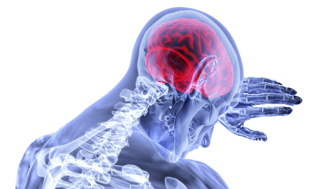
Source: CC0

A new study suggests that notion of the body and mind being inextricably intertwined is more than mere abstraction: parts of the brain area that control movement are plugged into networks involved in thinking and planning, and in control of involuntary bodily functions such as blood pressure and heartbeat. The findings represent a literal linkage of body and mind in the very structure of the brain, overturning decades of interpretations.

The research, published in the journal Nature, could help explain some baffling phenomena, such as why anxiety causes people to pace; why stimulating the vagus nerve, which regulates internal organ functions such as digestion and heart rate, may alleviate depression; and why people who exercise regularly report a more positive outlook on life.

“People who meditate say that by calming your body with, say, breathing exercises, you also calm your mind,” said first author Evan M. Gordon, PhD, an assistant professor of radiology at Washington University School of Medicine in St. Louis. “Those sorts of practices can be really helpful for people with anxiety, for example, but so far, there hasn’t been much scientific evidence for how it works. But now we’ve found a connection. We’ve found the place where the highly active, goal-oriented ‘go, go, go’ part of your mind connects to the parts of the brain that control breathing and heart rate. If you calm one down, it absolutely should have feedback effects on the other.”

Gordon and senior author Nico Dosenbach, MD, PhD, an associate professor of neurology, did not set out to answer age-old philosophical questions about the relationship between the body and the mind. They set out to verify the long-established map of the areas of the brain that control movement, using modern brain-imaging techniques.

In the 1930s, neurosurgeon Wilder Penfield, MD, mapped such motor areas of the brain by applying small jolts of electricity to the exposed brains of people undergoing brain surgery, and noting their responses. He discovered that stimulating a narrow strip of tissue on each half of the brain causes specific body parts to twitch. Moreover, the control areas in the brain are arranged in the same order as the body parts they direct, with the toes at one end of each strip and the face at the other. Penfield’s map of the motor regions of the brain – depicted as a homunculus, or “little man” – has become a staple of neuroscience textbooks.

Gordon, Dosenbach and colleagues set about replicating Penfield’s work with functional magnetic resonance imaging (fMRI). They recruited seven healthy adults to undergo hours of fMRI brain scanning while resting or performing tasks. From this high-density dataset, they built individualized brain maps for each participant. Then, they validated their results using three large, publicly available fMRI datasets – the Human Connectome Project, the Adolescent Brain Cognitive Development Study and the UK Biobank – which together contain brain scans from about 50 000 people.

To their surprise, they discovered that Penfield’s map wasn’t quite right. Control of the feet was in the spot Penfield had identified. Same for the hands and the face. But interspersed with those three key areas were another three areas that did not seem to be directly involved in movement at all, even though they lay in the brain’s motor area.

Moreover, the nonmovement areas looked different than the movement areas. They appeared thinner and were strongly connected to each other and to other parts of the brain involved in thinking, planning, mental arousal, pain, and control of internal organs and functions such as blood pressure and heart rate. Further imaging experiments showed that while the nonmovement areas did not become active during movement, they did become active when the person thought about moving.

“All of these connections make sense if you think about what the brain is really for,” Dosenbach said. “The brain is for successfully behaving in the environment so you can achieve your goals without hurting or killing yourself. You move your body for a reason. Of course, the motor areas must be connected to executive function and control of basic bodily processes, like blood pressure and pain. Pain is the most powerful feedback, right? You do something, and it hurts, and you think, ‘I’m not doing that again.'”

Dosenbach and Gordon named their newly identified network the Somato (body)-Cognitive (mind) Action Network, or SCAN. To understand how the network developed and evolved, they scanned the brains of a newborn, a 1-year-old and a 9-year-old. They also analysed data that had been previously collected on 9 monkeys. The network was not detectable in the newborn, but it was clearly evident in the 1-year-old and nearly adult-like in the 9-year-old. The monkeys had a smaller, more rudimentary system without the extensive connections seen in humans.

“This may have started as a simpler system to integrate movement with physiology so that we don’t pass out, for example, when we stand up,” Gordon said. “But as we evolved into organisms that do much more complex thinking and planning, the system has been upgraded to plug in a lot of very complex cognitive elements.”

Clues to the existence of a mind-body network have been around for a long time, scattered in isolated papers and inexplicable observations.

“Penfield was brilliant, and his ideas have been dominant for 90 years, and it created a blind spot in the field,” said Dosenbach, who is also an associate professor of biomedical engineering, of paediatrics, occupational therapy, radiology, and psychological & brain sciences. “Once we started looking for it, we found lots of published data that didn’t quite jibe with his ideas, and alternative interpretations that had been ignored. We pulled together a lot of different data in addition to our own observations, and zoomed out and synthesised it, and came up with a new way of thinking about how the body and the mind are tied together.”

Source: Washington University School of Medicine

Neurons in Developing Brains are Connected by Nanoscopic Tunnels

Example of 3D imaging of segmented granule cells shown in green and orange, with nuclei in blue and purple respectively, and mitochondria in yellow. A thin connection can be seen between the two cells in blue, with subcompartments attached to the tube containing the mitochondria, shown in pink. Credit: Diego Cordero / Membrane Traffic and Pathogenesis Unit, Institut Pasteur

Over a hundred years after the discovery of the neuron by neuroanatomist Santiago Ramón y Cajal, scientists continue to deepen their knowledge of the brain and its development. Now, scientists detail novel insights into how cells in the outer layers of the brain interact immediately after birth during formation of the cerebellum, the brain region towards the back of the skull. Publishing their results in in Science Advances, the scientists demonstrated a novel type of connection between neural precursor cells via nanotubes, even before synapses form.

In 2009, Chiara Zurzolo’s team from the Institut Pasteur identified a novel mechanism for direct communication between neuronal cells in culture via nanoscopic tunnels, known as tunnelling nanotubes. These are involved in the spread of various toxic proteins that accumulate in the brain during neurodegenerative diseases – but may also be tapped for the treatment of diseases or cancers.

In this new study, the researchers discovered nanoscopic tunnels that connect precursor cells in the brain, more specifically the cerebellum – an area that develops after birth and is important for making postural adjustments to maintain balance – as they mature into neurons. These tunnels, although similar in size, vary in shape from one to another: some contain branches while others don’t, some are enveloped by the cells they connect while others are exposed to their local environment. The authors believe these intercellular connections (ICs) may enable the exchange of molecules that help pre-neuronal cells physically migrate across various layers and reach their final destination as the brain develops.

Intriguingly, ICs share anatomical similarities with bridges formed when cells finish dividing. “ICs could derive from cellular division but persist during cell migration, so this study could shed light on the mechanisms allowing coordination between cell division and migration implicated in brain development. On the other hand, ICs established between cells post mitotically could allow direct exchange between cells beyond the usual synaptic connections, representing a revolution in our understanding of brain connectivity. We show that there are not only synapses allowing communication between cells in the brain, there are also nanotubes,” says Dr Zurzolo, senior author and head of the Membrane Traffic and Pathogenesis Unit (Institut Pasteur/CNRS).

To achieve these discoveries, the researchers used a three-dimensional (3D) electron microscopy method and brain cells from mouse models to study how the brain regions communicate between each other. Very high resolution neural network maps could thus be reconstructed. The 3D cerebellum volume produced and used for the study contains over 2000 cells. “If you really want to understand how cells behave in a three-dimensional environment, and map the location and distribution of these tunnels, you have to reconstruct an entire ecosystem of the brain, which requires extraordinary effort with twenty or so people involved over 4 years,” said the article’s first author Diego Cordero.

To meet the challenges of working with the wide range of cell types the brain contains, the authors used an AI tool to automatically distinguish cortical layers. Furthermore, they developed an open-source program called CellWalker to characterise morphological features of 3D segments. The tissue block was reconstructed from brain section images. This program being made freely available will enable scientists to quickly and easily analyse the complex anatomical information embedded in these types of microscope images.

The next step will be to identify the biological function of these cellular tunnels to understand their role in the development of the central nervous system and in other brain regions, and their function in communication between brain cells in neurodegenerative diseases and cancers.

Source: Institut Pasteur

Flipping the Switch on Osteoporosis with Epigenetic Discovery

Photo by Mehmet Turgut Kirkgoz on Unsplash

Van Andel Institute scientists have pinpointed a key driver of low bone density, a discovery that may lead to improved treatments with fewer side effects for women with osteoporosis. Their findings appear in the journal Science Advances.

Their research reveals that loss of an epigenetic modulator, KDM5C, preserves bone mass in mice. KDM5C works by altering epigenetic ‘marks’, switches that ensure the instructions written in DNA are read in the right time and place.

Several medications are approved to treat osteoporosis but fears of rare, severe side effects often are a barrier for their use. Treatments that leverage the hormone oestrogen also are available, but are only recommended for low-dose, short-term use due in part to associations with cancer risk.

It is well-established that women experience disproportionately lower bone mass than men throughout their lives. Loss of bone mass accelerates with menopause, increasing the risk of osteoporosis and associated fractures for women as they age.

To figure out why this happens, VAI Associate Professors Connie M. Krawczyk, PhD, and Tao Yang, PhD, and their teams looked at the differences in the ways bone is regulated in male and female mice, which share many similarities with humans and are important models for studying health and disease. They focused on osteoclasts, which help maintain bone health by breaking down and recycling old bone.

“Osteoporosis is a common disease that can have debilitating outcomes,” Yang said. “KDM5C is a promising target to treat low bone mass in women because it is highly specific. We’re hopeful that our findings will contribute to improved therapies.”

The researchers found reducing KDM5C disrupted cellular energy production in osteoclasts, which slowed down the recycling process and preserved bone mass. Importantly, KDM5C is linked to X chromosomes, which means it is more active in females than in males.

“Lowering KDM5C levels is like flipping a switch to stop an overactive recycling process. The result is more bone mass, which ultimately means stronger bones,” Krawczyk said. “We’re very excited about this work and look forward to carrying out future studies to refine our findings. At the end of the day, we hope these insights make a difference for people with osteoporosis.”

Source: Van Andel Research Institute

Processed Foods Increase Miscarriage Risk; Fruits Reduce it

A preconception and early-pregnancy diet that contains lots of fruit, vegetables, seafood, dairy, eggs and grain may be associated with reducing risk of miscarriage, suggests a new review of research in the journal Fertility and Sterility.

University of Birmingham researchers analysed 20 studies on eating habits in the months before and shortly after conceiving a baby to see whether these studies showed evidence of association with a lower or higher chance of miscarriage. The team conclude that there is evidence to suggest a diet rich in fruit, vegetables, seafood, dairy products, eggs and grain reduces miscarriage risk.

Not surprisingly, these are foods which typically make up ‘healthy’ well-balanced diets, with previous evidence showing that eating a well-rounded diet which is rich in vitamins and minerals during pregnancy is important.

The research review found that, when compared to low consumption, high intake of fruit may be associated with a 61% reduction in miscarriage risk. High vegetable intake may be associated with a 41% reduction in miscarriage risk. For dairy products it is a 37% reduction, 33% for grains, 19% for seafood and eggs.

Led by Dr Yealin Chung, researchers also looked at whether pre-defined dietary types, such as the Mediterranean Diet or Fertility Diet could also be linked to miscarriage risk. They could not find evidence that following any of these diets lowered or raised risk.

However, a whole diet containing healthy foods overall, or foods rich in antioxidant sources, and low in pro-inflammatory foods or unhealthy food groups may be associated with a reduction in miscarriage risk for women.

A diet high in processed food was shown to be associated with doubling of miscarriage risk.

The studies included in the analysis focused on the peri-conception period, which is before and during the first 3 months of pregnancy. Data collected from a total of 63 838 healthy women of reproductive age was included, with information on their diets typically collected through food frequency questionnaires for each study.

Dr Chung explains:

“Miscarriage is common, with estimates suggesting 1 in 6 pregnancies end in miscarriage, and there are many known causes, from problems with the baby’s chromosomes to infections in the womb.

“Yet nearly 50% of early pregnancy losses remain unexplained and in the absence of a cause, parents often turn to their healthcare providers for guidance on the best ways to be as healthy as possible and reduce the risk of future miscarriages.

“There’s a growing body of evidence to show that lifestyle changes – including changes to diet, stopping smoking and not drinking alcohol – before conceiving and in your pregnancy’s early stages – may have an impact.”

Source: University of Birmingham

New Vaccine Will Save Thousands of Children from Dying of Pneumonia

Scanning electron micrograph of human respiratory syncytial virus (RSV) virions (colourised blue) and labelled with anti-RSV F protein/gold antibodies (colourised yellow) shedding from the surface of human lung epithelial A549 cells. Credit: National Institute of Allergy and Infectious Diseases, National Institutes of Health (NIH)

By James Stent for GroundUp

Respiratory syncytial virus (RSV) is a dangerous early childhood viral infection, but results of a vaccine trial promise to change things radically.

A new study published in the New England Journal of Medicine, the world’s most prestigious medical journal, on 5 April that examined the effect of an RSV vaccine on pregnant women found that it reduced the risk of severe lower respiratory tract infections in newborns by 82%.

RSV is the most common cause of acute lower respiratory infection – or pneumonia – in infants. Globally, it was responsible for just over 100,000 deaths (with a lower bound of 84,000 deaths and an upper bound of 126,000 deaths) of children under five in 2019. Of these deaths 45% were infants (younger than six months), and nearly all deaths occurred in lower income countries (half in Africa alone). In an article in Spotlight in June 2022, Professor Cheryl Cohen, head of the Centre for Respiratory Diseases and Meningitis at the National Institute for Communicable Diseases (NICD), said that, pre-COVID, RSV led to 44 615 hospitalisations and 490 deaths in children under five each year in South Africa.

South Africa is currently experiencing an RSV epidemic, with 301 cases detected this year, according to the NICD surveillance programme.

RSV causes cold-like symptoms, but can lead to severe symptoms like pneumonia. At present, there is no licensed RSV vaccine, though the virus was first identified in the 1960s.

The study was a phase three, double-blind trial (which compares a new treatment to standard care, and leads the way to regulatory approval and production) conducted in 18 countries, led by Beate Kampmann, Professor of Paediatric Infection and Immunity at the London School of Hygiene and Tropical Medicine, Shabir Madhi, Dean of the Faculty of Health Sciences and Professor of Vaccinology at the University of the Witwatersrand, and Iona Munjal, Director of Clinical Research & Development at Pfizer. It builds on earlier work by Madhi and others.

Women who were between 24 and 36 weeks pregnant were given an injection of a protein–based vaccine (RSVpreF) and a placebo. Pregnant women can passively transfer their immunity to viruses and diseases to their foetuses in utero.

They were then monitored to see if they suffered a severe RSV-associated lower respiratory tract illness that required medical attention, and if their newborns required medical attention for RSV-associated lower respiratory tract illness up to six months after birth.

A total of 7,358 women participated across the two trial groups, and 7,128 babies were monitored, and no safety concerns were identified over the course of the trial.

In November last year, Pfizer announced that it planned to submit a licence application to the US Food and Drug Administration after trials showed that the vaccine was highly effective at reducing severe RSV cases in the first 90 days of an infant’s life.

In a Twitter thread announcing the results, Madhi said that the next challenge would be to ensure that the vaccine is licensed across lower income countries, where most infant RSV deaths occur. Madhi said that there is a “moral responsibility on pharma to licence [the RSV] vaccine in LMIC [Lower and Middle Income Countries] at [an] affordable price.” Governments in poorer countries, “need to act to protect children in their counties by funding and deploying the vaccine timeously,” he said.

Madhi also informed GroundUp that coincidentally in the same issue of the New England Journal of Medicine, a medicine called nirsevimab was found to protect infants against RSV-associated hospitalisation and severe lower respiratory tract infections. Madhi and his team at Wits also participated in this trial.

This medicine is “administered as a single dose at the onset of RSV season,” Madhi explained. “The two approaches [the vaccine and nirsevimab] will be complementary.”

Republished from GroundUp under a Creative Commons Attribution-NoDerivatives 4.0 International License.

Source: GroundUp