Day: February 7, 2023

A New Possibility for Non-hormonal Male Contraceptives

Photo by Reproductive Health Supplies Coalition on Unsplash

Thus far, very few contraceptive options being developed target the sperm cells. Researchers are now developing approaches that target testosterone or otherwise interrupt the sperm’s ability to fertilise an egg, yet these may not work for everyone. But now, researchers publishing in ACS’ Journal of Medicinal Chemistry have identified a new candidate molecule that could become an effective non-hormonal contraceptive for males.

Previously, Gunda I. Georg and colleagues investigated non-hormonal contraceptive options, as approaches targeting testosterone produced unwanted side effects. They developed a drug targeted at a specific vitamin A receptor and found that it worked as a highly effective contraceptive with no side effects. But numerous proteins are involved in forming sperm, and exploring multiple options would maximise chances for a drug that would eventually make it to market.

Another set of proteins involved in the cell cycle are the cyclin-dependent kinases, or CDKs, which play a role in sperm cell production and tumour development. Mice without the CDK2 receptor are sterile, so a drug that targets this protein could serve as an effective contraceptive. It also has potential as a cancer therapeutic because inhibiting the enzyme slowed tumour growth in previous studies. However, CDK2 has a very similar shape to other enzymes in its family, and currently available inhibitors tend to produce undesirable off-target effects by accidentally binding the others as well. So, Georg and her team wanted to develop a drug that could selectively inhibit CDK2 to serve as another contraceptive option.

The team previously discovered an unknown binding site in CDK2 and a commercially available dye molecule that successfully bound to it. Using the dye as a starting point, the researched screened tens of thousands of different compounds in their current work to find ones that also bound the pocket well. They narrowed the list down to just three, picking one to further optimize. The best version, named EF-4-177, demonstrated a long half-life and good diffusion into the testes of mice. After a 28-day exposure, the animals’ sperm counts decreased by about 45%. Additionally, EF-4-117 bound much more strongly to the CDK2 pocket than the dye, making it the highest affinity inhibitor for this site reported to date. The researchers say that this work proves the potential of this inhibitor for future therapeutic applications.

Source: Michigan State University

Boys can Also be at Risk for Eating Disorders

Depression, young man
Source: Andrew Neel on Unsplash

In the public mind, eating disorders are associated mainly with girls from wealthy backgrounds. Now, a new study on twins published in the Journal of Psychopathology and Clinical Science has found that boys living in disadvantaged circumstances are at an increased risk for disordered eating – particularly if they have underlying genetic risk factors.

“This is critical information for health care providers who might not otherwise screen for or recognize disordered eating in this population,” said Megan Mikhail, lead author of the study and Ph.D. candidate in the MSU Clinical Psychology program. “It is also important for the public to recognize that eating disorders can affect everyone, including people who do not fit the historical stereotypes.”

The study from Michigan State University, is the first to look at associations between multiple forms of disadvantage and risk for disordered eating in boys, as well as how disadvantage may interact with biological risks to impact disordered eating in boys.

Using a large population-based sample of male twins from the Michigan State University Twin Registry, the researchers found that boys from more disadvantaged backgrounds reported greater disordered eating symptoms and had earlier activation of genetic influences on disordered eating, which could lead to increased long-term risk.

By using population-based sample, the researchers could avoid overlooking those unable to afford mental health care. They examined factors such as parental income, education and neighbourhood disadvantage to see how those factors related to disordered eating symptoms in the boys. Since all the participants were twins, researchers were also able to study genetic influences on disordered eating.

“This research is particularly relevant following the COVID pandemic when many families experienced financial hardship,” said Kelly Klump, MSU Foundation Professor of Psychology and co-author of the study. “Those financial stressors are putting many young people at risk for an eating disorder, so it’s vital that there be increased screening and access to care for these young people.”

Source: Michigan State University

Prior COVID Infection Linked to New Autoimmune Conditions

Photo by Mufid Majnun on Unsplash

In a new entry to the growing list of lasting complications from COVID infection, a large German cohort study of over 600 000 COVID patients indicates that new autoimmune conditions may result from previous COVID infection. The findings, which are awaiting peer review on the MedRxiv preprint server, show that the odds of new autoimmune conditions appear to increase in line with the severity of COVID infection.

After the acute phase of infection, some people may develop long-lasting symptoms, known as post-COVID, which are consistent with COVID infection and last more than 12 weeks. Most studies to date have focused on symptoms that partly wane over time. Many studies examined a small selective sample of patients, and only a few studies included a control group or information on chronic health conditions, such as SARS-CoV-2 infection.

Compared to post-COVID emergence of cardiovascular and other diseases, autoimmune diseases are less discussed in the literature, although autoantibodies could be found in patients after SARS-CoV-2 infection. So far there is limited evidence on newly manifested autoimmune diseases after an infection based on several case reports and one recent cohort study using UK health record data. In addition, COVID itself has some similarities with systemic autoimmune rheumatic diseases, which could make diagnosis difficult.

The researchers selected a cohort from German routine health care data, identifying individuals with polymerase chain reaction (PCR)-confirmed COVID through December 31, 2020. Patients were matched 1:3 to control patients without COVID. Both groups were followed up until June 30, 2021. We used the four quarters preceding the index date until the end of follow-up to analyse the onset of autoimmune diseases during the post-acute period. The researchers calculated the incidence rates (IR) per 1000 person-years for each outcome and patient group, and estimated incidence rate ratios (IRRs) of developing an autoimmune disease conditional on a preceding COVID.

In total, 641 704 patients with COVID were included. When comparing the incidence rates in the COVID and matched control groups, the researchers found a 42.63% higher likelihood of acquiring autoimmunity for patients who had suffered from COVID. This estimate was similar for common autoimmune diseases, such as Hashimoto thyroiditis, rheumatoid arthritis, or Sjögren syndrome. The highest IRR was observed for autoimmune disease of the vasculitis group. Patients with a more severe course of COVID were at a greater risk for incident autoimmune diseases. These risk increases were as follows:

  • 41% higher risk of Grave’s disease
  • 42–45% higher risk of rheumatoid arthritis
  • 25% higher risk of type 1 diabetes
  • 27-29% higher risk of Crohn’s disease

The researchers concluded that SARS-CoV-2 infection is associated with an increased risk of developing new-onset autoimmune diseases after the acute phase of infection.

Preeclampsia Leads to 4x Higher Risk of MI in Decade after Delivery

Pregnant with ultrasound image
Source: Pixabay

Women with preeclampsia have a higher likelihood of heart attack and stroke than their peers within just seven years of delivery, with risks remaining elevated more than 20 years later. The study in more than one million pregnant women is published today in the European Journal of Preventive Cardiology, a journal of the ESC.

“The high risk of cardiovascular disease after preeclampsia manifests at young ages and early after delivery,” said study author Dr Sara Hallum of the University of Copenhagen. “This indicates that interventions to prevent heart attacks and strokes in affected women cannot wait until middle age when they become eligible for conventional cardiovascular screening programmes.”

Preeclampsia affects up to 8% of pregnancies worldwide, and signs include hypertension and albuminuria, which develop after 20 weeks of pregnancy or soon after delivery. Symptoms include severe headache, stomach pain and nausea. “Women may mistake these for ‘normal’ pregnancy symptoms and thus not seek medical help until the condition becomes severe,” said Dr Hallum. “Most cases are mild, but preeclampsia may lead to serious complications for the mother and baby if not treated in time.”

It is well established that preeclampsia predisposes women to an elevated likelihood of cardiovascular disease later in life. This was the first study to examine how soon after pregnancy these heart attacks and strokes manifest, and the magnitude of risk in different age groups.

National registers were used to identify all pregnant women in Denmark between 1978 and 2017. Women were grouped into those with one or more pregnancies complicated by preeclampsia and those with no preeclampsia. Participants were free of cardiovascular disease before pregnancy and with follow-up for heart attack and stroke up to 39 years later. Dr Hallum said: “This allowed us to evaluate exactly when cardiovascular disease occurs in women with and without pre-eclampsia, and to estimate risk in different age groups and at various durations of follow-up.”

Up to 2% of those with pre-eclampsia in their first pregnancy had a heart attack or stroke within 20 years of delivery, compared with up to 1.2% of unaffected women. Differences in risk became apparent seven years after delivery. “A 2% incidence of acute myocardial infarction and ischaemic stroke should not be accepted as the cost of a pregnancy complicated by preeclampsia, particularly considering the young age of these women when they fall ill (below 50 years of age),” states the paper.

Overall, women with pre-eclampsia were four times more likely to have a heart attack and three times more likely to have a stroke within 10 years of delivery than those without pre-eclampsia. The risk of heart attack or stroke was still twice as high in the preeclampsia group more than 20 years after giving birth compared to unaffected women.

When the researchers examined the risk of cardiovascular disease according to age, they found that women aged 30 to 39 years with a history of preeclampsia had five- and three-fold higher rates of heart attack and stroke, respectively, than those of similar age with no history of pre-eclampsia. The raised likelihood of cardiovascular disease in those with a history of pre-eclampsia persisted throughout adulthood, with women over 50 years of age still at doubled risk compared to their peers with no history of the pregnancy complication.

Dr Hallum said: “Women are often in contact with the healthcare system during and immediately after pregnancy, providing a window of opportunity to identify those at increased risk of cardiovascular disease. The number of women with previous pre-eclampsia is large, and routine follow-up could last years or even decades. Our study suggests that the women most likely to benefit from screening are those who had pre-eclampsia after age 35 and those who had it more than once. Prevention should start within a decade of delivery, for example by treating high blood pressure and informing women about risk factors for heart disease such as smoking and inactivity.”

Source: European Society of Cardiology

A New Understanding of Graft-versus-host Disease

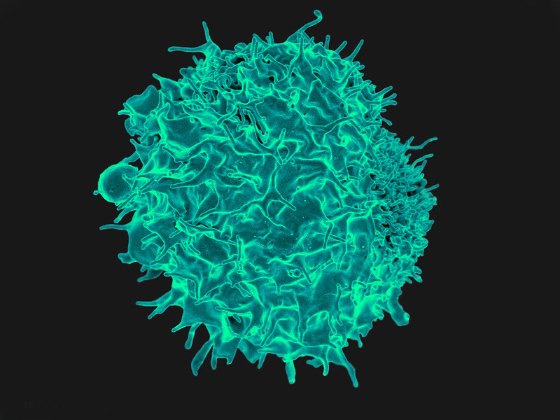
T cell
Scanning Electron Micrograph image of a human T cell. Credit: NIH/NIAID

New research published in the journal Immunity challenges the prevailing hypothesis for how donor stem cell grafts cause graft-versus-host disease (GVHD) and offers an alternative model that could guide development of novel therapies.

The study showed in a mouse model that GVHD, which often affects the skin, gut and liver, is maintained by donor T cells that seed those tissues soon after transplant and not by the continual recruitment of T cells from the blood as previously thought.

“This study changes the paradigm of how people think about GVHD,” said co-senior author Warren Shlomchik, MD, professor of medicine and immunology at the University of Pittsburgh School of Medicine. “It provides important mechanistic detail about what’s going on in the tissues affected by GVHD, which could ultimately inform the development of better therapeutics and lead to better outcomes for stem cell recipients.”

Allogeneic stem cell transplantation involves infusion of stem cells from a healthy donor’s blood or bone marrow to a recipient. While often lifesaving for patients with leukaemia and other blood disorders, the treatment also comes with a risk of developing GVHD, a life-threatening disease that occurs when donor alloreactive T cells attack the recipient’s healthy tissues.

According to a widely held theory, GVHD is maintained by T cells that continually migrate from secondary lymphoid organs throughout the body, including the spleen and lymph nodes, to affected tissues via the blood.

However, a different model posits that the disease is maintained locally by T cells in the tissues with little input from the blood. In the new study, Shlomchik, lead author Faruk Sacirbegovic, PhD, research assistant professor of surgery at Pitt, and their team investigated the two hypotheses for how GVHD is sustained in tissues.

The researchers developed a system to track alloreactive T cells in a mouse model of GVHD by labelling individual cells with unique tags to create different T cell “flavours.” By measuring the tags over time, they monitored where the T cells travelled and replicated.

The analysis showed that each tissue affected by GVHD had unique T cell populations with varying frequencies of each T cell flavour.

“This finding is strong evidence that the disease is locally maintained by T cells in each of the tissues,” explained Shlomchik. “If tissues were constantly getting T cells from circulating blood, then the frequencies of T cell flavors in each tissue should become more and more alike over time — but we didn’t see that.”

The team used mathematical models to predict that progenitor T cells seed out into recipient tissues early after transplant, differentiating there into disease-causing cells.

Next a series of experiments was conducted to confirm this prediction and identified these progenitors as T cells expressing a gene called Tcf7.

“We think that progenitor T cells are long-lived in target tissues and are critical for maintaining GVHD,” said co-senior author Thomas Höfer, PhD, professor of theoretical systems biology at the University of Heidelberg. “After the initial seeding phase, the disease is mostly sustained within the tissue itself without a lot of input from new T cells in the blood.”

Stem cell recipients are typically treated with immunosuppressants to prevent and treat GVHD. As these powerful drugs act systemically to suppress the immune system, they also lower immunity to infections and have other side effects.

According to the researchers, the study’s insights could eventually lead to new, targeted therapies for GVHD.

“Now that we know the identity of progenitor cells, we might be able to prevent them forming early post-transplant or target them directly after they’ve formed,” said Shlomchik. “The findings also suggest that treating GVHD in the tissues themselves would be effective – although targeting tissues beyond the skin remains a challenge.”

With better ways to minimise the risk of GVHD after stem cell transplantation, the procedure could become more widely used to treat a broader range of diseases, including blood disorders such as sickle cell anaemia and autoimmune diseases such as lupus and multiple sclerosis.

Source: University of Pittsburgh

FBI Disrupts Cybercrime Group Which Extorted Hospitals

Photo by Nahel Abdul Had on Unsplash

The Hive ransomware group that has targeted more than 1500 victims in over 80 countries around the world, including hospitals, has been disrupted in a months-long campaign against, the US Justice Department has announced.

Hive ransomware attacks have caused major disruptions in victim daily operations around the world and hindered responses to the COVID pandemic. In one case, a hospital attacked by Hive ransomware had to fall back to pen and paper to treat existing patients and could not take new admissions shortly after the attack. 

The Justice Department revealed that the FBI had penetrated Hive’s computer network and captured its decryption keys, which were then offered to victims around the world. This saved them $130 million in ransom they would have had to otherwise pay to get their networks back.

Finally, the department announced that, in coordination with German and Dutch law enforcement, it has seized control of the servers and websites that Hive uses to communicate with its members, disrupting Hive’s ability to attack and extort victims.

Since June 2021, the Hive ransomware group has targeted more than 1500 victims around the world and received over $100 million in ransom payments.  

Hive used a ransomware-as-a-service (RaaS) model featuring administrators, and affiliates. RaaS is a subscription-based model where the administrators develop an easy-to-use ransomware strain and then recruit affiliates to deploy the ransomware against victims. Affiliates identified targets and deployed this readymade malicious software to attack victims and then earned a percentage of each successful ransom payment.

Hive actors used a double-extortion model of attack: before encrypting the victim’s system, the affiliate would steal sensitive data. The affiliate then sought a ransom for both the decryption key necessary to decrypt the victim’s system and a promise to not publish the stolen data – usually the most sensitive, such as hospital patient data. After a victim pays, the affiliates and administrators split the ransom 80/20. Victims who do not pay on the Hive Leak Site. After Consulate Health Care was unable to pay the ransom, since its insurance did not cover such cyber crimes, Hive posted 550GB of personally identifiable information on its patients and employees online.

For more information about the malware, including technical information for organisations about how to mitigate its effects, is available from CISA, visit https://www.cisa.gov/uscert/ncas/alerts/aa22-321a.