Month: February 2023

A Link Between Head Injury and Increased Glioma Risk

Photo by John Simmons on Unsplash

Previous research has hinted at a possible link between head injury and increased rates of gliomas, rare but aggressive brain tumours. A University College London team has now identified a possible mechanism to explain this link, implicating genetic mutations acting in concert with brain tissue inflammation to change the behaviour of cells, making them more likely to become cancerous.

Publishing in Current Biology, the researchers have now identified a possible mechanism to explain this link, implicating genetic mutations acting in concert with brain tissue inflammation to change the behaviour of cells, making them more likely to become cancerous. Although this study was largely carried out in mice, it suggests that it would be important to explore the relevance of these findings to human gliomas.

The study was led by Professor Simona Parrinello (UCL Cancer Institute), Head of the Samantha Dickson Brain Cancer Unit and co-lead of the Cancer Research UK Brain Tumour Centre of Excellence. She said: “Our research suggests that a brain trauma may contribute to an increased risk of developing brain cancer in later life.”

Gliomas are brain tumours that often arise in neural stem cells. More mature types of brain cells, such as astrocytes, have been considered less likely to give rise to tumours. However, recent findings have demonstrated that after injury, astrocytes can exhibit stem cell behaviour again.

Professor Parrinello and her team therefore set out to investigate whether this property may make astrocytes able to form a tumour following brain trauma using a pre-clinical mouse model.

Young adult mice with brain injury were injected with a substance which permanently labelled astrocytes in red and knocked out the function of the p53 gene, known to have a vital role in suppressing many different cancers. A control group was treated the same way, but the p53 gene was left intact. A second group of mice was subjected to p53 inactivation in the absence of injury.

Professor Parrinello said: “Normally astrocytes are highly branched – they take their name from stars – but what we found was that without p53 and only after an injury the astrocytes had retracted their branches and become more rounded. They weren’t quite stem cell-like, but something had changed. So we let the mice age, then looked at the cells again and saw that they had completely reverted to a stem-like state with markers of early glioma cells that could divide.”

This suggested to Professor Parrinello and team that mutations in certain genes synergised with brain inflammation, which is induced by acute injury and then increases over time during the natural process of ageing to make astrocytes more likely to initiate a cancer. Indeed, this process of change to stem-cell like behaviour accelerated when they injected mice with a solution known to cause inflammation.

The team then looked for evidence to support their hypothesis in human populations. Working with Dr Alvina Lai in UCL’s Institute of Health Informatics, they consulted electronic medical records of over 20 000 people who had been diagnosed with head injuries, comparing the rate of brain cancer with a control group, matched for age, sex and socioeconomic status. They found that patients who experienced a head injury were nearly four times more likely to develop a brain cancer later in life, than those who had no head injury. It is important to keep in mind that the risk of developing a brain cancer is overall low, estimated at less than 1% over a lifetime, so even after an injury the risk remains modest.

Professor Parrinello said: “We know that normal tissues carry many mutations which seem to just sit there and not have any major effects. Our findings suggest that if on top of those mutations, an injury occurs, it creates a synergistic effect. In a young brain, basal inflammation is low so the mutations seem to be kept in check even after a serious brain injury. However, upon ageing, our mouse work suggests that inflammation increases throughout the brain but more intensely at the site of the earlier injury. This may reach a certain threshold after which the mutation now begins to manifest itself.”

Source: University College London

New US Report Rekindles ‘Lab Leak’ Debate over COVID Origins

Once again, the ‘lab leak’ theory of COVID’s origin has returned to the headlines. On Sunday, the Wall Street Journal revealed that a US Department of Energy report had determined that the origin of COVID was ” most likely” an accidental release from a laboratory, according to those who had read the report, though the assessment was with “low confidence”.

Ambassador Nicholas Burns told a US Chamber of Commerce event on Monday that China needs to “be more honest about what happened three years ago in Wuhan with the origin of the Covid-19 crisis”.

China’s foreign ministry countered that COVID’s origin “was about science and should not be politicised”.

The FBI assigned “moderate confidence” to a laboratory origin for the virus, while four other US agencies assigned a “low confidence” to a natural origin. Two others, including the CIA, remained undecided. An update on their views has been provided, apparently due to new information, but has not been made public.

To many scientists, the origin of SARS-CoV-2 has been settled as it has been traced to outbreaks in the Wuhan meat market two weeks before its first detection. A literature analysis published in PNAS concluded that the evidence overwhelmingly favoured a natural origin.

Many other scientists are not convinced by the zoonotic hypothesis. Virologist Jesse Bloom, at the Fred Hutchinson Cancer Center, said the PNAS review’s literature analysis was a good idea – but the zoonosis proponents haven’t provided much new data. “What we’ve seen is mostly reanalysis and reinterpretation of existing evidence.”

The PNAS review started out as a Lancet commission led by Jeffrey Sachs, who disbanded the task force due to a number of members with vested interests against the lab leak hypothesis. Their aim was to gather lessons learnt from the pandemic. The Lancet eventually published its own review, which concluded that there was equal probability for a laboratory or natural origin.

Even so, a continued lack of cooperation from China with international investigators has made it virtually impossible to definitively pinpoint the virus’s emergence. Ultimately, the lesson of past pandemics is that outbreaks can result from either zoonotic origins or from laboratory accidents, both of which are factors which need to be safeguarded against by humans.

Reproductive Factors in Women Linked to Cardiovascular Disease

Source: American Heart Association

An earlier first birth, a higher number of live births, and starting periods at a younger age are all linked to a higher risk of cardiovascular problems in women, according to new research published in the Journal of the American Heart Association. The study, led by Imperial College London researchers, provides evidence for a causal relationship between sex-specific factors and cardiovascular disease in women, and identifies potential ways to mediate this increased risk.

The study is the most comprehensive analysis to date of reproductive factors specific to women and their links to a range of cardiovascular diseases, including atrial fibrillation (irregular heart rate), coronary heart disease, heart failure, and stroke. The researchers hope it will help doctors to better understand and monitor women’s risk factors and intervene where appropriate.

Imperial College London researchers led a team that analysed genetic data linked to women’s age at first birth, their number of live births, age at menarche, and age at menopause. They looked at previous studies involving more than 100 000 women.

Observational research has previously identified that some reproductive factors are associated with cardiovascular disease for women in later life, but such studies are limited as they have been unable to support a causal relationship.

By using a statistical technique called Mendelian Randomization, the researchers were able to show a link between the genes that predict reproductive factors and the risk of multiple cardiovascular diseases. This type of analysis enables researchers to cut through the noise of factors such as diet, economic background and physical activity levels that can otherwise complicate the overall picture, and so it points to causal links.

The analysis showed that earlier first birth, a higher number of live births, and earlier menarche were associated with a higher risk of atrial fibrillation, coronary artery disease, heart failure, and stroke in women. However, it did not find an association between the age of menopause and cardiovascular disease.

The researchers also found that much of the increased risk for earlier menarche resulted from this factor being associated with women having a higher body mass index (BMI). This means that lowering a person’s BMI could help to reduce this risk. The increased risk for earlier first birth could be partly limited by acting on traditional cardiometabolic risk factors, such as BMI, high cholesterol and high blood pressure.

Dr Maddalena Ardissino, lead author of the study, from the National Heart and Lung Institute at Imperial College London, said: “Women are often mischaracterised as being at low risk for cardiovascular disease, leading to delays in diagnosis. Even when they are diagnosed, they tend to receive less targeted treatment than men.

“This study shows a clear link between reproductive factors and cardiovascular disease. This doesn’t mean that women should worry if they’ve had their period at a young age, or if they had an early first birth. Our research shows that the additional risk of cardiovascular disease can be minimised if traditional risk factors like BMI and blood pressure are well-controlled. These findings highlight the need for doctors to monitor these risk factors closely in women and intervene where needed.”

Dr Fu Siong Ng, senior author for the study, said: “Many of the previous studies on cardiovascular disease have focused on men, but our research shows that there are sex-specific factors that influence the risk for women.

“While we cannot say exactly how much these factors increase the risk of cardiovascular disease, our study shows that reproductive history is important and it points towards a causal impact. We need to understand more about these factors to make sure that women get the best possible care.”

Further research is needed to understand the extent of the relationship between reproductive factors and cardiovascular disease risk, such as whether there is a linear or non-linear relationship between a factor and increased risk.

Source: Imperial College London

Designs for Insulin Preparations may Have Been Miscalculated for Decades

Photo by Towfiqu Barbhuiya on Unsplash

For diabetics, the makeup of insulin doses – governed by the proportion of insulin molecule clusters present – are crucial for effective treatment. Getting too little or too much insulin can lead to hyperglycaemia or hypoglycaemia. A new study appearing in Communications Biology has discovered that though it is not a danger to patients, an assumption underlying the design of insulin preparations is well off the mark.

The absorption of insulin in the body is controlled by how insulin molecules assemble themselves in clusters. Whereas a single molecule provides rapid action in the body, clusters of six molecules – known as hexamers – are long-acting. For decades, it has been assumed that insulin assembles with a certain distribution of molecular clusters of either one, two or six molecules. Pharmaceuticals have been designed based upon this assumption – but now researchers have discovered that this important point has been wrong for years.

“It is now apparent to us that we’ve gotten things wrong by 200 percent. There are only half as many single molecules in insulin compared to what we thought. Conversely, there are far more six-molecule clusters than we assumed. These experiments were not on animals but were performed on a microscope slide and one should be careful how to interpret their direct application to humans,” says study lead author Professor Nikos Hatzakis of the University of Copenhagen.

He adds: “However, our results may mean that when we believe to be administering a certain dose, it may mean that insulin behaves in a different way than expected and that even better insulin therapeutics can be developed.”

This means that insulin taken by diabetics may not be getting absorbed as expected. Though the researchers stress that it is not outright dangerous for patients, there is potential for designing more precise medicines.

From a crude model to detailed view

“Insulin preparations have only gotten better and better over the years, and a great many diabetics are well regulated. However, the development of insulin preparations has been based on a certain assumption about how the molecules assemble. With the crude standard model, this process was never been appreciated at a detailed level. That’s what we can do,” says the study’s other lead author, Professor Knud J. Jensen, of the Department of Chemistry. 

“This doesn’t mean that current insulin medications are bad or that patients have been medicated wrongly. But we now have a basic understanding of how insulin behaves and how much could be available to the body as rapid-acting medication. We now have the right method for providing us with accurate figures. We hope that the industry will use this or a similar tool – both to check current insulin preparations and to develop new ones,” adds Nikos Hatzakis.

The research results were achieved through a mix of chemistry, machine learning, simulations and advanced microscopy. The Department of Chemistry researchers began by directly observing the process in which each insulin molecule joins forces with other molecules to assemble into clusters. This allowed them to see how fast each cluster forms. The researchers looked at about 50 000 clusters.

Knowing the exact distribution of different clusters in a given amount of insulin is fundamental when developing medications that need to have either short- or long-acting effects in the body:

“The clustering of insulin is incredibly important for how preparations work. Because the difference between a rapid- and slow-acting insulin preparation is dependent upon how quickly the molecules assemble in clusters and how quickly they disassemble. Access to highly advanced equipment makes it relatively simple and fast to gain insight into exact concentrations, knowledge that at the same time, is also quite sophisticated,” says lead author Freja Bohr, a PhD fellow in Nikos Hatzakis’ research group at the Department of Chemistry. 

Improving insulin preparations

In addition to the different distribution of molecular clusters, the observations also show that cluster formation is a much more complex process than once presumed. The clusters can both grow and shrink at far more different intervals than previously supposed.

“Without being able to say exactly how just yet, this should make it possible to expand the number of ways in which preparations are designed. This could lead to an insulin with a different effect profile that reduces the fluctuations in patients’ blood sugar – which remains a major challenge,” says Freja Bohr.

Source: University of Copenhagen

Tackling the Challenges of Lysosomal Storage Diseases Diagnosis and Treatment

Source: NCI

A disease is defined as ’rare’ when it affects fewer than 1 in 2000 people,and there are currently more than 7000 known rare diseases (lysosomal storage diseases), affecting more than 300 million people worldwide.1-2 Most (70–80%) lysosomal storage diseases are genetic and inherited, while some may be acquired, and 70% are exclusively paediatric in onset.2

Patients with lysosomal storage diseases present unique challenges to healthcare professionals (HCPs), including diagnostic delays and a lack of information, expertise, and treatment options for many lysosomal storage diseases. Appropriate referrals to specialists, timely diagnosis and treatment, coordinated cross-functional care, and assisting patients in obtaining the proper support are vital roles for HCPs in enhancing quality of life for lysosomal storage disease patients and their families.3,4

Monique Nel, Medical Advisor – Rare Diseases at Sanofi, says: “We understand that HCPs may face difficulties when it comes to the diagnosis of a lysosomal storage disease, and that a coordinated approach to diagnosis and care for people living with lysosomal storage diseases is needed. Lysosomal storage diseases deserve the same amount of time, resources and dedication to finding effective treatments and therapies as any other condition. This is a mission that Sanofi strives to uphold every day, to help HCPs to improve diagnosis, especially as we are starting to see more patients diagnosed with lysosomal storage diseases in both the public and private sectors.”

“Sanofi is focused on education around innovative treatment and research efforts that improve real-world outcomes, investing in education and research to better manage and understand these conditions, and identifying areas requiring more attention,” says Nel.

In the 10 years of its existence, patient advocacy group Rare Diseases SA has made great strides in advocacy for lysosomal storage disease patients. Founder and CEO, Kelly du Plessis, says: “We need to acknowledge that local doctors and healthcare practitioners may have limited knowledge and experience of lysosomal storage diseases. What we would like to see is that they are upskilled on the following three aspects: knowing that lysosomal storage diseases exist, knowing the impact that these have on the patient, and knowing where to refer a patient who they think may have a rare condition. If we can tick these three boxes, great strides will have been made for the diagnostic odyssey that patients with lysosomal storage diseases go through.”

Says du Plessis: “Most importantly, we need a lysosomal storage disease policy to be recognised and enforced in SA, and we need National Treasury to assign a budget to treat these patients so that once an official diagnosis is made, they can receive immediate care. There is also a need for mechanisms to escalate product registration where there are no existing products or alternatives available for lysosomal storage disease patients.”

Partnerships with various stakeholders are paramount in terms of bringing innovative medicines and access to treatment to lysosomal storage disease patients. Says Nel: “For more than 40 years, Sanofi has been a pioneer in science and innovation, rallying its people and resources to help improve the lives of those living with lysosomal storage diseases. Through its commitment to faster diagnoses, innovative treatments, sustainable access and integrated support along the patient journey, Sanofi strives to enable more fulfilling futures.”

Sanofi continues to build on its scientific understanding and strives to develop more therapies with the potential to improve the lives of those living with lysosomal storage diseases and beyond.Says Nel: “Sanofi’s lysosomal storage disease patient registries represent one of the largest collections of real-world data for lysosomal storage diseases amassed over the past 30 years. It has a presence in 65 countries, with more than 920 participating sites and over 18,000 patients enrolled. These registries help researchers to publish the latest information on real-world outcomes, showcasing innovative treatments and ongoing research for people living with lysosomal storage diseases.”

Sanofi also has a Rare Humanitarian Programme, which has been running for 32 years and provides humanitarian support to people living with lysosomal storage diseases.Says Nel: “This isan integral part of Sanofi’s mission to develop sustainable healthcare systems, increase access, and improve standards of care for lysosomal storage diseases worldwide. Over 1,000 people in over 70 countries are currently receiving access to free therapy.6

“By building meaningful connections with all stakeholders through various platforms, we continuously strive to transform the practice of medicine, sharing experiences and breaking down barriers,” says Nel.

A useful resource for HCPs and patients is the list of lysosomal storage diseases maintained by the Genetic and Rare Diseases Information Center (GARD) of the US National Institutes of Health.7           

References

1.   NIH. Genetic and Rare Disease Information Center. FAQs About Rare Diseases. Available at: https://rarediseases.info.nih.gov/diseases/pages/31/faqs-about-rare-diseases. Accessed January 2022.

2.   Nguengang Wakap S, Lambert DM, Olry A, et al. Estimating cumulative point prevalence of rare diseases: analysis of the Orphanet database. Eur J Hum Genet 2020;28:165–173.

https://doi.org/10.1038/s41431-019-0508-0.

3.    Elliott E, Zurynski Y. Rare diseases are a ‘common’ problem for clinicians. Aust Fam Physician. 2015 Sep;44(9):630. http://www.ncbi.nlm.nih.gov/pubmed/26488039.

4.    Dudding-Byth T. A powerful team: the family physician advocating for patients with a rare disease. Aust Fam Physician. 2015 Sep;44(9):634. http://www.ncbi.nlm.nih.gov/pubmed/26488040. NIH.

5.   Sanofi Your Health webpage. Rare Disease. https://www.sanofi.com/en/your-health/specialty-care/rare-diseases. Accessed February 2023.

6.   Sanofi. The Sanofi Genzyme Rare Humanitarian Program turns 30. Available at: https://www.sanofi.com/en/about-us/our-stories/the-sanofi-genzyme-rare-humanitarian-program-turns-30. Accessed February 2023.

7.   Genetic and Rare Disease Information Center. Browse by disease. Available at: https://rarediseases.info.nih.gov/diseases. Accessed February 2023.

Budget: Decision Not to Raise the Sugar Tax ‘Puts Profits Ahead of People’ Say Activists

The flash mob by HEALA featured a choreographed dance in which learners pretended to refuse sugary drinks. Photo: Ashraf Hendricks

By Daniel Steyn for GroundUp

Health activists demonstrating in Cape Town for a rise in the tax on sugary drinks were disappointed by Finance Minister Enoch Godongwana’s announcement in his Budget speech that the tax would be frozen for two years. Godongwana said this was “due to the difficult operating environment for the sugar industry from the impact of flooding and social unrest.”

The tax on sugary drinks was first introduced in 2018 to reduce consumption. The tax is imposed on drinks with more than 4g of sugar per 100ml. Research from the University of the Witwatersrand in 2021 showed that it has been effective in reducing the consumption of sugar-sweetened drinks.

HEALA, a coalition of organisations focused on nutrition, organised a flash mob in the Cape Town city centre ahead of the Finance Minister’s Budget Speech on Wednesday, advocating for an increase in the sugary drinks tax. They want the tax to be increased from 11% to 20%, following the guidance of the World Health Organisation.

The flash mob was part of HEALA’s “Less Sugar, More Life” campaign, and featured school pupils from Cape Town in a dance.

“We don’t even notice how much sugar we are drinking in sugary drinks and it’s harmful to our health. I want other young people to know that it’s dangerous,” said one of the dancers, Enkosi Stofile.

“The announcement by the Finance Minister, coupled with ineffective increases on other health taxes such as alcohol and tobacco, is a direct attack on the lives of millions of people at risk of serious health conditions such as diabetes, cardiovascular diseases and cancer,” said Nzama Mbalati, HEALA’s Programmes Manager.

Mbalati said there was no rationale for the decision to maintain the rate of tax on sugary drinks. “This decision is not in the interest of ordinary people. Instead, it puts profits ahead of people.”

About 10 000 new cases of diabetes are reported in South Africa each month, according to the International Diabetes Federation. Up to 70% of women and 39% of men are obese or overweight. Sugar is a cause of obesity and tooth decay, and is linked to a range of other non-communicable diseases. The national budget for 2023, tabled by Godongwana in parliament today, includes a R200-million reduction in health spending this year.

Before the budget speech, News24 reported that the South African Sugar Association said 6000 jobs could be lost if the tax was increased. SASA also said 9,000 jobs had already been lost since the levy was introduced.

However, in the aftermath of a fraud scandal at Tongaat Hulett, South Africa’s largest sugar producer, in 2018, 5,000 workers were served with retrenchment letters.

Disclosure: Community Media Trust does work for HEALA. GroundUp was once a project of Community Media Trust and still has a close relationship with Community Media Trust.

Republished from GroundUp under a Creative Commons Attribution-NoDerivatives 4.0 International License.

Source: GroundUp

Exercise for Managing Depression has Greater Efficacy than Meds or Counselling

Photo by Ketut Subiyanto on Pexels

University of South Australia researchers are calling for exercise to be a mainstay approach for managing depression as a new study shows that physical activity is 1.5 times more effective than counselling or the leading medications.

Published in the British Journal of Sports Medicine, the review is the most comprehensive to date, encompassing 97 reviews, 1039 trials and 128 119 participants. It shows that physical activity is extremely beneficial for improving symptoms of depression, anxiety, and distress.

Specifically, the review showed that exercise interventions that were 12 weeks or shorter were the most effective at reducing mental health symptoms, highlighting the speed at which physical activity can make a change.

The largest benefits were seen among people with depression, pregnant and postpartum women, healthy individuals, and people diagnosed with HIV or kidney disease.

According to the World Health Organization, one in every eight people worldwide (970 million people) live with a mental disorder

Lead UniSA researcher, Dr Ben Singh, says physical activity must be prioritised to better manage the growing cases of mental health conditions.

“Physical activity is known to help improve mental health. Yet despite the evidence, it has not been widely adopted as a first-choice treatment,” Dr Singh says.

“Our review shows that physical activity interventions can significantly reduce symptoms of depression and anxiety in all clinical populations, with some groups showing even greater signs of improvement.

“Higher intensity exercise had greater improvements for depression and anxiety, while longer durations had smaller effects when compared to short and mid-duration bursts.

“We also found that all types of physical activity and exercise were beneficial, including aerobic exercise such as walking, resistance training, Pilates, and yoga.

“Importantly, the research shows that it doesn’t take much for exercise to make a positive change to your mental health.”

Senior researcher, UniSA’s Prof Carol Maher, says the study is the first to evaluate the effects of all types of physical activity on depression, anxiety, and psychological distress in all adult populations.

“Examining these studies as a whole is an effective way for clinicians to easily understand the body of evidence that supports physical activity in managing mental health disorders.

“We hope this review will underscore the need for physical activity, including structured exercise interventions, as a mainstay approach for managing depression and anxiety.”

Source: University of South Australia

Lower Oestrogen Levels may Explain Migraine Increase During Menstruation

Woman feeling dizzy and kneeling
Photo by RODNAE Productions from Pexels

New research published in Neurology may explain why migraine attacks are more common during menstruation. The researchers found that, as oestrogen levels fluctuate, for female migraine sufferers, levels of the protein calcitonin gene-related peptide (CGRP) that plays a key role in starting the migraine process also fluctuate.

“This elevated level of CGRP following hormonal fluctuations could help to explain why migraine attacks are more likely during menstruation and why migraine attacks gradually decline after menopause,” said study author Bianca Raffaelli, MD, of Charité – Universitätsmedizin Berlin. “These results need to be confirmed with larger studies, but we’re hopeful that they will help us better understand the migraine process.”

The matched cohort study involved three groups of female participants with episodic migraine, all with least three days with migraine in the month before the study. The groups were those with a regular menstrual cycle, those taking oral contraceptives, and those who had gone through menopause. Each group had 30 people, for a total of 180, and were age-matched to women without migraine history.

Researchers collected blood and tear fluid to determine CGRP levels. In those with regular menstrual cycles, the samples were taken during menstruation when oestrogen levels are low and around the time of ovulation, when levels are the highest. In those taking oral contraceptives, samples were taken during the hormone-free time and the hormone-intake time. Samples were taken once from postmenopausal participants at a random time.

The study found that female participants with migraine and a regular menstrual cycle had higher CGRP concentrations during menstruation than those without migraine. Those with migraine had blood levels of 5.95 picograms per millilitre (pg/ml) compared to 4.61pg/ml for those without migraine. For tear fluid, those with migraine had 1.20ng/ml compared to 0.4ng/ml for those without migraine.

In contrast, those taking oral contraceptives or were postmenopausal had similar CGRP levels in the migraine and non-migraine groups.

“The study also suggests that measuring CGRP levels through tear fluid is feasible and warrants further investigation, as accurate measurement in the blood is challenging due to its very short half-life,” Raffaelli said. “This method is still exploratory, but it is non-invasive.”

Raffaelli noted that while hormone levels were taken around the time of ovulation, they may not have been taken exactly on the day of ovulation, so the fluctuations in oestrogen levels may not be fully reflected.

Source: American Academy of Neurology

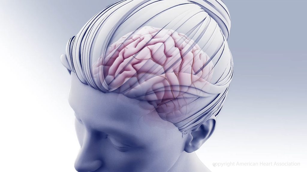
Sex Differences in Brain Glycogen After a Stroke may Yield New Treatments

Credit: American Heart Association

Although males and females are equally impacted by stroke, there are differences in recovery. Since oestrogen and progesterone have known neuroprotective effects, it is important to gauge their effects in stroke recovert. In a paper published in IBRO Neuroscience Reports, researchers have discovered differences between biomarkers such as glycogen levels in the brains of male and female mice.

“A stroke is caused by a loss of blood flow to brain cells. Without urgent intervention this may cause those cells to die because they constantly need energy and nutrients from the blood,” said Prof Nicole Sylvain, clinical research coordinator and lab manager at the University of Saskatchewan.

Sylvain and her colleagues are looking at treatments for post-stroke recovery that help supplement these energy losses. Using the Canadian Light Source (CLS) at the University of Saskatchewan (USask), the team was able to identify energy biomarkers in the brain, which could eventually inform clinicians about the effects of potential stroke treatments on brain recovery after a stroke.

The group’s recent study examined post-stroke differences between male and female mice, and found that female mice have higher amounts of glycogen in their brains. When the supply of glycogen is disrupted by stroke, the brain is severely impacted.

Most pre-clinical stroke research has been performed using male lab animals, with results usually generalised to both sexes. In clinical stoke cases, females have a higher incidence of ischaemic stroke and poorer outcomes, compared to males.

“We found that, for the most part, male data can be generalised for females, however, some of the metabolic markers we measured were actually different,” Sylvain said. “It’s really important to do the research on both sexes.”

It would be impossible for the team to detect the biomarkers without to the Mid-IR beamline.

“The only way to detect them in such an accurate way across the brain is with infrared imaging, so the CLS has been absolutely vital to our research.”

Source: University of Saskatchewan

Skipping Breakfast may Weaken Immune Cells

Photo by Malvestida on Unsplash

Fasting may be detrimental to fighting off infection, and could lead to an increased risk of heart disease, suggests a new study published in Immunity. The research in mouse models, shows that skipping meals triggers a response in the brain that negatively affects immune cells. The results could lead to a better understanding of how chronic fasting may affect the body long term.

“There is a growing awareness that fasting is healthy, and there is indeed abundant evidence for the benefits of fasting. Our study provides a word of caution as it suggests that there may also be a cost to fasting that carries a health risk,” says lead author Filip Swirski, PhD, Director of the Cardiovascular Research Institute at Icahn Mount Sinai. “This is a mechanistic study delving into some of the fundamental biology relevant to fasting. The study shows that there is a conversation between the nervous and immune systems.”

As part of a larger study on different fasting times affect the immune systems, researchers focused on the role of breakfast. They fed one group of mice breakfast right after waking up (breakfast is their largest meal of the day), and the other group had no breakfast. Researchers collected blood samples in both groups when mice woke up (baseline), then four hours later, and eight hours later.

Analysing the blood work, researchers saw a difference in the number of monocytes, which are white blood cells that are made in the bone marrow and travel through the body, where they play many critical roles, from fighting infections, to heart disease, to cancer.

At baseline, all mice had the same amount of monocytes. But after four hours, monocytes in mice from the fasting group were dramatically affected. Researchers found that 90% of these cells disappeared from the bloodstream, and the number further declined at eight hours. Meanwhile monocytes in the non-fasting group were unaffected.

In fasting mice, researchers discovered the monocytes travelled back to the bone marrow to hibernate. At the same time, production of new cells in the bone marrow diminished. The normally short-lived monocytes in the bone marrow survived longer as a consequence of staying in the bone marrow, and aged differently than the monocytes that stayed in the blood.

The researchers continued to fast mice for up to 24 hours, and then reintroduced food. The cells hiding in the bone marrow surged back into the bloodstream within a few hours, provoking heightened level of inflammation. Instead of protecting against infection, these altered monocytes were more inflammatory, making the body less resistant to fighting infection.

Researchers found that specific regions in the brain controlled the monocyte response during fasting. This study demonstrated that fasting elicits a stress response in the brain (feeling ‘hangry’) and this instantly triggers a large-scale migration of these white blood cells from the blood to the bone marrow, and then back to the bloodstream shortly after food is reintroduced.

Dr Swirski emphasised that while there is also evidence of the metabolic benefits of fasting, this new study is a useful advance in the full understanding of the body’s mechanisms.

“The study shows that, on the one hand, fasting reduces the number of circulating monocytes, which one might think is a good thing, as these cells are important components of inflammation. On the other hand, reintroduction of food creates a surge of monocytes flooding back to the blood, which can be problematic. Fasting, therefore regulates this pool in ways that are not always beneficial to the body’s capacity to respond to a challenge such as an infection,” explains Dr Swirski. “Because these cells are so important to other diseases like heart disease or cancer, understanding how their function is controlled is critical.”

Source: The Mount Sinai Hospital / Mount Sinai School of Medicine