Month: January 2023

Inpatient Blood Draws are Often Performed During Sleep Hours

Blood sample being drawn
Photo by Hush Naidoo Jade Photography on Unsplash

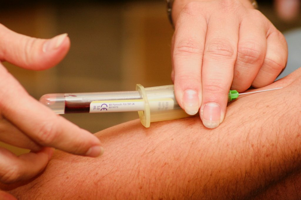
The sleep of hospitalised patients may be often interrupted due to non-urgent blood draws, according to findings from a Yale study published in JAMA. This may be exacerbating sleep deprivation, and putting them at greater risk for health events later on.

In an analysis of more than 5 million non-urgent blood draws collected at Yale New Haven Hospital from 2016 to 2019, a team of researchers found that a high proportion of them occurred during a three-hour window in the early morning.

“We found that nearly four in 10 of total daily blood draws were performed between 4am and 7am,” said César Caraballo-Cordovez, MD, a postdoctoral associate at Yale Center for Outcomes Research and Evaluation (CORE) and co-lead author of the study. “Importantly, we found that this occurred across patients with different sociodemographic characteristics, including older individuals who are at highest risk of adverse health events from sleep deprivation.”

Although early morning blood draws are often considered necessary to inform decisions during morning medical rounds, the authors suggest that sleep interruptions may increase the risk of delirium and other adverse events. “Patients who were recently hospitalised experience a period of generalised risk for myriad adverse health events, a condition named posthospital syndrome,” added Dr Caraballo-Cordovez. “The stress that patients experience during the hospitalisation – including stress from sleep deprivation – is a key contributor to this period of increased risk.”

“This is not an issue at just one hospital,” said Harlan M. Krumholz, MD, SM, professor of medicine and public health at Yale and CORE director. “Our findings reflect an aspect of how inpatient hospital care is being delivered in modern medicine. A more patient-centered care would limit nonurgent tests during sleep hours. However, these early morning blood draws are often considered necessary to make decisions during rounds.”

“We need to re-design our process to protect patients’ sleep, but major changes in our practice must be informed by solid studies that demonstrate the efficacy of strategies to do so without untoward effects,” added Krumholz.

Source: Yale School of Medicine

UK Study Reveals Doubling in Antipsychotics Prescriptions for Under-18s

Boy hanging from tree
Photo by Annie Spratt on Pexels

A cohort study on the prescription of antipsychotics to children and adolescents in the UK has found that they have doubled over the past two decades. The findings, published in The Lancet Psychiatry, depict a concerning tendency for more, longer prescriptions of antipsychotics for a wider array of indications, many of them off-label, and which may be influenced by US and European approvals.

Studies around the world have reported an increase in the prescription of antipsychotics for children and adolescents. While this may reflect actual changing clinical needs, most antipsychotics are not approved for use in under-18s due to lacking safety data, especially in the long term. There is also little evidence on indications for, and doses of, antipsychotic prescribing in children and adolescents.

The study used a cohort of over 7 million children and adolescents (age 3–18 years) assembled from a large English primary care database, and found a doubling in the proportion of prescribed antipsychotics between 2000 and 2019.

This increase resulted from the accumulation of repeated prescriptions to the same individuals combined with an increase in new prescriptions. The researchers found that antipsychotic prescribing was more frequent for children in more deprived areas, which reflected a previous UK study on adults.

The study also revealed multiple clinical indications for antipsychotics beyond their initial approvals, most commonly for anxiety and depression. Risperidone was the most prescribed antipsychotic for all indications apart from depression, for which the most prescribed antipsychotic was quetiapine, and eating disorders, for which it was olanzapine.

Prescribing trends for certain disorders could be though to reflect prevalence. The authors noted however that “the most common indications for antipsychotics were ASD, ADHD, anxiety, and depression. It could be the increasing prevalence of these disorders that causes higher prescribing rates. However, increasing ASD prevalence results primarily from patients with less severe ASD, who are unlikely to receive antipsychotics.”

They also observed that increases in prescribing appeared to be linked to new US and European approvals.

Limitations included the database not identifying whether a prescription was for a first time, and not tying indications directly to prescriptions. Dosage regimen information was also only available for a third of prescriptions. The database was also not necessarily nationally representative, and only reflected prescriptions issued in secondary care – referral to primary care means that the rate of prescribing may be underestimated.

Head Injury Doubles Long-term Mortality Risk

Photo by John Simmons on Unsplash

Adults who suffered any head injury during a 30-year study period had two times the rate of mortality than those who did not have any head injury, and mortality rates among those with moderate or severe head injuries were nearly three times higher, according to new research published in JAMA Neurology.

Head injury can be attributed to a number of causes, from motor vehicle crashes, unintentional falls, or sports injuries. Furthermore, head injury has been linked with a number of long-term health conditions, including disability, late-onset epilepsy, dementia, and stroke.

Previous studies have shown increased short-term mortality among hospitalised patients with head injuries. This longitudinal study evaluated 30 years of data from over 13 000 community-dwelling participants (ie not hospitalised or in nursing homes) to determine if head injury has an impact on mortality rates in adults over the long term. Of these, 18.4% reported one or more head injuries during the study period, and of those who suffered a head injury, 12.4% were recorded as moderate or severe. The median period of time between a head injury and death was 4.7 years.

Death from all causes was recorded in 64.6% of those individuals who suffered a head injury, and in 54.6% of those without any head injury. Accounting for participant characteristics, investigators found that the mortality rate from all-causes among participants with a head injury was 2.21 times the mortality rate among those with no head injury. Further, the mortality rate among those with more severe head injuries was 2.87 times the mortality rate among those with no head injury.

“Our data reveals that head injury is associated with increased mortality rates even long-term. This is particularly the case for individuals with multiple or severe head injuries,” explained the study’s lead author, Holly Elser, MD, PhD, MPH a Neurology resident at Penn. “This highlights the importance of safety measures, like wearing helmets and seatbelts, to prevent head injuries.”

Investigators also evaluated the data for specific causes of death among all participants. Overall, the most common causes of death were cancers, cardiovascular disease, and neurologic disorders (which include dementia, epilepsy, and stroke). Among individuals with head injuries, deaths caused by neurologic disorders and unintentional injury or trauma (like falls) occurred more frequently.

When investigators evaluated specific neurologic causes of death among participants with head injury, they found that nearly two-thirds of neurologic causes of death were attributed to neurodegenerative diseases, like Alzheimer’s and Parkinson’s disease. These diseases composed a greater proportion of overall deaths among individuals with head injury (14.2%) versus those without (6.6%). Further research into this association is recommended.

Source: University of Pennsylvania School of Medicine

In Older Women, Physical Activity Reduces Risk of Dementia

Photo by Bennett Tobias on Unsplash

Senior women were less likely to develop mild cognitive impairment or dementia if they did more daily walking and moderate-to-vigorous physical activity, according to a new study published in of Alzheimer’s & Dementia: The Journal of the Alzheimer’s Association.

The University of California San Diego-led team reported that among women aged 65 or older, each extra 31 minutes per day of moderate-to-vigorous physical activity was linked to a 21% lower risk of developing mild cognitive impairment or dementia. Risk was also reduced by 33% with each extra 1865 daily steps.

“Given that the onset of dementia begins 20 years or more before symptoms show, the early intervention for delaying or preventing cognitive decline and dementia among older adults is essential,” said senior author Andrea LaCroix, PhD, MPH, UC San Diego professor.

Dementias are a debilitating neurological condition that can cause loss of memory, the ability to think, problem solve or reason. Mild cognitive impairment is an early stage of memory loss or thinking problems that is not as severe as dementias.

More women live with and are at higher risk of developing dementia than men.

“Physical activity has been identified as one of the three most promising ways to reduce risk of dementia and Alzheimer’s disease. Prevention is important because once dementia is diagnosed, it is very difficult to slow or reverse. There is no cure,” said LaCroix.

However, because few large studies have examined device measures of movement and sitting in relation to mild cognitive impairment and dementia, much of the published research on the associations of physical activity and sedentary behavior with cognitive decline and dementia is based on self-reported measures, said first author, Steven Nguyen, Ph.D., M.P.H., postdoctoral scholar at the Herbert Wertheim School of Public Health.

For this study, the researchers sampled data from 1,277 women as part of two Women’s Health Initiative (WHI) ancillary studies – the WHI Memory Study (WHIMS) and the Objective Physical Activity and Cardiovascular Health (OPACH) study. The women wore research-grade accelerometers and went about their daily activities for up to seven days to obtain accurate measures of physical activity and sitting.

The activity trackers showed the women averaged 3,216 steps, 276 minutes in light physical activities, 45.5 minutes of moderate-to-vigorous physical activity and 10.5 hours of sitting per day. Examples of light physical activity could include housework, gardening or walking. Moderate-to-vigorous physical activity could include brisk walking.

The study findings also showed that higher amounts of sitting and prolonged sitting were not associated with higher risk of mild cognitive impairment or dementia.

Together, this information has clinical and public health importance as there is little published information on the amount and intensity of physical activity needed for a lower dementia risk, said Nguyen.

“Older adults can be encouraged to increase movement of at least moderate intensity and take more steps each day for a lower risk of mild cognitive impairment and dementia,” said Nguyen.

“The findings for steps per day are particularly noteworthy because steps are recorded by a variety of wearable devices increasingly worn by individuals and could be readily adopted.”

The authors said further research is needed among large diverse populations that include men.

Source: University of California – San Diego

Schizophrenia Associated with 12-hour Gene Cycles in the Brain

Photo by Alex Green on Pexels

In the open-access journal PLOS Biology, researchers present the first evidence of 12-hour cycles of gene activity in the human brain. Led by Madeline R. Scott, the study also reveals that some of those 12-hour rhythms are missing or altered in the postmortem brains of patients with schizophrenia.

Schizophrenia patients are known to have disturbances in several types of 24-hour bodily rhythms, including sleep/wake cycles, hormone levels, and gene activity in the prefrontal cortex of the brain. However, virtually nothing is known about gene activity in the brain for cycles that are shorter than the usual 24-hour circadian rhythm. A few years ago, researchers discovered that certain genes in the body were associated with 12-hour bodily rhythms, which may have an origin in the 12-hour cycle of ocean tides.

As it is not possible to measure gene transcript levels in living brains, the new study instead used a time-of-death analysis to search for 12-hour rhythms in gene activity within postmortem brains. They focused on the dorsolateral prefrontal cortex as it is associated with cognitive symptoms and other abnormalities in gene expression rhythms that have been observed in schizophrenia.

Numerous genes in the normal dorsolateral prefrontal cortex were found to have 12-hour rhythms in activity. Among them, gene activity levels related to building connections between neurons peaked in the afternoon/night, while those related to mitochondrial function (and therefore cellular energy supply) peaked in the morning/evening.

In contrast, postmortem brains from patients with schizophrenia contained fewer genes with 12-hour activity cycles, and those related to neural connections were missing entirely. Additionally, although the mitochondria-related genes did maintain a 12-hour rhythm, their activity did not peak at the normal times. Whether these abnormal rhythms underlie the behavioural abnormalities in schizophrenia, or whether they result from medications, nicotine use, or sleep disturbances should be examined in future studies.

Co-author Colleen A. McClung adds: “We find that the human brain has not only circadian (24 hour) rhythms in gene expression but also 12-hour rhythms in a number of genes that are important for cellular function and neuronal maintenance. Many of these gene expression rhythms are lost in people with schizophrenia, and there is a dramatic shift in the timing of rhythms in mitochondrial-related transcripts which could lead to suboptimal mitochondrial function at the times of day when cellular energy is needed the most.”

Source: ScienceDaily

A Severe Form of Dementia may in Fact be Caused by a Cerebrospinal Fluid Leak

MRI images of the brain
Photo by Anna Shvets on Pexels

A new study suggests that some patients diagnosed with behavioural-variant frontotemporal dementia (bvFTD) – a presently incurable, mentally debilitating condition – may instead have a cerebrospinal fluid leak, which is detectable on MRI scans and often treatable. The researchers say these findings, published in the peer-reviewed journal Alzheimer’s & Dementia: Translational Research and Clinical Interventionscould lead to a cure.

“Many of these patients experience cognitive, behavioural and personality changes so severe that they are arrested or placed in nursing homes,” said Wouter Schievink, MD, professor of Neurosurgery at Cedars-Sinai. “If they have behavioural-variant frontotemporal dementia with an unknown cause, then no treatment is available. But our study shows that patients with cerebrospinal fluid leaks can be cured if we can find the source of the leak.”

When cerebrospinal fluid (CSF) leaks into the body, the brain can sag, causing dementia symptoms. Schievink said many patients with brain sagging, detectable in MRI, go undiagnosed, and he advises clinicians to take a second look at patients with telltale symptoms.

“A knowledgeable radiologist, neurosurgeon or neurologist should check the patient’s MRI again to make sure there is no evidence for brain sagging,” Schievink said.

Clinicians can also ask about a history of severe headaches that improve when the patient lies down, significant sleepiness even after adequate night-time sleep, and whether the patient has ever been diagnosed with a Chiari brain malformation, a condition in which brain tissue extends into the spinal canal. Brain sagging, Schievink said, is often mistaken for a Chiari malformation.

Even when brain sagging is detected, the source of a CSF leak can be difficult to locate. When the fluid leaks through a tear or cyst in the surrounding membrane, it is visible on CT myelogram imaging with the aid of contrast medium.

Schievink and his team recently discovered an additional cause of CSF leak: the CSF-venous fistula. In these cases, the fluid leaks into a vein, making it difficult to see on a routine CT myelogram. To detect these leaks, technicians must use a specialized CT scan and observe the contrast medium in motion as it flows through the cerebrospinal fluid.

In this study, investigators used this imaging technique on 21 patients with brain sagging and symptoms of bvFTD, and they discovered CSF-venous fistulas in nine of those patients. All nine patients had their fistulas surgically closed, and their brain sagging and accompanying symptoms were completely reversed.

“This is a rapidly evolving field of study, and advances in imaging technology have greatly improved our ability to detect sources of CSF leak, especially CSF-venous fistula,” said Keith L. Black, MD, chair of the department of Neurosurgery at Cedars-Sinai. “This specialised imaging is not widely available, and this study suggests the need for further research to improve detection and cure rates for patients.”

The remaining 12 study participants, whose leaks could not be identified, were treated with nontargeted therapies designed to relieve brain sagging, such as implantable systems for infusing the patient with CSF. However, only three of these patients experienced relief from their symptoms.

“Great efforts need to be made to improve the detection rate of CSF leak in these patients,” Schievink said. “We have developed nontargeted treatments for patients where no leak can be detected, but as our study shows, these treatments are much less effective than targeted, surgical correction of the leak.”

Source: Cedars-Sinai Medical Center

Neuronal Molecule Drives Prostate Cancer Aggressiveness

Cancer-associated fibroplasts surrounding a prostate tumour. Credit: Moscat and Diaz Meco labs

Prostate cancer is the second most common cancer and the second leading cause of cancer death among American men. Now, researchers have discovered key molecular players that drive prostate cancer to progress into a highly aggressive form of the disease called neuroendocrine prostate cancer that currently has no effective treatment. The finding, published in in the journal Scientific Reports, opens new avenues to therapeutics to treat neuroendocrine prostate cancer.

“We have found novel pathways that promote neuroendocrine prostate cancer,” says senior author Lucia R. Languino, PhD, a professor in the department of Pharmacology, Physiology and Cancer Biology and director of the Genetics, Genomics, and Cancer Biology PhD Program at Thomas Jefferson University.

Most prostate cancers are a type of disease called prostate adenocarcinoma. Other types of prostate cancer, including neuroendocrine tumours, are rare. However, unlike prostate adenocarcinoma, neuroendocrine prostate cancer is very aggressive and can quickly spread to other parts of the body. Treatments that are effective for adenocarcinomas in the prostate do not work against neuroendocrine prostate cancers.

Adenocarcinoma prostate cancers can progress into neuroendocrine prostate cancer. Until now, how this transition occurs has been a mystery.

To better understand how neuroendocrine prostate cancer develops, Dr Languino and colleagues looked for biomarkers of the disease. In previous work, they discovered that a molecule known as aVb3 integrin is abundant in mice and humans with neuroendocrine prostate cancer, but missing in prostate adenocarcinoma.

To look for molecules unique to neuroendocrine prostate cancer, the researchers found that aVb3 integrin expression in prostate cancer cells bumped up the expression of a known marker of neuroendocrine prostate cancer and significantly increased the expression of a molecule called Nogo receptor 2 (NgR2).

The finding “was a big discovery,” Dr Languino says. That’s because NgR2 is a protein found in nerve cells, where it contributes to neuronal functions. It has never before been studied in cancer, of any kind.

Dr Languino and her colleagues wanted to find out what this molecule, a neuronal protein, is doing in cancer.

An initial experiment revealed that NgR2 binds the aVb3 integrin. The scientists also saw that in mice with neuroendocrine prostate tumors, aVb3 integrin and NgR2 were both present in the primary tumor and in cancerous lesions that had formed in the lungs of the animals. A follow-up experiment made it clear that both aVb3 integrin and NgR2 are necessary for neuroendocrine prostate cancers.

When Dr Languino and her team lowered the amount of NgR2 in neuroendocrine prostate cancer cells, neuroendocrine markers also decreased. The results suggest that NgR2 plays a role in the development of neuroendocrine prostate cancer. Lowering the amount of NgR2 also reduced the ability of cancer cells to grow and move, indicating that NgR2 may have a hand in cancer spreading to other parts of the body, in a process known as metastasis. Metastases are often what makes cancers fatal.

“These two molecules, aVb3 integrin and NgR2, seem to create a combination that is lethal,” Dr Languino says.

She and her colleagues are now looking for a molecule or antibody that would block the effect of NgR2, or the aVb3 integrin/NgR2 complex, to inhibit their ability to promote neuroendocrine prostate cancer growth and development, and make the cancer more susceptible to therapy.

Source: Thomas Jefferson University

A New Mosquito Repellent Alternative to DEET

Mosquito, a malaria parasite vector
Photo by Егор Камелев on Unsplash

The chemical DEET has proven effective at keeping disease-carrying mosquitoes at bay, but the repellent is smelly and its protection is short-lived. Now, researchers report in the Journal of Agricultural and Food Chemistry that they have designed safe alternatives with some advantages over DEET, including a nice smell and much longer protection.

DEET disrupts a mosquito’s ability to locate humans. Until recently, it was considered the gold standard among topical repellents, but some find its strong odor offensive. It has to be reapplied frequently, and at high concentrations, it can damage synthetic fabrics and plastics. Another popular repellent known as picaridin is now regarded as a better alternative, since its protective effect lasts longer, and it doesn’t have an odor or damage items. However, like DEET, it has to be reapplied after swimming or sweating.

So, Francesca Dani and colleagues wanted to look for alternatives to these established products. In prior work, the team used as starting materials two plant-based natural repellents that offered only short-term protection from mosquitoes. The researchers converted these terpenoids into cyclic acetals and hydroxyacetals, thereby extending their protective timespan beyond that of DEET. But the researchers wanted to improve on these initial products.

In the current work, the team synthesised additional cyclic hydroxyacetals from inexpensive, commercially available carbonyls. The new cyclic compounds had pleasant, much fainter odors and were easier to dissolve in water, meaning they can be formulated without high concentrations of alcohol. Some were as effective as DEET and picaridin at repelling Asian tiger mosquitoes, which have spread widely in the U.S. and carry diseases, including encephalitis, dengue and dog heartworm. And like picaridin, they provided human volunteers more than 95% protection from bites for at least eight hours, while DEET’s protection rapidly declined below that level after just two hours.

Toxicity of some of the most active new compounds was comparable to or lower than the traditional repellents. Two hydroxyacetals were also less likely to cause immune reactions or to penetrate cell layers than picaridin. The researchers conclude that their compounds represent a new class of promising mosquito repellents that can compete favorably with DEET and picaridin in terms of efficacy and safety.

Source: American Chemical Society

For Women, Lymphoma Chemotherapy more Effective in Afternoon

Photo by Malvestida on Unsplash

While chemotherapy is highly effective at killing cancer cells, it also kills healthy cells, something which medical research is trying minimise. Recently, ‘chronochemotherapy’ has garnered growing attention in the research community. As the name suggests, the aim is timing the delivery of the drug when the body is least vulnerable to the harmful effects of the drug, while the cancer cells are at their most vulnerable.

Chronochemotherapy exploits the fact that human physiological processes, including cell proliferation and differentiation, are regulated by the circadian clock. However, it is not yet widely exploited in real-world clinical settings because there at present is no systematic method to find the optimal chemotherapy delivery time.

This problem was tackled by an interdisciplinary team of researchers from the Institute for Basic Science (IBS), South Korea. They were led by the principal investigators, mathematician Kim Jae Kyoung at IBS and oncologist Koh Youngil at Seoul National University Hospital. The researchers studied a group of patients suffering from diffuse large B-cell lymphoma (DLBCL), which accounts for about 30 to 40% of non-Hodgkin’s lymphoma. Their findings are published in the journal JCI Insights.

The research team noticed that DLBCL patients at Seoul National University Hospital received chemotherapy at two different schedules, with some patients receiving morning treatment (8:30), while others taking the drugs in the afternoon (14:30). All patients received R-CHOP, which is a combination of targeted therapy and chemotherapy, 4 to 6 times in the morning or afternoon at intervals of about 3 weeks.

Figure 1. Chemotherapy in the afternoon can improve treatment outcomes
The daily fluctuation of proliferative activity of bone marrow is larger in females than in males, and it becomes higher in the morning (left). Thus, chemotherapy in the morning strongly inhibits proliferative activity in female lymphoma patients, resulting in a higher incidence of adverse events such as neutropenia and infections. This forces the clinicians to reduce the dose intensity (center). Consequently, female patients undergoing the morning treatment show a lower survival probability than those undergoing the afternoon treatment (right). Specifically, only ~13% of female patients treated in the afternoon had a worse outcome and ~2% of them died while ~37% of female patients treated in the morning had a worse outcome and ~25% of them died. Male patients did not show any difference in treatment outcomes depending on the chemotherapy delivery time.

They analysed 210 patients to investigate differences between morning and afternoon treatment. They found that female patients who received afternoon treatment had 12.5 times reduced mortality rate (25% to 2%), while the cancer recurrence after 60 months was decreased by 2.8 times (37% to 13%). In addition, chemotherapy side effects such as neutropenia were more common in female patients who received morning treatment.

Surprisingly, there was no difference in treatment efficiency depending on the treatment schedule in the case of male patients.

To understand the cause of gender differences, the research team analysed ~14 000 blood samples from the Seoul National University Hospital Health Examination Center. It was found that in females, white blood cell count tends to decrease in the morning and increase in the afternoon. This indicates that the bone marrow proliferation rate is higher in the morning than in the afternoon because there is a ~12hr delay between the bone marrow proliferation and blood cell production.

This means that if a female patient receives chemotherapy in the morning when bone marrow is actively producing blood cells, the possibility of adverse side effects becomes greater. These results are consistent with the findings from recent randomised clinical trials that showed female colorectal cancer patients treated with irinotecan in the morning suffered from higher drug toxicities.

One confounding variable was the drug dose. Since the morning female patients suffered from greater adverse side effects, oftentimes the dose had to be reduced in these patients. On average, the drug dose was reduced by ~10% compared to the dose intensity given to female patients receiving the afternoon treatment.

Unlike female patients, it was found that male patients did not show a significant difference in white blood cell count and bone marrow cell proliferation activity throughout the day, which is the reason why the timing of the treatment had no impact.

Professor Koh Young-il said, “We plan to verify the conclusions of this study again with a large-scale follow-up study that completely controls confounding variables, and to confirm whether chemotherapy has similar effects in other cancers.”

Ci Kim Jae Kyung said, “Because the time of the internal circadian clock can vary greatly depending on the individual’s sleep-wake patterns, we are currently developing a technology to estimate the time of the circadian clock from the patient’s sleep pattern. We hope that it can be used to develop an individualised anti-cancer chronotherapy.”

Source: Institute for Basic Science

New Microneedle Patch Rapidly Stops Bleeding

Haemostatic microneedle technology can be applied like a typical adhesive bandage to quickly stop bleeding. The biocompatible and biodegradable microneedle arrays (MNAs) on the patch increase its surface contact with blood to accelerate the clotting process and also increase the adhesive properties of the patch via mechanical interlocking to promote wound closure. Credit: Designed by Amir Sheikhi and Reihaneh Haghniaz/Executed by Natan Barros. All Rights Reserved.

In the US, secondary, uncontrolled bleeding from traumatic injury is the leading cause of death from ages one to 46. Amir Sheikhi, assistant professor of chemical engineering and of biomedical engineering at Penn State, has a plan to change that with a novel microneedle patch that can immediately stop bleeding after injury.

He described his technology in a new paper in the journal Bioactive Materials.

“Excessive bleeding is a serious challenge for human health,” Sheikhi said. “With haemorrhaging injuries, it is often the loss of blood – not the injury itself – that causes death. There is an unmet medical need for ready-to-use biomaterials that promote rapid blood coagulation.”

Sheikhi’s haemostatic microneedle technology can be applied like a typical adhesive bandage to quickly stop bleeding. The biocompatible and biodegradable microneedle arrays (MNAs) on the patch increase its surface contact with blood, accelerating the clotting process. The needles also increase the adhesive properties of the patch via mechanical interlocking to promote wound closure.

In vitro, the engineered MNAs reduced clotting time from 11.5 minutes to 1.3 minutes; and in a rat liver bleeding model, they reduced bleeding by more than 90%,” Sheikhi said. “Those 10 minutes could be the difference between life and death.”

The MNA patch is comparable hydrogel technology that is currently used to treat bleeding wounds in hospitals, but hydrogel applications require preparation and medical expertise. The microneedle patch is pre-engineered for immediate application that anyone can use to stop bleeding, Sheikhi said, much like a typical over-the-counter adhesive bandage.

Microneedles – which are already in use to deliver biologics, such as cells or drugs, through the skin or for cosmetic procedures to stimulate collagen production – are tiny, making their application pain-free, according to Sheikhi.

The researchers are now working to commercialise the patch, with more testing plans.

Source: Penn State