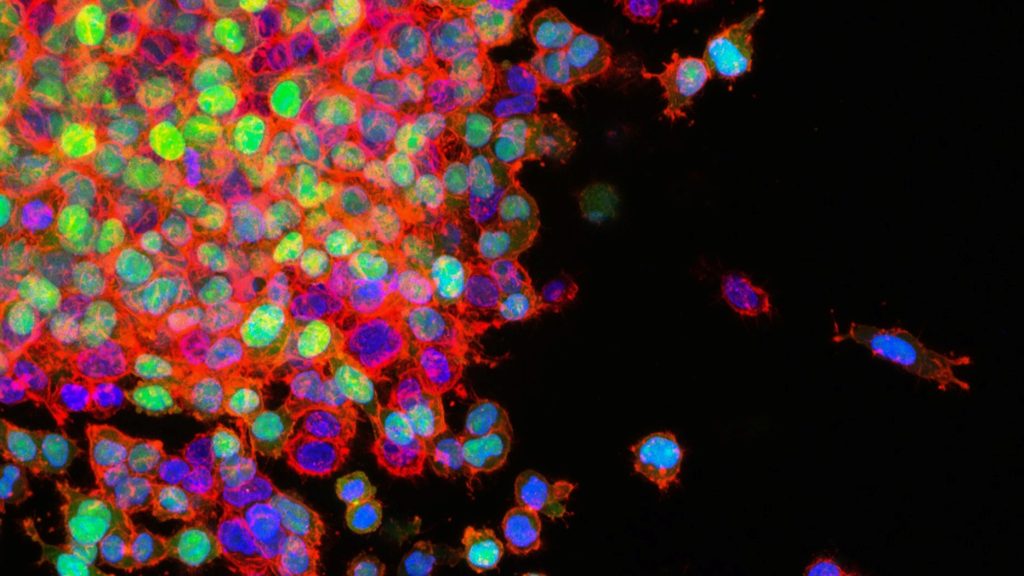
When cancer cells metastasise, they have to break connections with neighbouring cells and migrate to other tissues. Both processes are promoted by signalling molecules released by the cancer cells, which thereby increase the malignancy of tumours. Researchers found that the release of these ‘prometastatic’ factors is influenced by the cellular skeleton – specifically, actin filaments. The study was published in Advanced Science.
Actin’s multiple role functions in cancer propagation
Actin filaments are part of the cell skeleton and essential for stability and motility. They form a network that dynamically builds up and gets broken down by the addition or detachment of building blocks at the filaments’ ends. These processes are precisely regulated by other molecules, such as formins. The dynamics of the actin network enable the movement of cells, for example during development or wound closure, but also that of spreading cancer cells. Actin also plays a role in the transport of substances within the cell. However, this is less well understood than that of other intracellular transport mechanisms.
The research team led by Prof Dr Robert Grosse and Dr Carsten Schwan from the University of Freiburg, now found that the actin network also enables the release of prometastatic factors, such as ANGPTL4 which is an important prometastatic factor that promotes the formation of metastases in various types of cancer. For their study, they used high-resolution microscopy to track the movement of individual transport vesicles within living cancer cells.
“We observed that ANGPTL4-loaded vesicles are conveyed to the periphery of the cell by means of dynamic and localised polymerisation of actin filaments,” says Grosse, who is a member of the Cluster of Excellence CIBSS – Centre for Integrative Biological Signalling Studies at the University of Freiburg.
Transportation along actin filaments
Based on microscopic observations and genetic analyses, the scientists conclude that the vesicles’ movement is controlled by the formin-like molecule FMNL2 by initiating polymerisation (ie elongation) of actin filaments directly at the vesicle. “We already knew that increased FMNL2 activity has prometastatic effects in many types of tumours,” says Grosse. “In our current work we could now demonstrate an important underlying process and a connection to the TGFbeta signalling pathway.” According to the scientist, this knowledge could be used for tumour diagnostics or therapy. for example, by developing an antibody that indicates the presence of active FMNL2 or pharmacologically targets active, phosphorylated FMNL2.
Source: University of Freiburg

