Day: November 21, 2022

More Metabolic Imbalances in Paediatric T1D Diagnoses in the Pandemic

Photo by Towfiqu Barbhuiya on Unsplash

During the COVID pandemic, significantly more children and young people had already developed diabetic ketoacidosis when diagnosed with type 1 diabetes (T1D) than in previous years. These findings were reported in The Lancet Diabetes & Endocrinology,

If children and young people have already developed metabolic imbalances (diabetic ketoacidosis) at the time of diagnosis of T1D, this can result in complications such as extended stays in hospital, poorer long-term control of blood sugar levels, brain enema, or even a higher mortality rate.

During the COVID pandemic, diabetes centres around the world saw an increased prevalence of diabetic ketoacidosis in diagnosed cases of T1D. DZD researchers, together with international colleagues, investigated whether the number of diabetic ketoacidosis cases associated with the diagnosis of paediatric T1D increased more than expected. To achieve this, they analysed the number of diabetic ketoacidosis cases before and during the pandemic.

The team evaluated data from 13 national diabetes registers, with 104 290 children and young people aged between 6 months and 18 years old who were diagnosed with T1D between 1 January 2006 and 31 December 2021. The observed prevalence of diabetic ketoacidosis during 2020 and 2021 was compared with predictions based on the years before the pandemic (2006–2019).

Increase greater than expected

Between 2006 and 2019, 23 775 of 87 228 children had diabetic ketoacidosis when diagnosed with T1D (27.3%). The mean annual increase in the prevalence of diabetic ketoacidosis for the entire cohort between 2006 and 2019 was 1.6%. During the pandemic, the numbers were significantly above the predicted prevalence. In 2020, the adjusted observed prevalence of diabetic ketoacidosis was 39.4% (predicted prevalence 32.5%) and 38.9% in 2021 (predicted prevalence 33.0%).

“The increasing prevalence of diabetic ketoacidosis associated with the diagnosis of type 1 diabetes in children is a global problem. There was already an increase in prevalence before the COVID-19 pandemic. During the pandemic, this increase was even greater,” notes DZD scientist Prof. Reinhard W. Holl from Ulm University.

The authors suggest that providing a comprehensive explanation of the classic symptoms of T1D in childhood to the general public, those active in the childcare or daycare settings, and primary care physicians could help raise awareness of the symptoms of T1D. Furthermore, public health measures could be used, eg, implementing a general islet-cell autoantibodies screening program for children to reduce the number of dangerous metabolic imbalances.

Source: Deutsches Zentrum fuer Diabetesforschung DZD

Temporary Low-protein Diet could Enhance Colon Cancer Treatment

Photo by Ella Olsson on Pexels

A brief switch to a low-protein diet could be a key to enhancing colon cancer treatment, say researchers investigating cancer metabolism. They reported their findings in the journal Gastroenterology.

Like all cells, cancer cells need nutrients to survive and grow. One of the most important nutrient sensing molecules in a cell is called mTORC1. Often called a master regulator of cell growth, it lets cells sense different nutrients, thereby growing and proliferating. When nutrients are limited, cells dial down nutrient sensing cascade and turn off mTORC1.

While mTORC1 is known to be hyperactive in colon cancer, the key question is whether colon tumours hijack nutrient sensing pathways to fire up the master regulator.

“In colon cancer, when you decrease the nutrients available in the tumours, the cells don’t know what to do. Without the nutrients to grow, they undergo a kind of crisis, which leads to massive cell death,” said senior author Yatrik M. Shah, PhD, professor at Michigan Medicine.

Researchers found in cells and in mice that a low-protein diet blocked the nutrient signalling pathway that fires up a master regulator of cancer growth.

The regulator, mTORC1, controls how cells use nutritional signals to grow and multiply. It’s highly active in cancers with certain mutations and is known to cause cancer to become resistant to standard treatments. A low-protein diet, and specifically a reduction in two key amino acids, changed the nutritional signals through a complex called GATOR.

GATOR1 and GATOR2 work together to keep mTORC1 in business. When a cell has plenty of nutrients, GATOR2 activates mTORC1. When nutrients are low, GATOR1 deactivates mTORC1. Limiting certain amino acids blocks this nutrient signalling.

Previous efforts to block mTORC have focused on inhibiting its cancer-causing signals. But these inhibitors cause significant side effects — and when patients stop taking it, the cancer comes back. The study suggests that blocking the nutrient pathway by limiting amino acids through a low-protein diet offers an alternative way to shut down mTORC.

“We knew that nutrients were important in mTORC regulation but we didn’t know how they directly signal to mTORC. We discovered the nutrient signalling pathway is just as important to regulate mTORC as the oncogenic signalling pathway,” said study first author Sumeet Solanki, Ph.D., a research investigator at the Rogel Cancer Center.

Researchers confirmed their findings in cells and mice, where they saw that limiting amino acids stopped the cancer from growing and led to increased cell death. They also looked at tissue biopsies from patients with colon cancer, which confirmed high markers of mTORC correlated with more resistance to chemotherapy and worse outcomes. Solanki said this could provide an opportunity to direct treatment for patients with this marker.

“A low-protein diet won’t be standalone treatment. It has to be combined with something else, such as chemotherapy,” Solanki said.

The risk with a low-protein diet is that people with cancer often experience muscle weakness and weight loss, which limiting protein could exacerbate.

“Putting cancer patients on a protein-deficient diet long-term is not ideal. But if you can find key windows – like at the start of chemotherapy or radiation – when patients could go on a low protein diet for a week or two, we could potentially increase the efficacy of those treatments,” Shah said.

Further research will refine this concept of a therapeutic window to limit amino acids. Researchers will also seek to understand how these pathways are creating resistance to treatment and whether an inhibitor could block the GATOR complexes.

Source: Michigan Medicine – University of Michigan

Metastases Survive by Adapting to Different Tissues

Source: National Cancer Institute on Unsplash

In a breakthrough for understanding metastases, researchers have found that, as metastatic cancers spread to different parts of the body, they adapt their metabolism to the tissue in which they grow. The findings, which help further break down the puzzle of metastasis, are published in PNAS.

Metabolism in the body is an important target for cancer treatments, where the focus is on stopping the progress of cancer cells.

“Obviously, the local environment affects the cancer cells more than previously known. The metastatic tumours should show the same metabolic properties no matter where in the body they are located, but we discovered that the cancer cells largely adapted their metabolism to the new tissue in order to continue to develop and grow. This is important knowledge, which shows that we cannot consider the metastases as their original tumours,” says Fariba Roshanzamir, PhD in Systems and Synthetic Biology at Chalmers and the study’s lead author.

Cutting off cancer metabolism

Fariba Roshanzamir works in Professor Jens Nielsen’s research group at Chalmers and has, together with Swedish and international colleagues, been able to establish the groundbreaking results. The study focused primarily on triple-negative breast cancer but the conclusions can, according to the researchers, be applied to all types of metastatic cancer. This opens new doors to develop more effective treatments.

“If we manage to shut down the metabolism in a tumour, it will stop working and this study provides important keys to better understand what to target. Selecting metabolic inhibitors that specifically target the metastases in the organs to which the tumour has spread, rather than treating them as their original tumours, is of great importance to be able to find good strategies for treatments in the future,” she says.

Source: Chalmers University of Technology

Alzheimer’s Prions also Appear in Down Syndrome

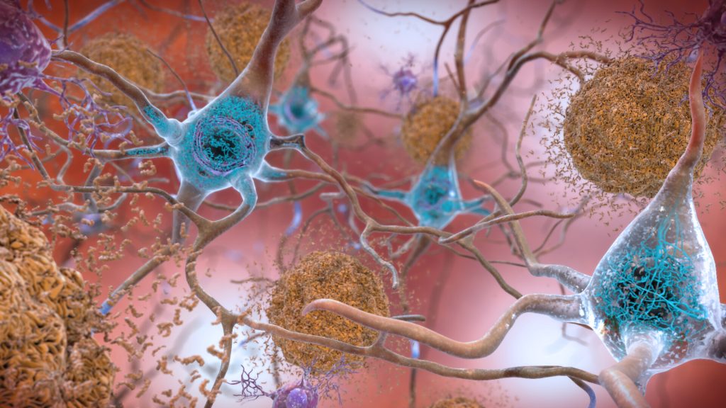
Plaques and neurons. Source: NIAH

The brains of people with Down syndrome develop the same neurodegenerative tangles and plaques associated with Alzheimer’s disease and they frequently demonstrate signs of the neurodegenerative disorder in their 40s or 50s. A new study in the journal PNAS shows that these tangles and plaques are driven by the same amyloid beta (Aß) and tau prions that they showed are behind Alzheimer’s disease.

Prions begin as normal proteins that become misshapen and self-propagate. They spread through tissue like an infection by forcing normal proteins to adopt the same misfolded shape. In both Alzheimer’s and Down syndrome, as Aß and tau prions accumulate in the brain, they cause neurological dysfunction that often manifests as dementia.

Tau tangles and Aß plaques are evident in most people with Down syndrome by age 40, according to the National Institute on Aging, with at least 50% of this population developing Alzheimer’s as they age.

The new study highlights how a better understanding of Down syndrome can lead to new insights about Alzheimer’s, as well.

“Here you have two diseases – Down syndrome and Alzheimer’s disease – that have entirely different causes, and yet we see the same disease biology. It’s really surprising,” said Stanley Prusiner, MD, the study’s senior author, who was awarded the Nobel Prize in 1997 for his discovery of prions.

Down syndrome is the most common neurodegenerative disease among younger people in the United States, while Alzheimer’s is the most common among adults.

Down syndrome occurs because of an extra copy of chromosome 21. Among the many genes on that chromosome is one called APP, which codes for one of the major components of amyloid beta. With an extra copy of the gene, people with Down syndrome produce excess APP, which may explain why they develop amyloid plaques early in life.

A clearer picture in young brain

It’s been known for some time that Aß plaques and tau tangles are present in both Down syndrome and Alzheimer’s. Having shown earlier that these neurodegenerative features are provoked by prions in Alzheimer’s, the researchers wanted to know whether the same aberrant proteins were present in the brains of people with Down syndrome.

While these plaques and tangles in the brains of people with Alzheimer’s disease have been well-studied, it can be challenging to discern which changes in the brain are from old age and which are from prion activity, said Prusiner.

“Because we see the same plaques-and-tangles pathology at a much younger age in people with Down syndrome, studying their brains allows us to get a better picture of the early process of disease formation, before the brain has become complicated by all the changes that go on during aging,” he said. “And ideally, you want therapies that address these early stages.”

Employing a variation on the novel assay they used in the Alzheimer’s study, the team looked at donated tissue samples from deceased people with Down syndrome, which they obtained from biobanks around the world. Of the 28 samples from donors aged 19 to 65 years old, the researchers were able to isolate measurable amounts of both Aß and tau prions in almost all of them.

New insights could yield preventative measures

The results confirm not only that prions are involved in the neurodegeneration seen in Down syndrome, but that Aß drives the formation of tau tangles as well as amyloid plaques, a relationship that has been suspected but not proven.

“The field has long tried to understand what the intersection is between these two pathologies,” said lead author Carlo Condello, PhD, also a member of the UCSF Institute for Neurodegenerative Diseases. “The Down syndrome case corroborates the idea; now you have this extra chromosome that’s driving the Aß, and there’s no tau gene on the chromosome. So, it’s truly by increasing the expression of Aß that you kick off production of the tau.”

These and other insights gained from studying the brains of people with Down syndrome will lead to a much better picture of how prions begin to form in the first place, said Condello.

Whether the Down syndrome brain tissue will prove to be the ultimate model for developing treatments for Alzheimer’s remains to be seen, the researchers said. While the two disorders share many similarities in their prion pathobiology, there are some differences that may be limiting.

Still, the researchers said, studying the plaques and tangles in Down syndrome is a promising route to identifying the specific prions that arise at the very earliest stages of the disease process. That insight could open new vistas on not only treating but perhaps even fending off Alzheimer’s disease.

“If we can understand how this neurodegeneration begins, we are one big step closer to being able to intervene at a meaningful point and actually prevent these large brain lesions from forming,” Condello said.

Source: University of California – San Francisco

ADHD Drug for Amphetamine Addiction Linked to Reduced Risks

Photo by Towfiqu barbhuiya on Unsplash

In a large registry-based study investigating medication use in people with substance use disorders, the ADHD medication lisdexamfetamine was associated with the lowest risk of hospitalisation and death in people with amphetamine addiction. The findings, which also showed drugs which worsened outcomes, were published in JAMA Psychiatry.

“Our results suggest that lisdexamfetamine is associated with the best outcomes, and encourage the conduction of randomised controlled trials to explore this further, says first author Jari Tiihonen, professor at Karolinska Institutet.

Worldwide, amphetamines are the second most used illicit drugs and hospitalisations related to its use are rising.

At present, there are no approved pharmacological interventions available for treating amphetamine or methamphetamine addiction. While certain medications have shown promising results, the studies so far have often been small and convincing evidence is lacking.

Registry-based study

In the present study, the researchers investigated the association between generally used medications among persons with substance use disorder and the risk of two primary outcomes in people with amphetamine or methamphetamine use disorder: 1) hospitalisation due to substance use disorder or 2) hospitalisation due to any cause, or death.

The study enrolled nearly 14 000 individuals aged 16 to 64 years in Sweden with a registered first-time diagnosis of amphetamine or methamphetamine use disorder from July 2006 to December 2018. Individuals with schizophrenia or bipolar disorder were excluded.

Patients were followed from diagnosis until they died, emigrated, were diagnosed with schizophrenia or bipolar disorder or the study ended. The median follow-up time was 3.9 years.

Comparing effects in the same individual

The researchers looked at how the risk of hospitalisation or death for each individual differed depending on whether they were on or off the medication at that time.

”Our results show that lisdexamfetamine, a medication approved for treating ADHD and in some countries also for binge eating, was the only specific medication associated with reduced risk of hospitalisation and death,” says first author Milja Heikkinen, researcher at the University of Eastern Finland and Niuvanniemi Hospital.

The risk of hospitalisation due to substance use disorder was 18% lower and the risk of hospitalisation due to any cause or death was 14% lower during periods of lisdexamfetamine use, compared to periods without the ADHD medication.

The combination of two or more different medications for substance use disorder was also associated with a lower risk of hospitalisation or death.

Some medications linked to worse outcomes

Use of benzodiazepines was associated with poorer outcomes; 17% higher risk of hospitalisation due to substance use disorder and 20% higher risk of hospitalisation due to any cause or death, during periods of use compared to periods of non-use. The use of antidepressants was also associated with slightly worse outcomes than non-use.

The researchers note that pharmacological treatments are often discontinued when the clinical state has improved, and are started when the clinical state deteriorates. Therefore, the results may underestimate the putative beneficial effect of treatments. To control for this phenomenon, the researchers conducted analyses by omitting the first 30 days of use. The results were then in line with the main analyses.

Source: Karolinska Institutet