Day: November 3, 2022

Pregnancy Permanently Alters Skeletal Composition
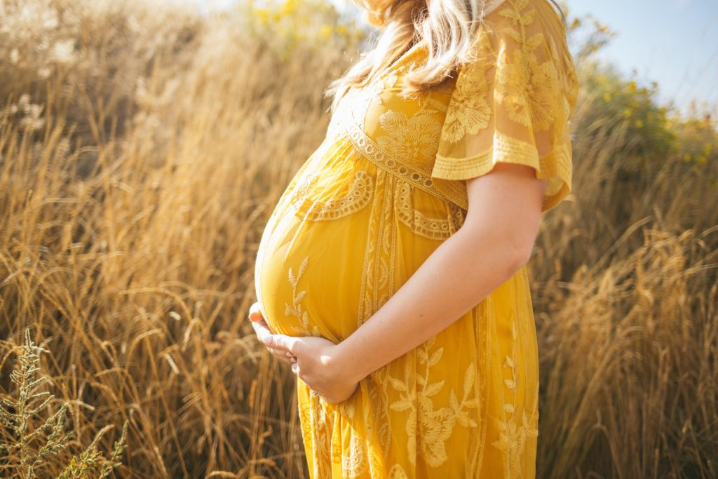
pregnant woman holding her belly
Source: Anna Hecker on Unsplash

Reproduction permanently alters the skeletons of females in ways not previously known, a team of anthropologists has concluded from research findings published in PLOS ONE. This discovery, based on an analysis of primates, sheds new light on how giving birth can permanently change the body.

“Our findings provide additional evidence of the profound impact that reproduction has on the female organism, further demonstrating that the skeleton is not a static organ, but a dynamic one that changes with life events,” explains Paola Cerrito, who led the research as a doctoral student in NYU’s Department of Anthropology and College of Dentistry.

Specifically, the researchers found that calcium, magnesium, and phosphorus concentrations are lower in females who have experienced reproduction. These changes are linked to giving birth itself and to lactation.

However, they caution that while other clinical studies show calcium and phosphorus are necessary for optimal bone strength, the new findings do not address overall health implications for either primates or humans. Rather, they say, the work illuminates the dynamic nature of our bones.

“A bone is not a static and dead portion of the skeleton,” notes NYU anthropologist Shara Bailey, one of the study’s authors. “It continuously adjusts and responds to physiological processes.”

It’s been long-established that menopause can have an effect on females’ bones. Less clear is how preceding life-cycle events, such as reproduction, can influence skeletal composition. To address this, the researchers studied the primary lamellar bone, the main type of bone in a mature skeleton. This aspect of the skeleton is an ideal part of the body to examine because it changes over time and leaves biological markers of these changes, allowing scientists to monitor alterations during the life span.

The researchers examined the growth rate of lamellar bone in the femora, or thigh bones, of both female and male primates who had lived at the Sabana Seca Field Station in Puerto Rico and died of natural causes. Veterinarians at the field station had monitored and recorded information on these primates’ health and reproductive history, allowing the researchers to match bone-composition changes to life events with notable precision.

Cerrito and her colleagues used electron microscopy and energy-dispersive X-ray analysis to calculate changes in concentrations of calcium, phosphorus, oxygen, magnesium, and sodium in the primates’ bones.

Their results showed different concentrations of some of these elements in females who gave birth compared males as well as females who did not give birth. Specifically, in females who gave birth, calcium and phosphorus were lower in bone formed during reproductive events. Moreover, there was a significant decline in magnesium concentration during these primates’ breastfeeding of infants.

“Our research shows that even before the cessation of fertility the skeleton responds dynamically to changes in reproductive status,” says Cerrito, now a research fellow at ETH Zurich. “Moreover, these findings reaffirm the significant impact giving birth has on a female organism – quite simply, evidence of reproduction is ‘written in the bones’ for life.”

Source: New York University

Oxygen Deficiency in Newborns may Increase Later Cardiovascular Risk

Photo by Christian Bowen on Unsplash

A population-based observational study has shown that babies suffering oxygen-deficiency complications at birth are almost twice as likely to develop cardiovascular disease in childhood or early adulthood, though such conditions are rare in youth. The findings are published in the journal The Lancet Regional Health – Europe.

According to the Karolinska Institutet researchers, the study could be the first of its kind to examine how complications related to asphyxiation at birth, which affects four million babies annually, affects the risk of cardiovascular disease later in life. Previous research has mostly concentrated on the association between asphyxia in the neonatal period and brain development.

Despite the relatively high risk, the absolute number of babies who suffer from cardiovascular disease despite asphyxiation at birth is very low. After the 30-year follow-up period, only 0.3% of those with asphyxia-related complications had a cardiovascular diagnosis, compared with 0.15% of those without complications.

Since the study was observational, the researchers are unable to establish any causality or propose any underlying mechanisms.

Largest risk increase for stroke and heart failure

The study followed over 2.8 million individuals born in Sweden between 1988 and 2018, of whom 31 419 suffered asphyxia-related complications at birth. A total of 4165 cases of cardiovascular disease were identified during the follow-up period. The increase in risk was particularly salient for stroke and heart failure, as well as for atrial fibrillation. The researchers took into account potential confounders such as birth weight and maternal lifestyle.

“Even if the absolute risk of cardiovascular disease is low at a young age, our study shows that asphyxia-related complications at birth are associated with a higher risk of cardiovascular disease later in life,” says the study’s corresponding author Neda Razaz, assistant professor at the Department of MedicineSolna, Karolinska Institutet.

Source: Karolinska Institutet

Morning Blue Light Exposure Eases PTSD Symptoms and Aids Sleep

Photo by Stormseeker on Unsplash

After being treated with a course of blue light exposure in the morning, people with post-traumatic stress disorder (PTSD) experienced better sleep, a reduction in the severity of PTSD symptoms and had more effective treatments overall, according to a new study recently published in Frontiers in Behavioral Neuroscience.

Sleep is crucial for maintaining physical and mental health, and inadequate sleep over time can impact all aspects of life with serious implications for long-term health, relationships, cognitive abilities such as learning, and healing.

The influence of sleep disruption on PTSD symptom severity is well established. Those who seek treatment to allay their PTSD symptoms often face a vicious cycle where poor sleep interferes with the effectiveness of treatments, negating any lessening of symptoms, which in turn contributes to sleep disruptions. To reduce and eliminate the emotional impact of traumatic memories, the patient needs quality sleep to integrate healing mechanisms achieved through cognitive or exposure therapy treatments.

“This research is exciting and unique because it points to an easy-to-use method for helping those with PTSD to retain the benefits of therapy long after the treatment ends,” said psychiatry professor William “Scott” Killgore, PhD, senior author on the paper, “Morning blue light treatment improves sleep complaints, symptom severity, and retention of fear extinction memory in post-traumatic stress disorder.”

Dr Killgore and the SCAN Lab team conducted a comprehensive assessment of daily morning blue-wavelength light exposure on individuals with clinically significant levels of PTSD. The goal was to ascertain if blue light therapy would help improve sleep and PTSD symptoms and sustain learned fear extinction memories, an analogue of therapeutic treatment for trauma.

Study participants committed to 30 minutes of morning light exposure daily for six weeks, with half of the participants using blue-wavelength light and half using amber light. Researchers examined the neurobiological, autonomic and behavioural outcome changes during the study.

The 43 participants who received blue light therapy not only demonstrated significant improvements in the severity of their PTSD symptoms, but also reported improvements in sleep and showed an increased retention of fear extinction memories. The control 39 controls receiving amber light did not show the same retention of the extinction memories, but rather showed a return of the original fear memories.

“While the limitations of the research include its modest sample size and difficulties monitoring compliance, the possibilities of utilising a treatment that is relatively simple, drug-free and inexpensive can offer hope for the large population of people living with the intense challenges of post-traumatic stress disorder,” Dr Killgore said.

Source: University of Arizona Health Sciences

Scientists Witness the Creation of a Hybrid Virus

In a world first, scientists have witnessed the fusion two viruses, influenza A virus (IAV) and respiratory syncytial virus (RSV), forming a single, hybrid virus particle (HVP). The discovery was published in Nature Microbiology.

Viruses often share tropism for the same system, such as respiratory viruses preferentially infecting the respiratory system. Coinfections by more than one virus represent between ~10–30% of all respiratory viral infections and are common among children. The clinical impact of viral coinfections is unclear: while some studies indicate that coinfections do not alter the outcome of disease, others report increased incidence of viral pneumonia.

Though evidence suggests virus–virus interactions play an important role in virus dynamics and transmission, viruses are typically studied in isolation. Recent work showed that interactions among respiratory viruses occur and have impacts at multiple levels, from populations, to individuals and tissues. However, studies characterising direct virus–virus interactions within cells are scarce. Here we report previously unknown interactions between IAV and RSV, two clinically important respiratory viruses that belong to different taxonomical families.

To investigate virus–virus interactions, the researchers infected human lung cells with both influenza A virus (IAV) and respiratory syncytial virus (RSV). Using super-resolution microscopy, live-cell imaging, scanning electron microscopy and cryo-electron tomography, the researchers found extracellular and membrane-associated filamentous structures consistent with hybrid viral particles (HVPs).

The researchers found that HVPs harbour surface glycoproteins and ribonucleoproteins of IAV and RSV. HVPs use the RSV fusion glycoprotein to evade anti-IAV neutralising antibodies and infect and spread among cells lacking IAV receptors. Finally, we show that IAV and RSV coinfection in primary cells of the bronchial epithelium results in viral proteins from both viruses latching on together at the apical cell surface.

“Our observations define a previously unknown interaction between respiratory viruses that might affect virus pathogenesis by expanding virus tropism and enabling immune evasion,” the researchers wrote.

“This kind of hybrid virus has never been described before,” virologist and senior author Pablo Murcia told The Guardian. “We are talking about viruses from two completely different families combining together with the genomes and the external proteins of both viruses. It is a new type of virus pathogen.”

When IAV and RSV coinfect, IAV becomes more infectious, infecting a wider array of human cells. Carrying the RSV surface proteins, IAV was able to better evade the immune system. The HVP also spread into cells lacking influenza receptors, letting it progress further down the respiratory tract.

The relationship is not mutually beneficial for the viruses as RSV loses potency. Overall though, pilfering another virus’s tools could play a role in viral pneumonia.

“RSV tends to go lower down into the lung than the seasonal flu virus, and you’re more likely to get more severe disease the further down the infection goes,” said Dr Stephen Griffin, a virologist at the University of Leeds who was not involved in the study.

“It is another reason to avoid getting infected with multiple viruses, because this [hybridisation] is likely to happen all the more if we don’t take precautions to protect our health,” he added.

The researchers also found that the combination of viruses was important; IAV did not form an effective hybrid with rhinovirus.

Iron Drives Chronic Heart Failure in Myocardial Infarction Survivors

Photo by Freestocksorg on Pexels

A multi-institution study has found that iron drives the formation of fatty tissue in the heart and leads to chronic heart failure in about 50% of myocardial infarction (MI) survivors. The discovery, recently published in Nature Communications, paves the way for treatments that have the potential to prevent heart failure.

“For the first time, we have identified a root cause of chronic heart failure following a heart attack,” said study leader Rohan Dharmakumar, PhD, of Indiana University School of Medicine.

“While advances across populations have made survival after a heart attack possible for most, too many survivors suffer long-term complications like heart failure,” said Subha Raman, MD, who is physician director of the Cardiovascular Institute. “Dr. Dharmakumar’s breakthrough science illuminates who is at risk and why and points to an effective way to prevent these complications.”

The study followed large animal models over six months. In MI with bleeding complications, scar tissue is slowly replaced by fat. Fatty tissue can’t push blood from the heart effectively, and this is what leads to heart failure and eventually to death in many survivors of haemorrhagic MI, Dharmakumar said.

“Using noninvasive imaging, histology and molecular biology techniques, and various other technologies, we have shown that iron from red blood cells is what drives this process,” he explained. “When we removed the iron, we reduced the amount of fat in the heart muscle. This finding establishes a pathway for clinical investigations to remedy or mitigate the effects associated with iron in haemorrhagic myocardial infarction patients.”

Dharmakumar’s team is currently testing iron chelation therapy to do just that in a just-launched clinical trial.

“Thanks to a clinical trial underway being led by his team at Indiana University, I’m excited to see this treatment improve the lives of millions of heart attack survivors worldwide,” said Raman.

Source: Indiana University School of Medicine