In new research published in the Journal of Bone and Mineral Research, elevated blood levels of a certain chemokine, or small signalling protein, that promotes osteoclast formation were linked with a higher risk of hip fracture in men.
To maintain bone health, a balanced activity of various bone cell types including bone-forming osteoblasts and bone-resorbing osteoclasts has to take place. When osteoclasts dominate without adequate bone formation to compensate, osteoporosis results.
The study included 55 men and 119 women who had experienced a hip fracture an average of 6.3 years after their blood was collected. The participants were matched individually to controls who did not develop hip fractures.
The researchers found higher levels of the chemokine CXCL9 in the pre-fracture blood samples of men with subsequent hip fractures compared with their non-fracture controls. In women, the researchers saw no such.
“The unexpected difference in the results between men and women in our study may be explained by how changes in sex hormone levels during aging could influence the level and effects of CXCL9 differently in older men and women,” explained corresponding author Woon-Puay Koh, MBBS, PhD, from the National University of Singapore (NUS).
“Our findings open the exciting possibility that early interventions targeting CXCL9 or CXCL9-CXCR3 signalling could be beneficial in preventing hip fractures in older men,” added co-corresponding author Christoph Winkler, PhD, also from NUS.
In a study in the Journal of Public Health, participants who exchanged 30 minutes of social media use per day for exercise felt happier, more satisfied, less stressed by the COVID pandemic and less depressed than a control group. These effects persisted even six months after the study had ended.
The downside of social media
While it helped people stay connected during the COVID pandemic, social media consumption has also its drawbacks. Heavy use can lead to addictive behaviour that manifests itself in, for example, a close emotional bond to the social media. In addition, fake news and conspiracy theories can spread uncontrollably on social channels and trigger even more anxiety.
“Given that we don’t know for certain how long the coronavirus crisis will last, we wanted to know how to protect people’s mental health with services that are as free and low-threshold as possible,” explained assistant professor Dr. Julia Brailovskaia, who lead a team from the Mental Health Research and Treatment Center at Ruhr-Universität Bochum. To find out whether the type and duration of social media use can contribute to this, she and her team conducted an experimental study, with a total of 642 participants randomised to one of four groups.
A two-week experiment
The first group reduced the daily social media consumption by 30 minutes during an intervention period of two weeks. Since previous studies had shown that physical activity can increase well-being and reduce depressive symptoms, the second group increased the duration of physical activity by 30 minutes daily during this period, while continuing to use social media as usual. The third group combined both, reducing social media use and increasing physical activity. A control group didn’t change the behaviour during the intervention phase.
Before, during and up to six months after the two-week intervention phase, the participants responded to online surveys on the duration, intensity and emotional significance of their social media use, physical activity, their satisfaction with life, their subjective feeling of happiness, depressive symptoms, the psychological burden of the COVID pandemic and their cigarette consumption.
Healthy and happy in the age of digitalisation
The findings clearly showed that both reducing the amount of time spent on social media each day and increasing physical activity have a positive impact on people’s well-being. The combination of the two interventions in particular increases one’s satisfaction with life and subjective feeling of happiness and reduces depressive symptoms. Even six months after the two-week intervention phase had ended, participants in all three intervention groups spent less time on social media than before: about a half hour in the groups that had either reduced social media time or increased their daily exercise, and about three-quarters of an hour in the group that had combined both measures. Six months after the intervention, the combination group engaged one hour and 39 minutes more each week in physical activity than before the experiment. The positive influence on mental health continued throughout the entire follow-up period.
“This shows us how vital it is to reduce our availability online from time to time and to go back to our human roots,” Julia Brailovskaia concluded. “These measures can be easily implemented into one’s everyday life and they’re completely free — and, at the same time, they help us to stay happy and healthy in the digital age.”
The High Court in Gqeberha has found that the Eastern Cape MEC for Health and Livingstone Hospital are liable to pay damages to the widow of a man who died after falling from the fifth floor of the hospital.
In the ruling last week, Acting Judge Ivana Bands found that the patient, George Williams, had not been properly medicated or monitored. She said that had this been done, Williams would not have been “pacing up and down the ward, in confused, restless and disoriented state”, and would not have fallen to his death from the window.
Judge Bands said the conduct of the medical and nursing personnel who treated Williams after he was admitted to the hospital on 3 October 2013, “fell far short of what is regarded as sound practice” in dealing with patients suffering from alcohol withdrawal – delirium tremens which involves sudden and severe mental or nervous system changes – and secondary schizophrenia.
“Had he been properly medicated, it cannot be gainsaid [denied] that he would have been reduced to a calm and lightly dozing state. This would have enabled the medical and nursing staff to monitor his vital signs and his condition appropriately until such time that delirium tremens had abated,” Judge Bands said.
Judge Bands’s finding of negligence means that Williams’s widow Jeanine can now pursue a monetary damages claim against the MEC and hospital. This could be determined at another trial or through negotiation.
Jeanine Williams, in her papers, contended that the hospital staff were under a legal duty to provide her husband with adequate and timeous medical treatment; that they had not properly sedated him, restricted his movements and monitored his condition.
The defendants, however, argued that Williams had been treated with sedatives, including diazepam (Valium) and that he had been put in an “enclosed locked ward” close to the nurses’ station.
Bands said Wiliams was a known alcoholic who was admitted to the hospital late on 3 October 2013. In the early hours of the morning, he had been given diazepam, with little effect. During the evening of 4 October, he was given more sedatives and an antipsychotic agent, also with no effect.
Soon after, at about 10:30pm, Williams broke the outside entrance glass door of the nurses’ tearoom and fell from the fifth floor. He died about an hour later.
Two key witnesses during the trial were Dr Candice Harris, a professional nurse and general practitioner, who testified for Williams, and Dr Michelle Walsh, a general surgeon, who testified for the MEC and the hospital.
In her evidence, Harris had said delirium tremens was a “medical emergency” and, according to guidelines, immediate management of the condition was necessary. She had stressed the importance of re-orientating the patient and said it was the nurse’s duty to inform the doctor if the patient was not responding to medication.
The judge said Walsh’s evidence was that it was not that the hospital was doing nothing – “they were doing something”.
“She said the sedation prescribed is usually based on what the assessing doctor thinks will have the desired effect to calm the patient to the extent that they would sit calmly in a chair. It is common cause that this desired state was never reached,” the judge said.
“Not only was he under-sedated, there is no evidence that the initial dose, which had no effect, was ever increased as per the published guidelines, in spite of multiple entries in the hospital records that he remained confused, disorientated, restless and walking up and down – and that he had become so agitated that the nursing staff feared he would assault them,” the judge said.
Bands said Williams had not been treated according to the guidelines, thus the MEC and the hospital are liable for any proven damages.
Although bones are considered a mostly structural component of the human body, bones are in fact active living tissues that can respond to applied mechanical forces. For example, martial arts training, with its kicks, punches and throws, has been shown to increase bone mineral density in the arms, legs and spines of practitioners.
At present, it is unclear whether this thickening of the skull is beneficial or detrimental: theoretically, a thicker skull is a stronger skull, suggesting that this may be the bone’s attempt to protect the brain from subsequent impacts.
“This is a bit of a conundrum,” Assoc Prof Semple said. “As we know, repeated concussions can have negative consequences for brain structure and function. Regardless, concussion is never a good thing.”
The team hopes that the microstructural skull alterations caused by concussion are now considered by researchers in the field to better understand how concussions affect the whole body.
A form of mild traumatic brain injury, concussion have been linked to long-term neurological consequences if they happen repetition.
While most studies focus on its effect on the brain and its function, they largely ignore the overlying skull bones.
Study collaborator Professor Melinda Fitzgerald, from Curtin University and the Perron Institute in Western Australia, has previously shown that repeated concussive impacts lead to subtle problems with memory, and evidence of brain damage.
In this new study, high-resolution neuroimaging and tissue staining techniques were used in a pre-clinical animal model, and revealed an increase in bone thickness and density, in close proximity to the site of injury.
“We have been ignoring the potential influence of the skull in how concussive impacts can affect the brain,” Associate Professor Semple said. “These new findings highlight that the skull may be an important factor that affects the consequences of repeated concussions for individuals.”
Future studies are planned, with collaborator and bone expert Professor Natalie Sims from St Vincent’s Institute of Medical Research in Melbourne, to understand if a thickened skull resulting from repeated concussions alters the transmission of impact force through the skull and into the vulnerable brain tissue underneath.
A serious neonatal health threat, preterm labour has long mystified researchers – and how does the uterus normally stay dormant, letting it stretch and expand during the 40 weeks it takes a foetus to grow? New research published in The Journal of Physiology suggests that a protein called Piezo1 keeps the uterus relaxed throughout gestation.
Preterm birth is a major cause of neonatal mortality and morbidity. The identification of Piezo1 in the uterus, and its role to maintain relaxation of uterus through stretch-activation during pregnancy, paves the way for drugs and therapies to be developed that could one day treat or delay preterm labour.
The muscular outer layer of the uterus is peculiar because it is the only muscle that it is not regulated by nerves and it must remain dormant for the 40 weeks despite significant expansion and stretch as the foetus develops into a baby. The researchers from University of Nevada USA studied tissue samples of the smooth muscle of the uterus to explore the mechanistic pathways to better understand the dynamics controlling the uterus, how pregnancy is maintained and what maintains quiescence until labour.
Stretching the uterus tissue, to mimic what happens during pregnancy, activates Piezo1 channels. This drives the flow of calcium molecules generating a signalling cascade that activates the enzyme nitric oxide synthase to produce the molecule nitric oxide. This Piezo1 cascade promotes and maintains the dormant state of the uterus.
Piezo1 controls the uterus by working in a dose-dependent manner, where channel activity is stimulated by the chemical Yoda1 and inhibited by a chemical called Dooku1 (Star Wars fans will no doubt recognise the inspiration behind these two names). When Piezo1 is upregulated, the uterus remains in a relaxed state. However, in preterm tissue, the expression of Piezo1 is significantly downregulated, ‘switching off’ the dormant signalling to the muscle, so the uterus contracts and initiates labour.
Professor Iain Buxton at the Myometrial Research Group at the University of Nevada said: “Pregnancy is the most impressive example of a human muscle enduring mechanical stress for a prolonged period. Finding Piezo1 in the muscular layer of the uterus means the uterus is controlled locally and is coordinated by a stretch-activated mechanism rather than hormonal influence from the ovaries or the placenta, which has been the assumption.
“It is troubling that there are still no drugs available to stop preterm labour. Thanks to the Nobel Prize winning discovery of Piezo proteins, which are responsible for how the body responds to mechanical force, and our investigation we are now closer to developing a treatment. Piezo1 and its relaxation mechanism provide a target for us which we could potentially activate with drugs. We need to test this with further studies and we hope to carry out clinical trials in the future.”
Contraction and relaxation were assessed in tissue samples compared for the following gestational periods: non-pregnant, term non-labouring, term labouring, preterm non-labouring and preterm labouring. The presence of Piezo1 channels was discovered using molecular tools while pregnant tissues contracting in a muscle bath were stimulated with Piezo1 channel activator and inhibitor to characterize the regulation of quiescence.
More research is needed to understand just how all the molecular signals and steps involved in the Piezo1 channel regulate uterus relaxation, and to identify other chemicals that may be involved.
With more than 64% of women suffering from premenstrual mood swings and anxiety, they represent a “key public health issue globally,” according to a new study in Archives of Women’s Mental Health.
The UVA Health study found that most women have premenstrual symptoms every menstrual cycle, with one of the most common symptoms, regardless of age, being mood swings or anxiety. At least 61% of women in all age groups surveyed reported mood-related symptoms every menstrual cycle, which the researchers say suggests “that premenstrual mood symptoms are a key public health issue globally.”
“Our study demonstrates that premenstrual mood symptoms are incredibly common worldwide,” said senior author Jennifer L Payne, MD. “More important, a majority of women reported that their premenstrual symptoms interfered with their everyday life at least some of the time.”
Better understanding premenstrual symptoms
To better understand the type of premenstrual symptoms women experience and how those symptoms affect their daily lives, the researchers analysed more than 238 000 survey responses from women ages 18–55 from 140 countries on the Flo app, which helps women track their menstrual cycle or track their mood or physical symptoms during and after pregnancy.
Food cravings topped the most common symptoms (85.28%), followed by mood swings or anxiety (64.18%) and fatigue (57.3%). Among the study respondents, 28.61% said their premenstrual symptoms interfered with their everyday life during every menstrual cycle, while an additional 34.84% said their premenstrual symptoms interfered with their everyday life sometimes.
“The incidence of reported premenstrual mood and anxiety symptoms varied significantly by country from a low of 35.1% in Congo to a high of 68.6% in Egypt,” Payne said. “Understanding whether differences in biology or culture underlie the country level rates will be an important future research direction.”
A group of symptoms — absentmindedness, low libido, sleep changes, gastrointestinal symptoms, weight gain, headaches, sweating or hot flashes, fatigue, hair changes, rashes and swelling — was significantly more frequent among older survey respondents, the researchers found. The increase in physical symptoms among older survey respondents “makes sense,” the researchers said, as many of these symptoms are associated with perimenopause, a transition period to menopause marked by irregular menstrual cycles.
Payne is hopeful that this survey data will help women get better care by making healthcare providers more aware of the frequency of these symptoms, especially anxiety and mood-related symptoms.
“There are a number of treatment strategies that are available to treat premenstrual symptoms that interfere with a woman’s every day functioning,” she said. “Increasing awareness of how common these symptoms are, and that if they impact functioning that there are treatments available, will help women improve their quality of life.”
Schematic diagram of the alveolar chip (upper left), photograph of the chip (upper middle), CAD drawing of the multi-generation alveolar structure (upper right), and two typical flow patterns in the alveolar chip (bottom). CREDIT: Yonggang Zhu
Understanding how air and particulates through the alveoli is important to better treat respiratory disease. In Biomicrofluidics, researchers detail a model alveolar system that they built to mimic the breathing action of the human lung and allows visualisation of flow patterns within the alveoli. They observed that flow changes after the 20th branching of the alveoli.
The scientists, from Harbin Institute of Technology in China, designed a chip that includes tubes arranged like the structure of a bifurcation point in the bronchial network. The upper layer of the chip is made of a flexible polymer moulded into small tubes that mimic the alveolar structure. The lower layer is glass, which allows the authors to visualise fluid flow through the tubes.
To mimic inhalation and exhalation, the scientists devised a system in which gas was pressurised in a sinusoidal fashion and pumped around the flexible tubes. Small red polystyrene spheres were added to the fluid flowing through tubes. These spheres allowed them to photograph movement of the fluid as it was pushed through the tubes by the artificial breathing apparatus.
Subsequent branches in the bronchial network are termed ‘generations’, and the team found different flow patterns for different generations. In the human lung, alveoli appear at the 15th generation and remain present for generations up to 23. The researchers found a change in flow pattern between the 19th–20th and the 21st–22nd generations.
“The alveolar flow pattern of the 19th generation is dominated by vortex flow,” author Yonggang Zhu said. “Alveolar flow patterns in the 20th generation are similar to those in the 19th, but somewhat compressed.”
The investigators observed a change in the next generation.
“The alveolar flow pattern in the 21st generation has both vortex flow and radial flow. The vortex region is much smaller than the radial flow region. By the time the flow reaches the 22nd generation, vortex flow disappears completely, and we observe only radial flow,” Zhu said.
The authors also found evidence of chaotic behaviour near the vortex centre. They said more research is needed to fully understand this, but they felt the current study provides a good baseline for deeper investigations.
With the model, researchers will be able to study changes in flow patterns in the alveoli due to diseases such as emphysema and COPD.
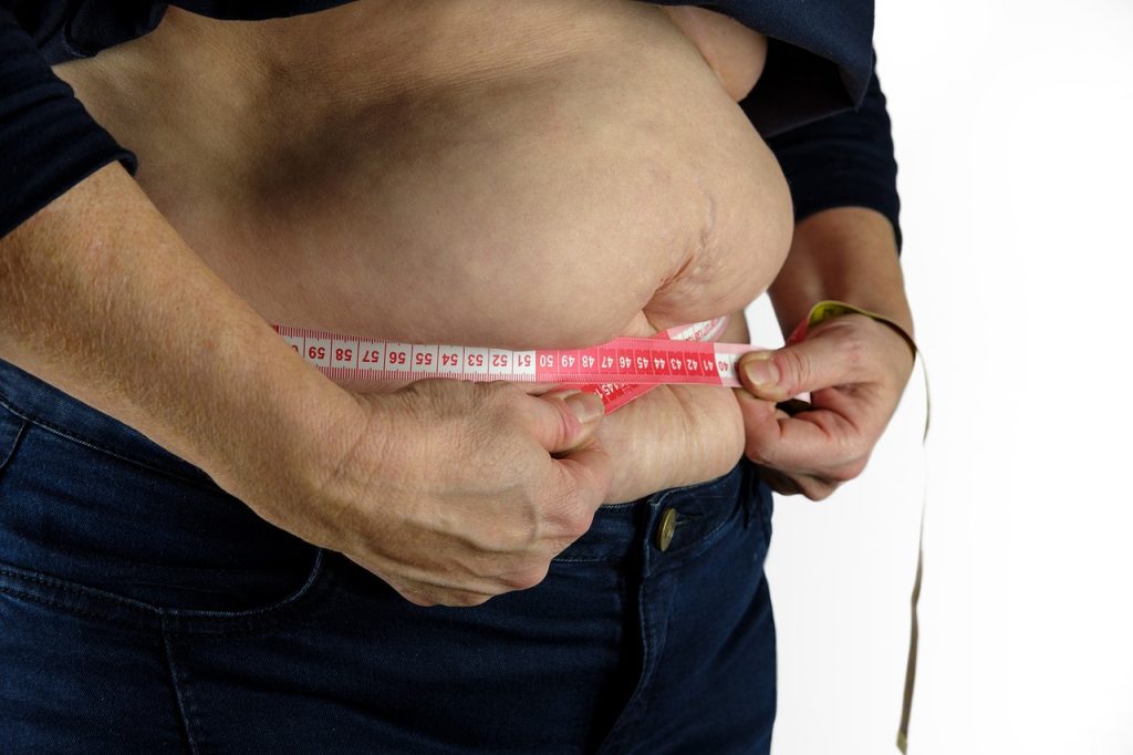
Lifestyle leading to overweight increases the risk of metabolic diseases such as diabetes – but the relationship also works in reverse, according to a new study. If insulin production is compromised, as is the case in the early stages of type 2 diabetes, this can contribute to overweight. The researchers report their findings in the journal Nature Communications.
When hormone activation goes awry
The research group, led by Dr Daniel Zeman-Meier of the University Hospital of Basel, focused on protease PC1/3 – a key enzyme in the body that transforms various inactive hormone precursors into the final, active forms. Sever endocrine disorders can result if PC1/3 does not function properly. The consequences include a feeling of uncontrollable hunger and severe overweight.
“Until now, it was assumed that this dysregulation is caused by a lack of activation of satiety hormones,” explained Dr Zeman-Meier. “But when we turned off PC1/3 in the brains of mice, the animals’ body weight did not change significantly.” The researchers concluded from this that something other than a brain malfunction must be responsible.
Incorrect activation of insulin leads to hunger and overweight
In their next step, they tested whether overweight could be caused by incorrect activation of other hormones. Among other things, PC1/3 activates insulin. “Investigating the role of insulin production as a cause of overweight was obvious,” said Dr Zeman-Meier. The researchers shut off PC1/3 specifically in the insulin-producing beta cells of the pancreas in mice. The animals consumed significantly more calories and soon became overweight and diabetic.
An important mechanism in humans
“These results are also interesting because PC1/3 is reduced in the pancreas of patients with prediabetes,” says Professor Marc Donath, research leader and final author of the study. This indicates that incorrect insulin activation could cause overweight as well as result from it.
But PC1/3 is also important in the weight regulation of healthy individuals, Prof Donath stressed. The researchers were able to show that the gene expression of PC1/3 in the pancreas is negatively correlated with body weight in the general population — meaning that sufficient PC1/3 promotes a healthy body weight.
The finding that a defect in the beta cells is a trigger of overweight promises new therapeutic possibilities. For example, it is conceivable that medications could be used to reduce the production of immature insulin precursors, creating a new tool in the fight against overweight and diabetes.
There is a view being promoted that COVID didn’t hit Africa as badly as the rest of the world. The reason for this, as recently expressed in an article by Boniface Oyugi in The Conversation, was the effective and well-coordinated response of African governments.
We understand the desire to find good news on the continent. But, on balance, the very little evidence available shows that COVID has hit Africa hard. The continent is highly diverse with over 50 states, so broad generalisations should be treated cautiously but, with an exception or two, there is little evidence of an effective response to the COVID pandemic. For one thing, Africa has the lowest vaccination rate of any continent.
Oyugi uses the WHO’s official COVID infection and death statistics to claim that the continent fared better than elsewhere. These state that as of late July, less than 2% of global cases and less than 3% of global deaths occurred in Africa, which has about 17% of the world’s population. (Oyugi also cites a study which pretty much says the same thing.)
COVID test statistics and confirmed COVID deaths don’t paint an accurate picture of how seriously the pandemic has hit a country (see here). If you don’t measure something properly, you can’t conclude that it’s a small problem. COVID tests are typically only administered with any regularity to a small, predominantly better off, part of a country’s population, and countries that test more tend to find more cases. Official COVID death tolls typically count people who have died in hospital with a confirmed positive test result. But it often doesn’t happen this way, especially on a continent with large rural populations and under-resourced hospitals.
Excess deaths: a vital measure
This is why the most important measure of how hard COVID has hit a country is the excess death toll. By excess deaths, we mean the number of deaths that occurred above what you’d expect given recent historical mortality. In sub-Saharan Africa, the only country that has a system capable of reliably estimating this is South Africa. Every week since the beginning of the epidemic, the Medical Research Council (MRC), using death certificate data provided by Home Affairs, has diligently analysed excess deaths. (Many countries wealthier than South Africa do not have as good a system, so it’s something to be proud of.)
The MRC researchers calculate that there have been over 320 000 excess deaths in South Africa since May 2020 (as of July 2022). As they’ve explained, conservatively 85% of these are COVID deaths. It may be as high as 95%. We can conclude that close to 300 000 people have died of COVID in South Africa. Over the past two years about 1 in 200 people in the country have died of this new infection.
The Economist has been reporting excess deaths by country. It states: “Among developing countries that do produce regular mortality statistics, South Africa shows the grimmest picture, after recording three large spikes of fatalities.”
Official deaths are much lower than excess deaths
But if you look at South Africa’s official, and much less accurate, COVID death toll you get a very different picture: Then we’re only 65th worst in the world (source: Worldometer deaths per million people). Lesotho is in 167th place, suggesting it has had a very small epidemic. Is it plausible that an area with a porous border entirely surrounded by South Africa has a completely different epidemic? (See this set of tweets – by one of the authors of South Africa’s weekly mortality report – that explains how the little mortality data we have from Lesotho suggests it had a serious pandemic.)
What about Namibia at position 74 in the Worldometer list, Botswana at 89, Zimbabwe at position 143 and Mozambique at position 190? Is it plausible that this ordering, almost in reverse order of industrial development, accurately reflects how these countries were affected by COVID?
Depending on your bias, you can approach these statistics in two ways. You can be very optimistic and see this as evidence of a smaller epidemic in sub-Saharan Africa. Or you can be realistic and acknowledge that the official numbers are likely very badly undercounted.
We can’t know for sure though because nearly all African governments did not have the systems in place to count excess deaths.
Most African countries need much better death registration systems
Attempts to estimate excess mortality in most African countries are based on almost no data. To the extent that there is data, it supports the view that the numbers have been badly undercounted. For example, a study published in the British Medical Journal, albeit with many caveats, found death rates in developing countries were twice those of rich countries.
During the height of the AIDS pandemic in the 2000s there was much optimism that the massive influx of foreign aid in response could be used to build better health systems. Bits and pieces of evidence do suggest health on the continent has improved. But it’s very disappointing that most countries on the continent still do not have the vital registration systems in place to measure mortality with decent accuracy. This is one of the most important measures of how a population is doing.
By claiming that African governments have responded well to COVID, when there’s no proper evidence to support this, we fail to hold politicians accountable. We also create the impression that institutions like the World Health Organisation and the African Union’s African Centre for Disease Control are more successful than they’ve actually been. This is a disservice to the vast majority of people living in Africa.
Geffen is GroundUp’s editor. Professor Venter is an infectious diseases clinician and head of Ezintsha at Wits University.
Blood type may be associated with early-onset stroke risk, according to a new meta-analysis, which was published in the journal Neurology. The meta-analysis included all available data from genetic studies focusing on ischaemic strokes in adults under age 60.
“The number of people with early strokes is rising. These people are more likely to die from the life-threatening event, and survivors potentially face decades with disability. Despite this, there is little research on the causes of early strokes,” said study co-principal investigator Steven J. Kittner, MD, MPH, Professor of Neurology at University of Maryland School of Medicine (UMSOM).
He and his colleagues performed a meta-analysis of 48 studies on genetics and ischaemic stroke that included 17 000 stroke patients and nearly 600 000 healthy controls. They looked for genetic variants associated with a stroke and found a link between early-onset stroke (before age 60) and the area of the chromosome that includes the gene that determines whether a blood type is A, AB, B, or O.
The study found that people with early stroke were more likely to have blood type A and less likely to have blood type O, compared to people with late stroke and people who never had a stroke. Both early and late stroke were also more likely to have blood type B compared to controls. After adjusting for sex and other factors, researchers found that, compared to those with other blood types, blood type A had a 16% higher risk while blood type O had a 12% lower risk.
“Our meta-analysis looked at people’s genetic profiles and found associations between blood type and risk of early-onset stroke. The association of blood type with later-onset stroke was much weaker than what we found with early stroke,” said study co-principal investigator Braxton D. Mitchell, PhD, MPH, Professor of Medicine at UMSOM.
The researchers emphasised that the increased risk was very modest and that those with type A blood should not worry about having an early-onset stroke or engage in extra screening or medical testing based on this finding.
“We still don’t know why blood type A would confer a higher risk, but it likely has something to do with blood-clotting factors like platelets and cells that line the blood vessels as well as other circulating proteins, all of which play a role in the development of blood clots,” said Dr Kittner. Previous studies suggest that those with an A blood type have a slightly higher risk of developing blood clots in the legs known as deep vein thrombosis. “We clearly need more follow-up studies to clarify the mechanisms of increased stroke risk,” he added.
A limitation of the study was the relative lack of diversity among participants, with only 35% of the participants having non-European ancestry.