Month: September 2022

With Warfarin, Dropping Aspirin Reduces Bleeding Complications for Some
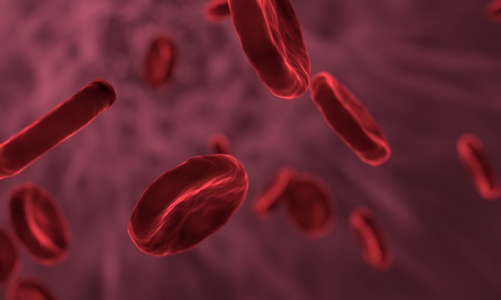
Red blood cells
Source: Pixabay

Research from Michigan Medicine suggests that, for venous thromboembolism (VTE) or atrial fibrillation (AF) patients without a history of heart disease who are taking warfarin, stopping aspirin use causes their risk of bleeding complications to drop significantly.

For the study, which is published in JAMA Network Open, researchers analysed over 6700 people treated at anticoagulation clinics across Michigan for VTE as well as AF. Patients were treated with warfarin but also took aspirin despite not having history of heart disease.

“We know that aspirin is not a panacea drug as it was once thought to be and can in fact lead to more bleeding events in some of these patients, so we worked with the clinics to reduce aspirin use among patients for whom it might not be necessary,” said senior study author and cardiologist Geoffrey Barnes, MD.

Over the course of the study, aspirin use among patients fell by 46.6%. With aspirin used less commonly, the risk of a bleeding complication dropped by 32.3% – equivalent to preventing one major bleeding event per every 1000 patients who stop taking aspirin.

“When we started this study, there was already an effort by doctors to reduce aspirin use, and our findings show that accelerating that reduction prevents serious bleeding complications which, in turn, can be lifesaving for patients,” said Dr Barnes. “It’s really important for physicians and health systems to be more cognisant about when patients on a blood thinner should and should not be using aspirin.”

Several studies had found concerning links between concurrent use of aspirin and different blood thinners, which prompted this aspirin de-escalation.

One study reported that patients taking warfarin and aspirin for AF and VTE experienced more major bleeding events and had more ER visits for bleeding than those taking warfarin alone. Similar results were seen for patients taking aspirin and direct oral anticoagulants – who were found more likely to have a bleeding event but not less likely to have a blood clot.

“While aspirin is an incredibly important medicine, it has a less widely used role than it did a decade ago,” Dr Barnes said. “But with each study, we are seeing that there are far fewer cases in which patients who are already on an anticoagulant are seeing benefit by adding aspirin on top of that treatment. The blood thinner they are taking is already providing some protection from clots forming.”

For some people, aspirin can be lifesaving. Many patients who have a history of ischaemic stroke, heart attack or a stent placed in the heart to improve blood flow — as well as those with a history of cardiovascular disease — benefit from the medication.

The challenge comes when some people take aspirin without a history of cardiovascular disease and are also prescribed an anticoagulant, said first author Jordan Schaefer, MD.

“Many of these people were likely taking aspirin for primary prevention of heart attack or stroke, which we now know is less effective than once believed, and no one took them off of it when they started warfarin,” Dr Schaefer said. “These findings show how important it is to only take aspirin under the direction of your doctor and not to start taking over-the-counter medicines like aspirin until you review with your care team if the expected benefit outweighs the risk.”

Source: Michigan Medicine – University of Michigan

Is Protein Restriction the Best Option after Kidney Transplant?

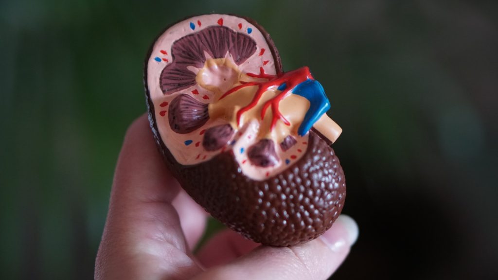
Anatomic model of a kidney
Photo by Robina Weermeijer on Unsplash

Scientists at Osaka Metropolitan University have challenged the conventional wisdom that low protein intake is essential for kidney disease patients with their recent study on the relationship between protein intake and skeletal muscle mass in kidney transplant recipients. Their findings were published in Clinical Nutrition.

Chronic kidney disease patients are known to have induced sarcopenia due to chronic inflammation, hypercatabolism, decreased nutrient intake, and decreased physical activity associated with impaired kidney function. A successful kidney transplantation is able to correct or improve many of those physiological and metabolic abnormalities, with the transplant recipients increasing skeletal muscle mass after receiving their new kidney. Since excessive protein intake worsens kidney function, it is commonly believed that patients with chronic kidney disease, including kidney transplant recipients, should limit protein intake to protect their kidneys. On the other hand, it has been suggested that severe protein restriction may worsen sarcopenia and adversely affect prognosis.

Since nutrition and exercise therapy are recommended to improve sarcopenia, protein intake is suspected to relate to recovery of skeletal muscle mass after kidney transplantation. However, few studies have examined the relationship between skeletal muscle mass and protein intake in kidney transplant recipients.

In order to fill this knowledge gap, a research group led by Drs Akihiro Kosoku and Tomoaki Iwai, and Professor Junji Uchida at Osaka Metropolitan University followed 64 kidney transplant recipients for 12 months after their procedure. They investigated the relationship between changes in skeletal muscle mass, as measured by bioelectrical impedance analysis, and protein intake from urine sample. The results showed that changes in skeletal muscle mass during this period were positively correlated with protein intake, and that insufficient protein intake resulted in decreased muscle mass.

Drs Iwai and Kosoku commented, “To improve the life expectancy of kidney transplant recipients, further research is needed to clarify the optimal protein intake to prevent either deterioration in kidney function or sarcopenia. We hope that nutritional guidance, including protein intake, will lead to improved life expectancy and prognosis.”

Source: Osaka Metropolitan University

7 Lifestyle Habits may Reduce Dementia Risk in Diabetes

Old man jogging
Photo by Barbra Olsen on Pexels

A combination of seven healthy lifestyle habits including sleeping seven to nine hours daily, regular exercise and frequent social contact was associated with a lower risk of dementia in people with type 2 diabetes, according to a study published online in the journal Neurology.

“Type 2 diabetes is a worldwide epidemic that affects one in 10 adults, and having diabetes is known to increase a person’s risk of developing dementia,” said study author Yingli Lu, MD, PhD, of Shanghai Jiao Tong University School of Medicine in China. “We investigated whether a broad combination of healthy lifestyle habits could offset that dementia risk and found that people with diabetes who incorporated seven healthy lifestyle habits into their lives had a lower risk of dementia than people with diabetes who did not lead healthy lives.”

For the study, researchers looked at a health care database in the United Kingdom and identified 167 946 people 60 or older with and without diabetes who did not have dementia at the start of the study. Participants completed health questionnaires, provided physical measurements and gave blood samples. For each participant, researchers calculated a healthy lifestyle score of 0 to 7, with one point for each of 7 healthy habits. Habits included no current smoking, moderate alcohol consumption of up to one drink a day for women and up to two a day for men, regular weekly physical activity of at least 2.5 hours of moderate exercise or 75 minutes of vigorous exercise, and 7 to nine hours of sleep daily.

Another factor was a healthy diet including more fruits, vegetables, whole grains and fish and fewer refined grains, processed and unprocessed meats. The final habits were being less sedentary, which was defined as watching television less than four hours a day, and frequent social contact, which was defined as living with others, gathering with friends or family at least once a month and participating in social activities at least once a week or more often. Researchers followed participants for an average of 12 years. During that time, 4,351 people developed dementia. A total of 4% of the people followed only zero to two of the healthy habits, 11% followed three, 22% followed four, 30% followed five, 24% followed six and 9% followed all seven. People with diabetes who followed two or fewer of the seven healthy habits were four times more likely to develop dementia than people without diabetes who followed all seven healthy habits. People with diabetes who followed all of the habits were 74% more likely to develop dementia than those without diabetes who followed all the habits. For people with diabetes who followed all the habits, there were 21 cases of dementia for 7474 person-years, or 0.28%.

For people with diabetes who followed only two or fewer habits, there were 72 cases of dementia for 10 380 person years or 0.69%. After adjusting for factors like age, education and ethnicity, people who followed all the habits had a 54% lower risk of dementia than those who followed two or fewer. Each additional healthy habit people followed was associated with an 11% decreased risk of dementia. The association between healthy lifestyle score and dementia risk was not affected by medications people took or how well they controlled their blood sugar.

“Our research shows that for people with type 2 diabetes, the risk of dementia may be greatly reduced by living a healthier lifestyle,” Dr Lu said. “Doctors and other medical professionals who treat people with diabetes should consider recommending lifestyle changes to their patients. Such changes may not only improve overall health, but also contribute to prevention or delayed onset of dementia in people with diabetes.” A limitation of the study was that people reported on their lifestyle habits and may not have remembered all details accurately. Lifestyle changes over time were also not captured.

Source: American Academy of Neurology

Schwann Cells Turn Nerve Tumours Benign

Microscopy image of mouse sciatic nerves showing axons (red) wrapped by Schwann cells (green) with their nuclei depicted in blue. Credit: A. Alvarez-Prats and T. Balla, Eunice Kennedy Shriver National Institute of Child Health and Human Development/NIH

In addition to forming the myelin sheath along peripheral nerves and supporting neighbouring neurons, Schwann cells have also been found to play an important immune modulating function, starting and shutting off inflammation. This function not only helps nerve repair, but may also turn nerve tumours benign. These new findings were reported in the journal Glia.

The research has revealed that Schwann cells produce signalling molecules that can activate other immune cells. In particular, however, they are able to stop inflammatory reactions in order to prevent excessive tissue damage and allow the nerve to regenerate.

“This is essential, because inflammation releases free radicals against which nerve fibers cannot protect themselves. Therefore, the inflammation must be cleared quickly, which is precisely what Schwann cells do,” explained study designer Dr. Sabine Taschner-Mandl, who designed the study and heads a research group at St. Anna CCRI.

Do Schwann cells protect against malignancy?

These findings also have implications for protection against malignancy After nerve injury, Schwann cells engage a ‘repair’ mode that is also found in benign infantile nerve tumours. There, it causes the tumour cells to mature and thus reach a stage where they lose their aggressive properties and no longer divide unchecked

“Based on the current results, we now suspect that the immune cell functions of Schwann cells also become effective in childhood nerve tumours. This is because in cancer, there is always a kind of inflammation bubbling away that never comes to a halt. In benign nerve tumours, ganglioneuromas, the accompanying chronic inflammation could be stopped by Schwann cells similar to nerve healing, because unlike malignancies, ganglioneuromas have many Schwann cells in their microenvironment. We also see that a lot of immune cells migrate into these tumours, for which the Schwann cells could also be responsible,” said Dr Taschner-Mandl.

Healthy Inflammation: First Activate, Then Shut Down

In particular, the current study shows that Schwann cells can influence T cells, which are key in cancer defence. Schwann cells – both those in nerve regeneration and those in benign tumours – carry MHC-I and MHC-II molecules on their surface that are important for T-cell regulation. Via these molecules, Schwann cells present recognition features of material they have previously taken up from their environment.

“We mimicked an inflammatory response in the laboratory and detected a whole range of additional stimulatory and inhibitory surface molecules that are also necessary for T cell activation,” explained Jakob Berner, MSc, co-first author of the study and interim PhD student at St. Anna CCRI. “Our experiments show that Schwann cells are able to take up large amounts of material via phagocytosis.”

As the first immune response to a nerve cut, Schwann cells secrete substances that attract T cells, macrophages and other immune cells. Now it turned out that not only a reaction between the classical immune cells takes place, but also between Schwann cells and T cells.

While Schwann cells initially fuel the inflammatory response by releasing interferon-gamma, they can later shut it down by up-regulating the T-cell inhibitory PD-L1 molecule.

“First activate, then shut down – that’s the normal process of an inflammatory response. If this were also the case in cancer, then it could curb cancer growth,” Dr Taschner-Mandl theorised. Researchers are now investigating whether and how these findings could be applied to cancer treatment.

Source: St. Anna Children’s Cancer Research Institute

Patient Safety Incidents Doubled for Docs Suffering from Burnout

Photo by Mulyadi on Unsplash

Doctors experiencing burnout are twice as likely to be involved in patient safety incidents and four times more likely to be dissatisfied with their job, suggests research published today in The BMJ.

The scale of burnout amongst clinicians and the serious impact it can have on patient safety and staff turnover has been revealed in the largest and most comprehensive systematic review and analysis of studies on the subject to date.

Evidence is showing that burnout is is reaching global epidemic levels among physicians. Representatives have warned that spare capacity in the field of medicine is nearing what they call crisis point.

Burnout is defined as emotional exhaustion, cynicism and detachment from the job, and a feeling of reduced personal accomplishment. In the UK, a third of trainee doctors report that they experience burnout to a high or very high degree, while in the US, four in 10 physicians report at least one symptom of burnout. And in a recent review of low and middle income countries the overall single-point prevalence of burnout ranged from 2.5% to 87.9% among 43 studies.

Yet there is a lack of evidence about the association of burnout with a physician’s career engagement and how that potentially impacts on the quality of patient care.

To address this, a team of researchers based in the UK and Greece set out to examine the association of burnout with the career engagement of physicians and the quality of patient care globally.

To do this, they selected and analysed the results of 170 observational studies on the subject involving nearly 240 000 physicians.

Their analysis showed that physicians with burnout were up to four times more likely to be dissatisfied with their job and more than three times as likely to have thoughts or intentions to leave their job (turnover) or to regret their career choice.

Equally worrying was the finding that physicians with burnout were twice as likely to be involved in patient safety incidents and show low professionalism, and over twice as likely to receive low satisfaction ratings from patients.

The analysis also found that burnout and poorer job satisfaction was greatest in hospital settings, physicians aged 31–50 years, and those working in emergency medicine and intensive care, while burnout was lowest in general practitioners.

The association with burnout and patient safety incidents was strongest among physicians aged 20–30 years and emergency medicine workers.

The study authors acknowledge some limitations in their research including the fact that precise definitions of terms, such as patient safety, professionalism, and job satisfaction, varied between the studies analysed so may have led to some overestimation of their association with burnout.

The assessment methods varied widely between the 170 studies, and the design of the original studies imposed limits on their ability to establish causal links between physician burnout and patient care or career engagement.

Nevertheless, the authors concluded: “Burnout is a strong predictor for career disengagement in physicians as well as for patient care. Moving forward, investment strategies to monitor and improve physician burnout are needed as a means of retaining the healthcare workforce and improving the quality of patient care.”

“Healthcare organisations should invest more time and effort in implementing evidence-based strategies to mitigate physician burnout across specialties, and particularly in emergency medicine and for physicians in training or residency,” they added.

This research adds to growing evidence that the poor mental health of healthcare providers jeopardises the quality and the safety of patient care, says Matthias Weigl, Professor of Patient Safety at Bonn University, in a linked editorial.

“The pervasive nature of physician burnout indicates a defective work system caused by deep societal problems and structural problems across the sector,” Prof Weigl warned. 

“Urgent action is imperative for the safety of physicians, patients, and health systems, including interventions that are evidence based and system oriented, to design working environments that promote staff engagement and prevent burnout,” he concluded.

Source: The British Medical Journal

New Way to Improve Outcomes in Kids with Eosinophilic Oesophagitis

Children
Photo by Ben Wicks on Unsplash

Researchers have determined the threshold for a new measure of early scarring in the oesophagus of children with eosinophilic oesophagitis (EoE), which allows immediate intervention during endoscopy to halt further damage and prevent food from getting stuck. Their findings were published in the journal Clinical Gastroenterology and Hepatology.

EoE is a chronic immune-mediated disorder of the oesophagus that affects adults and children with a prevalence of 0.5 to 1 per 1000. Left untreated, chronic inflammation promotes scarring of the oesophagus, and the development of oesophageal rings and stricture, which interferes with passage of solid food and can cause impaction (when food is stuck in the oesophagus and cannot dislodge).

The researchers used the Endoscopic Functional Luminal Impedance Probe (EndoFLIP) in the study to measure the “distensibility index,” which is a functional measure of how much force is required to stretch open the oesophagus. Previously, the extent of scar tissue in the oesophagus could only be evaluated visually during endoscopy, making it challenging to detect the early changes and intervene before the damage becomes more extensive.

“This is a gamechanger in how we care for kids with EoE,” said senior author Joshua Wechsler, MD, MSCI. “Now, if distensibility is low, we can dilate the oesophagus during the same procedure, and because we can pinpoint exactly where the scarring is, our intervention is more targeted and takes much less time. We are seeing improvements in symptoms, which is incredibly exciting.”

Source: Ann & Robert H. Lurie Children’s Hospital of Chicago

Unlocking the Complex Neurological Puzzle of Depression

Source: Pixabay

By studying the brains of fruit flies, which share similar mechanisms to human ones, scientists at Johannes Gutenberg University Mainz (JGU) are attempting to gain a better understanding of depression-like states and thus improve means of treating them. Their findings include the effect of Asian traditional medicine and its mode of preparation, and the effect of timing, such as getting a reward in the evening as opposed to other times of the day. The results were published recently in the journal Current Biology.

One aspect of their research “We have been looking at the effects of natural substances used in traditional Asian medicine, such as in Ayurveda, in our Drosophila fly model,” explained Professor Roland Strauss at JGU. “Some of these could have an anti-depressive potential or prophylactically strengthen resilience to chronic stress, so that a depression-like state might not even develop.”

The researchers intend to demonstrate efficacy, find optimal formulations, and isolate the active substances from the plant, which could lead to new drugs.

“In the Drosophila model we can pinpoint exactly where these substances are active because we are able to analyse the entire signalling chain,” Strauss pointed out. “Furthermore, every stage in the signalling pathway can also be proven.” The researchers subject the flies to a mild form of recurrent stress, such as irregular phases of vibration of the substrate. This treatment results in the development of a depression-like state (DLS) in the flies, ie, they move more slowly, do not stop to examine unexpectedly encountered sugar, and, unlike their more relaxed counterparts, are less willing to climb wide gaps. Whether or not the natural substances have an effect depends on the preparation of each natural substance, eg, whether it has been extracted with water or alcohol.

The research team has also discovered that if they reward the flies for 30 minutes on the evening of a stressful day, by offering them food with a higher sugar content than usual, or by activating the reward signalling pathway, this can prevent DLS developing. Flies have sugar receptors on their tarsi (the lower part of their legs) and their proboscis, while the end of the signalling pathway at which serotonin is released onto the mushroom body (equivalent to the human hippocampus) have also been located.

The researchers’ investigations showed that the pathway was considerably more complex than anticipated. Three different neurotransmitter systems have to be activated until the serotonin deficiency at the mushroom body, which is present in flies in a DLS, is compensated for by reward. One of these three systems is the dopaminergic system, which also signals reward in humans. Humans might obtain a reward through something other than sugar.

Boosting resilience by preventing depression

In addition, the researchers decided to look for resilience factors in the fly genome. The team intends to find out whether and how the genomes of flies that are able to better cope with stress differ from those that develop a DLS in response to exposure to recurrent mild stress. The hope is that in the future it will be possible to diagnose genetic susceptibility to depression in humans – and then treat this with the natural substances that are also being investigated during the project.

Source: Johannes Gutenberg Universitaet Mainz

Third of Unvaccinated People Lose SARS-CoV-2 Antibodies A Year after Infection

Image from Pixabay

Findings from a prospective seroprevalence study in Spain reveal falling SARS-CoV-2 antibody levels in unvaccinated but infected individuals a year after their infection. This reinforces the necessity of vaccination even after infection, and confirms that hybrid immunity – gained from both infection and vaccination – is the most robust. The study findings appears in BMC Medicine.

Both infection and vaccination against SARS-CoV-2 contribute to population immunity, an important factor for deciding when and to whom booster shots should be offered. Although immunity against a pathogen is more than antibodies, the easiest strategy for assessing population immunity is to perform seroepidemiological studies.

“Most of the serological studies performed after COVID vaccination focused on specific groups such as healthcare workers, did not distinguish between people with or without previous infection, or did not have clinical and immunological data of the infection,” explained senior co-author Manolis Kogevinas, ISGlobal researcher.

In this study, the research team performed a second measurement in a population-based cohort from Catalonia six months after the start of the vaccination campaign (the first one was just after the first confinement), to monitor the level and type of antibodies against five viral antigens (the whole Spike (S) protein, the RBD receptor binding domain, the S2 fragment, the full nucleocaspid (N) protein, or the N-terminal fragment). They also used information from a questionnaire and health records to identify potential factors that determine the magnitude and duration of the antibody response in unvaccinated, vaccinated, or vaccinated and infected persons. A total of 1076 people, aged 43 to 72 years, were included in the analysis.

The results yielded three main conclusions: First, that in 36% of infected but unvaccinated persons, antibodies were no longer detectable almost a year after the infection, particularly in those older than 60 years and smokers.

The second conclusion was that vaccination induced significantly higher antibody levels in people who had a prior infection, as compared to those without prior infection; and that these levels were strongly associated with the magnitude of the response during the infection. “Our data underscore the importance of vaccinating people even if they have been previously infected, and confirm that hybrid immunity is superior and more durable. This means that people who have been vaccinated but have not been infected would need a booster earlier than those who have,” pointed out Marianna Karachaliou, first author of the study together with Gemma Moncunill.

The third was that the factor most strongly associated with the level of antibodies is the type of vaccine, with Moderna’s Spikevax generating the highest levels of antibodies. Other factors also appear to play a role: people older than 60 or with mental illness had lower antibody levels post-vaccination. “The association between mental health and antibody responses requires further investigation, but it is known that people with disorders such as depression, chronic stress or schizophrenia have a lower response to vaccination in general,” explained co-author Carlota Dobaño.

Among those vaccinated, only 2.1% had no antibodies at the time of testing and approximately 1% had a breakthrough infection. “However, it should be noted that this study was done before the Omicron variant became dominant,” warned Kogevinas.

Source: Barcelona Institute for Global Health (ISGlobal)

Algorithm Rapidly Assesses Level of Consciousness in ICU Patients

Source: Pixabay CC0

Neurological assessment of an ICU patient’s level of consciousness is an important but time-consuming task that may take up to an hour. Now, researchers have developed an algorithm that can accurately track patients’ level of consciousness based on simple physiological markers that are already routinely monitored in hospital settings.

The work, published in Neurocritical Care, may eventually yield a way to reduce the strain on medical staff, and could also provide vital new data to guide clinical decisions and enable the development of new treatments.

“Consciousness isn’t a light switch that’s either on or off – it’s more like a dimmer switch, with degrees of consciousness that change over the course of the day,” said Associate Prof Samantha Kleinberg at Stevens Institute of Technology. “If you only check patients once per day, you just get one data point. With our algorithm, you could track consciousness continuously, giving you a far clearer picture.”

To develop their algorithm, A/Prof Kleiberg’s team gathered a variety of data, simple heart rate monitors up to sophisticated devices that measure brain temperature, and used them to forecast the results of a clinician’s assessment of a patient’s level of consciousness. Yet, even using just the simplest physiological data, the algorithm proved as accurate as a trained clinical examiner, and only slightly less accurate than more sophisticated tests such as MRI.

“That’s hugely important, because it means this tool could potentially be deployed in virtually any hospital setting – not just neurological ICUs where they have more sophisticated technology,” A/Prof Kleinberg explained. The algorithm could be installed as a simple software module on existing bedside patient-monitoring systems, she noted, making it relatively cheap and easy to roll out at scale.

Besides giving doctors better clinical information, and patients’ families a clearer idea of their loved ones’ prognosis, continuous monitoring could help to drive new research and ultimately improve patient outcomes.

“Consciousness is incredibly hard to study, and part of the reason is that there simply isn’t much data to work with,” said A/Prof Kleinberg. “Having round-the-clock data showing how patients’ consciousness changes could one day make it possible to treat these patients far more effectively.”

More work will be needed before the team’s algorithm can be rolled out in clinical settings. The team’s algorithm was trained based on data collected immediately prior to a clinician’s assessment, and further development will be needed to show that it can accurately track consciousness around the clock. Additional data will also be required to train the algorithm for use in other clinical settings such as paediatric ICUs.

A/Prof Kleinberg also hopes to improve the algorithm’s accuracy by cross-referencing different kinds of physiological data, and studying the way they coincide or lag one another over time. Some such relationships are known to correlate with consciousness, potentially making it possible to validate the algorithm’s consciousness ratings during periods when assessments by human clinicians aren’t available.

Source: Stevens Institute of Technology

African Scientists Show How COVID Variants Spread across Africa

Source: Fusion Medical Animation on Unsplash

A major scientific report from Africa is featured in the journal Science today. This scientific report shows how the rapid expansion of genomics surveillance in Africa allowed the continent to describe the introduction and spread of the SARS-CoV-2 variants in African countries in real time during the COVID pandemic.

The scientific report includes over 300 authors from Africa and abroad who worked together to describe and analyse over 100 000 genomes and characterise SARS-CoV-2 variants in real time. This was the largest consortium of African scientists and public health institutions ever to work together to support data-driven COVID response in Africa.

This report shows how the large investment, collaboration and capacity building in genomic surveillance on the African continent enabled real-time public health response. Particularly it describes the setting up of the Africa Centres for Disease Control (CDC) – Africa Pathogen Genomics Initiative (Africa PGI) and the continental network by the Africa CDC and World Health Organisation (WHO) Regional Office for Africa (WHO AFRO) to expand access to sequencing and cover surveillance blind spots, in parallel with the growth of the number of countries that are able to sequence SARS-CoV-2 within their own country.

The publication highlights that sustained investment for diagnostics and genomic surveillance in Africa was needed to not only combat SARS-CoV-2 on the continent, but establish a platform to address the emerging, re-emerging, endemic infectious disease threats, such as Ebola, HIV/AIDS, TB and Malaria. These investments are crucial for pandemic preparedness and response and will serve the health of the continent well into the 21st century,” said Dr. Yenew Kebede, Head Division of Laboratory Systems and Acting Head: Surveillance and Disease Intelligence at the Africa CDC.

African Scientists receiving training in genomics surveillance at the KwaZulu-Natal Research Innovation and Sequencing Platform (KRISP), South Africa.

This study was led by two labs that setup the network for genomics surveillance in South Africa – the Centre for Epidemic Response and Innovation (CERI) at Stellenbosch University and the KwaZulu Natal Research and Innovation Sequencing Platform (KRISP) at the University of KwaZulu-Natal, in close coordination with the Africa CDC, WHO AFRO and 300 other institutions across the continent.
 
“The enormous leap Africa made in genomic surveillance during the past two years is the silver lining in the COVID pandemic,” said Dr Matshidiso Moeti, WHO Regional Director for Africa. “The continent is now better prepared to face down both old and emerging pathogens. This is a model of how when Africans are in the driving seat we can come up with lasting change and stay a step ahead of dangerous diseases.”
 
“It has been an inspiring experience to continuously share knowledge, support and learn from colleagues all over the continent during the pandemic. We witnessed small countries with no previous genomics experience become empowered in sequencing and bioinformatics methods, and how they started to actively participate in regular pathogen genomic surveillance for SARS-CoV-2. I think it will be a real model of how scientists and public health officials across countries can form a unified front against infectious diseases in the future,” says Houriiyah Tegally, Bioinformatician at KRISP and CERI and first author on this report.
 
The results also highlight the early warning capacity that genomic surveillance in Africa has had for the rest of the world with the detection of new lineages and variants, the most relevant being the detection of the Beta and various Omicron subvariants. The report highlights that most SARS-CoV-2 variants, which caused  an epidemic in Africa, were introduced from abroad.

The scientists proceeded carefully in analysing genomic and epidemiological data collected in over 50 countries that experienced quite heterogenous epidemics in order to reconstruct transmission dynamics of the virus in the most accurate way. “The phylogeographic methods that we employ to investigate the movement of the SARS-CoV-2 virus and its variants into, out of, and within the African continent account for uneven testing and sampling proportions across countries, arising from the realities of doing genomic sequencing in the middle of a pandemic, often in low resourced settings,” explains Dr Eduan Wilkinson, head of bioinformatics at CERI at Stellenbosch University and senior author on this report.
 
The initial waves of infections in Africa were primarily seeded by multiple introductions of viral lineages from abroad (mainly Europe). The Alpha variant that emerged in Europe at the end of 2020, was responsible for infections in 43 countries with evidence of community transmission in Ghana, Nigeria, Kenya, Gabon and Angola. For the Delta variant, the bulk of introductions were attributed to India (~72%), mainland Europe (~8%), the UK (~5%), and the US (~2.5%). Viral introductions of Delta also occurred between African countries in 7% of inferred introduction. For Omicron, the scientific results indicate more reintroductions of the variant back into Africa, with at least 69 (95% CI: 60 – 78) from Europe and 102 (95% CI: 92 – 112) from North America than from other African countries. This was amplified for Omicron BA.2; the results suggest at least 99 separate introduction or reintroduction events of BA.2 into African countries, ~65% of which are from Europe and ~30% from Asia.
 
“The ironical part of these results is that most of the introductions of variants in Africa were from abroad, but Africa was the most discriminated and penalized continent in the world with travel bans imposed. Instead of unscientific and inappropriate reactions, we should be building on the infrastructure established in Africa so that the continent can rapidly pivot to other epidemics without the fear of being punished,” says Prof Tulio de Oliveira, director of CERI and KRISP, which lead the consortium analysis with the Africa CDC and WHO AFRO.
 
“This study is a testament of the Africa CDC – Africa PGI efforts to expand access to sequencing to member states and create a platform of coordination and collaboration among institutions within and outside of the continent,” said Dr. Ahmed Ogwell, Acting Director of the Africa CDC.

Provided by Stellenbosch University