Month: September 2022

Shields Up: Tumour Cells Form Temporary Structures to Resist Immunotherapy

Shown here is a pseudo-colored scanning electron micrograph of an oral squamous cancer cell (white) being attacked by two cytotoxic T cells (red), part of a natural immune response. Photo by National Cancer Institute on Unsplash

Scientists have shown how tumour cells evade immunotherapy by forming temporary structures, where cells on the inside remain intact and can return to an individual state. These findings, published today in eLife, provide a novel theory as to how tumour cells avoid destruction by the immune system. They could also inform new treatments that combine immunotherapy with the timed inhibition of relevant signalling pathways in tumour cells.

“Cancer immunotherapy harnesses the body’s immune system to fight cancer. Despite its remarkable success, the majority of patients who receive immunotherapy will only see their tumours shrink in size temporarily before returning, and these relapsed tumours will likely be resistant to immunotherapy treatment,” said first author Amit Gutwillig, who was a PhD student at Tel Aviv University when the study was done.

To identify how tumours relapse after immunotherapy, Carmi and colleagues began by comparing the genetic sequences of whole genomes in primary and relapsed tumours in the same patient. Their analysis suggested that relapsed tumours do not change dramatically following immunotherapy.

Next, the team studied this process in breast cancer and melanoma, using mouse models in which immunotherapy-resistant tumours had relapsed. They administered the mice with cells from treated tumours and allowed these cells to reach a palpable size. The team found that the cells were equally susceptible to the same immunotherapy approach as the parent tumour, although they relapsed sooner.

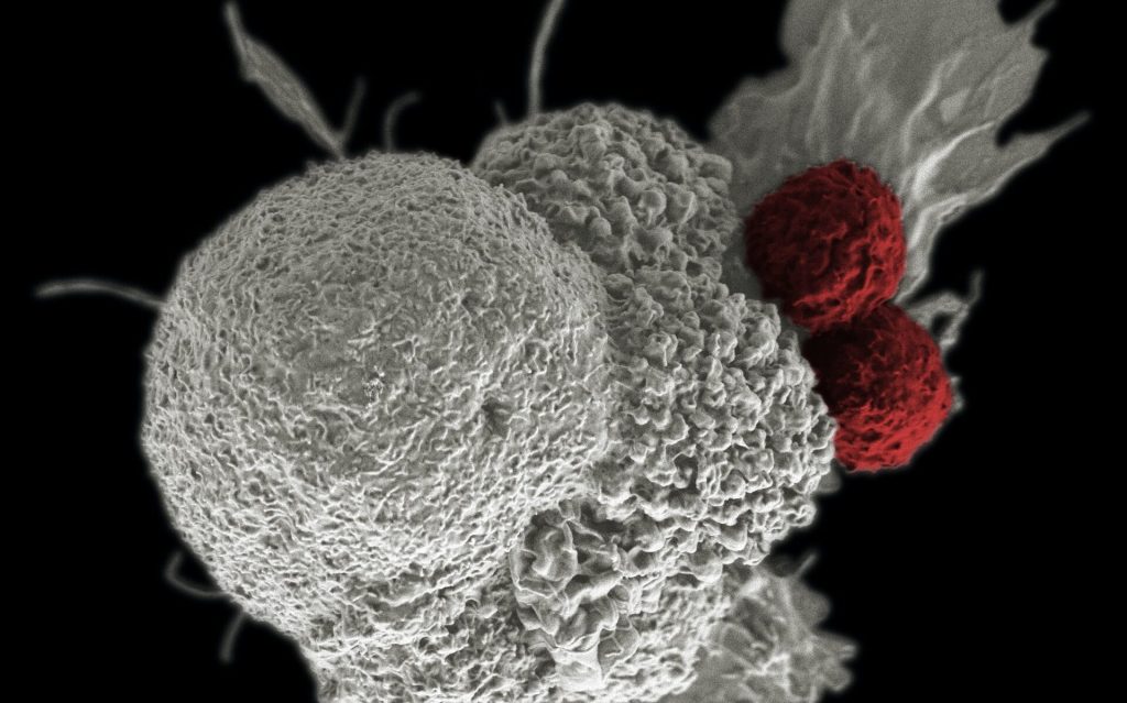
To better characterise the tumour cells that survived in mice following immunotherapy, the researchers isolated and studied the live tumour cells. They found that most of the cells responded to the presence of T cells by organising into temporary formations. These were made up of clusters of several tumour cell nuclei, which are surrounded by a single, multilayered membrane and a meshwork of cortical actin filaments. The inner cell of the formation was dense and appeared to be compacted within another cell.

To show that this result was not due to the isolation of the melanoma cells, the team also analysed tumours with fluorescently labelled cell nuclei and membranes. They found that the cell-in-cell formation was more prevalent in immunotherapy-treated tumours, particularly in sites associated with tumour cell death. Further analysis indicated that roughly half of the tumour cells that survived immunotherapy were arranged in the cell-in-cell formation. Over time, these cells returned to a single-cell state, with similar structural features to those of the parental cell line.

The team next tested whether this phenomenon occurs in human cancers. To do this, they incubated tumour cell lines with pre-activated T cells from healthy donors. They discovered that the vast majority of breast, colon and melanoma tumour cells that survived T cell killing organised into the cell-in-cell structure. A three-day observation of T cells interacting with tumour cells showed that these structures were dynamic, with individual tumour cells constantly forming and disseminating from the structure.

Finally, they tested the clinical relevance of this discovery by analysing cancerous tissues from multiple organs of four stage 4 melanoma patients. These patients were undergoing surgical removal of primary and metastatic lymph nodes. The researchers found that in all four patients, the cell-in-cell formation was highly abundant in the T cell zone of the draining lymph nodes, but not in the primary tumours. Furthermore, in a patient with untreated recurrent melanoma, most of the cells in the primary tumour were single cells, whereas the recurrent tumours had an abundance of the cell-in-cell formations.

“This previously unknown mechanism of tumour resistance highlights a current limitation of immunotherapy,” said senior author Yaron Carmi at Tel Aviv University. “Over the past decade many clinical studies have used immunotherapy followed by chemotherapy. But our findings suggest that timed inhibition of relevant signalling pathways needs to occur alongside immunotherapy to prevent the tumour becoming resistant to subsequent treatments.”

Source: eLife

Simple, Painless Microneedle Tattoos

Photo by Benjamin Lehman on Unsplash

Researchers from the Georgia Institute of Technology have developed a low-cost patch of microneedles that can be applied just by pressing it into the skin, with none of the pain and blood of traditional tattooing. The team development presented their research in the journal iScience.

These tattoos, which can be self-administered, have many potential applications, from medical alerts to tracking neutered animals to cosmetics. 

“We’ve miniaturised the needle so that it’s painless, but still effectively deposits tattoo ink in the skin,” said principal investigator Mark Prausnitz “This could be a way not only to make medical tattoos more accessible, but also to create new opportunities for cosmetic tattoos because of the ease of administration.” 

Medical applications of tattoos include covering up scars, guiding radiotherapy, or restoring nipples after breast surgery. Tattoos also serve instead of bracelets as medical alerts to communicate serious medical conditions such as diabetes, epilepsy, or allergies.  

<p>Medical alert tattoo: microneedle patch (above) and tattoo on skin (below).</p><p>Credit: Song Li, Georgia Tech</p>
Medical alert tattoo: microneedle patch (above) and tattoo on skin (below). Credit: Song Li, Georgia Tech

Various cosmetic products using microneedles are already on the market – mostly for anti-ageing – but developing microneedle technology for tattoos is new. Prausnitz, a veteran in this area, has studied microneedle patches for years to painlessly administer drugs and vaccines to the skin without the need for hypodermic needles. 

“We saw this as an opportunity to leverage our work on microneedle technology to make tattoos more accessible,” Prausnitz said. “While some people are willing to accept the pain and time required for a tattoo, we thought others might prefer a tattoo that is simply pressed onto the skin and does not hurt.”   

Transforming tattooing 

Tattoos typically use large needles to puncture repeatedly into the skin to get a good image, a time-consuming and painful process. The Georgia Tech team has developed microneedles that are smaller than a grain of sand and are made of tattoo ink encased in a dissolvable matrix.  

“Because the microneedles are made of tattoo ink, they deposit the ink in the skin very efficiently,” said former Georgia Tech postdoctoral fellow Song Li, the lead author of the study. 

In this way, the microneedles can be pressed into the skin just once and then dissolve, leaving the ink in the skin after a few minutes without bleeding.   

Creating the tattoo 

Although most microneedle patches for pharmaceuticals or cosmetics have dozens or hundreds of microneedles arranged in a square or circle, microneedle patch tattoos imprint a design that can include letters, numbers, symbols, and images. By arranging the microneedles in a specific pattern, each microneedle acts like a pixel to create a tattoo image in any shape or pattern.  

The researchers start with a mold containing microneedles in a pattern that forms an image. They fill the microneedles in the mold with tattoo ink and add a patch backing for convenient handling. The resulting patch is then applied to the skin for a few minutes, during which time the microneedles dissolve and release the tattoo ink. Tattoo inks of various colors can be incorporated into the microneedles, including black-light ink that can only be seen when illuminated with ultraviolet light.  

Prausnitz’s lab has been researching microneedles for vaccine delivery for years and realised they could be equally applicable to tattoos. Prausnitz’s team started working on tattoos to identify spayed and neutered pets, but then realised the technology could be effective for people, too. 

The tattoos were also designed with privacy in mind. The researchers even created patches sensitive to environmental factors such as light or temperature changes, where the tattoo will only appear with ultraviolet light or higher temperatures. This provides patients with privacy, revealing the tattoo only when desired. 

The study showed that the tattoos could last for at least a year and are likely to be permanent, which also makes them viable cosmetic options for people who want an aesthetic tattoo without risk of infection or the pain associated with traditional tattoos. Microneedle tattoos could alternatively be loaded with temporary tattoo ink to address short-term needs in medicine and cosmetics.  

Microneedle patch tattoos can also be used to encode information in the skin of animals. Rather than clipping the ear or applying an ear tag to animals to indicate sterilisation status, a painless and discreet tattoo can be applied instead.  

However, the technology does not aim to put tattoo artists out of business.

“The goal isn’t to replace all tattoos, which are often works of beauty created by tattoo artists,” Prausnitz said. “Our goal is to create new opportunities for patients, pets, and people who want a painless tattoo that can be easily administered.”  

Source: Georgia Institute of Technology

A New Seated Exercise Using the Calf Muscle Boosts Metabolism

Photo by TheStandingDesk on Unsplash

A simple, groundbreaking exercise developed by researchers at the University at Houston can help boost metabolism in the sedentary office-based lifestyle that causes so many health problems. By using the soleus muscle in the calf, though accounting for only 1% of the body’s weight, the metabolic health of the rest of the body can be boosted – if this muscle activated in a very specific way.

Marc Hamilton, professor of Health and Human Performance at the University of Houston, has discovered such an approach for optimal activation: the “soleus pushup” (SPU) which effectively elevates muscle metabolism for hours, even while sitting. The soleus, one of the human body’s 600 muscles, is a posterior leg muscle that runs from just below the knee to the heel.

Prof Hamilton’s research, published in the journal iScience, suggests the soleus pushup’s ability to sustain an elevated oxidative metabolism to improve the regulation of blood glucose is more effective than any popular methods currently touted as a solution including exercise, weight loss and intermittent fasting. Oxidative metabolism burns metabolites like blood glucose or fats, but it partly depends on the immediate energy needs of the muscle when it’s working.

“We never dreamed that this muscle has this type of capacity. It’s been inside our bodies all along, but no one ever investigated how to use it to optimise our health, until now,” said Prof Hamilton. “When activated correctly, the soleus muscle can raise local oxidative metabolism to high levels for hours, not just minutes, and does so by using a different fuel mixture.”

Muscle biopsies had revealed that the soleus used minimal glycogen – the predominant carbohydrate for fuelling muscular exercise. Instead of breaking down glycogen, the soleus can use blood glucose and fats.

“The soleus’s lower-than-normal reliance on glycogen helps it work for hours effortlessly without fatiguing during this type of muscle activity, because there is a definite limit to muscular endurance caused by glycogen depletion,” he added. “As far as we know, this is the first concerted effort to develop a specialised type of contractile activity centred around optimising human metabolic processes.”

When the SPU was tested, the whole-body effects on blood chemistry included a 52% improvement in the excursion of blood glucose and 60% less insulin requirement over three hours after ingesting a glucose drink.

This new approach of keeping the soleus muscle metabolism going also doubles the normal rate of fat metabolism in the fasting period, reducing levels of VLDL triglyceride.

The soleus pushup

Building on years of research, Hamilton and his colleagues developed the soleus pushup, which activates the soleus muscle in a different way than standing or walking does. The SPU targets the soleus to increase oxygen consumption more than what’s possible with these other types of soleus activities, while also being resistant to fatigue.

While seated with feet flat on the floor and muscles relaxed, a soleus pushup is performed by the heel rising while the front of the foot stays put. When the heel gets to the top of its range of motion, the foot is passively released to come back down. The aim is to simultaneously shorten the calf muscle while the soleus is naturally activated by its motor neurons.

While the SPU movement might look like walking (though performed while seated) it is the exact opposite, the researchers say. The body is designed to minimise the amount of energy used in walking, because of how the soleus moves. Prof Hamilton’s method reverses that and makes the soleus use as much energy as possible for a long duration.

However, the method is very specific, and if you are trying this while seated at your desk right now, you may not be doing it in the right way.

“The soleus pushup looks simple from the outside, but sometimes what we see with our naked eye isn’t the whole story. It’s a very specific movement that right now requires wearable technology and experience to optimise the health benefits,” said Prof Hamilton.

Additional publications are in the works focused on how to instruct people to properly learn this singular movement, but without the sophisticated laboratory equipment used in this latest study.

The researchers are quick to point out that this is not some new fitness tip or diet of the month. It’s a potent physiological movement that capitalises on the unique features of the soleus.

Potential first step toward a health care breakthrough

Prof Hamilton said it is the “most important study” ever completed at his lab, and could be a solution to a variety of health problems caused by spending hours each day living with insufficient muscle metabolism caused by inactivity. The average American sits about 10 hours a day.

Inactivity is a major health risk, and a low low metabolic rate while seated is especially troublesome for people who are at high risk for age-associated metabolic diseases such as metabolic syndrome and type 2 diabetes.

Prof Hamilton said inactive muscles require less energy than most people seem to understand, saying it’s “one of the most fundamental, yet overlooked issues” guiding the way toward discovering metabolic solutions to assist in preventing some age associated chronic diseases.

“All of the 600 muscles combined normally contribute only about 15% of the whole-body oxidative metabolism in the three hours after ingesting carbohydrate. Despite the fact that the soleus is only 1% the body weight, it is capable of raising its metabolic rate during SPU contractions to easily double, even sometimes triple, the whole-body carbohydrate oxidation.

“We are unaware of any existing or promising pharmaceuticals that come close to raising and sustaining whole-body oxidative metabolism at this magnitude.”

Source: University of Houston

Elevated Cardiovascular Disease Risk in Adults with ADHD

Photo by Freestocksorg on Pexels

Adults with ADHD have a greater risk of developing a range of cardiovascular diseases than those without the condition, according to a large observational study. The study researchers say that these findings, published in the journal World Psychiatry, underscore the need to monitor cardiovascular health in people with ADHD.

Attention deficit/hyperactivity disorder (ADHD) is one of the most common neurodevelopmental disorders, with a global prevalence of around 2.5% in adults. It often exists in parallel with other psychiatric and physical conditions, some of which have been linked to an increased risk of cardiovascular disease (CVD). But whether ADHD is independently associated with overall and specific cardiovascular diseases has not received as much attention.

In the current study, led by Karolinska Institutet and Örebro University, the researchers investigated associations between ADHD and some 20 different cardiovascular diseases when separated from other known risk factors such as smoking and diabetes.

A doubled risk

“We found that adults with ADHD were more than twice as likely to develop at least one cardiovascular disease, compared with those without ADHD,” says the study’s first author Lin Li, postdoctoral researcher at Karolinska Institutet. “When we accounted for other well-established risk factors for CVDs, the association weakened but still remained significant, which indicates that ADHD is an independent risk factor for a wide range of cardiovascular diseases.”

The study accessed data of more than five million Swedish adults, of which some 37 000 had ADHD. After an average 11.8 years of follow-up, 38% of individuals with ADHD had at least one diagnosis of cardiovascular disease, compared with 24% of those without ADHD.

Risks were elevated for all types of cardiovascular diseases and especially high for cardiac arrest, haemorrhagic stroke and peripheral vascular diseases, with somewhat stronger associations in men than in women. Some psychiatric comorbidities, especially eating and substance use disorders, significantly increased the risk of cardiovascular disease in people with ADHD. Pharmacological treatments for ADHD, such as anti-anxiety drugs, did not significantly affect the association between ADHD and cardiovascular disease. A causal link could not be established due to the observational nature of the study, and limitations included a lack of information about confounding factors such as lifestyle.

Important information for clinicians

“Clinicians needs to carefully consider psychiatric comorbidity and lifestyle factors to help reduce the CVD risk in individuals with ADHD, but we also need more research to explore plausible biological mechanisms, such as shared genetic components for ADHD and cardiovascular disease,” said Henrik Larsson, the study’s last author, a professor at the School of Medical Sciences, Örebro University, and affiliated researcher at Karolinska Institutet.

Source: Karolinska Institutet

COVID Damages Placenta’s Immune Response

Photo by Shvets Productions on Pexels

In a study published in the American Journal of Obstetrics & Gynecology, researchers found that, if a woman is infected by SARS-CoV-2 during her pregnancy, the infection damages the placenta’s immune response to further infections – even if the infection was mild.

“This is the largest study to date of placentas from women who had COVID during their pregnancies,” said Professor Kristina Adams Waldorf, at the University of Washington School of Medicine and senior author of the study. “We were surprised to find that women who had COVID during their pregnancies had placentas with an impaired immune response to new infection.”

This finding, Prof Adams Waldorf added, “was the tip of the iceberg” in how COVID might affect foetal or placental development.

During the early stages of the pandemic, many thought that COVID did not appear to harm the developing foetus because there were so few babies born with COVID infection, she noted.

“But what we’re seeing now is that the placenta is vulnerable to COVID-19, and the infection changes the way the placenta works, and that in turn is likely to impact the development of the foetus,” Prof Adams Waldorf said.

“To date, the studies about how COVID might affect foetal or child development are very limited as the children are still very young,” noted co-author Dr Helen Feltovich, professor at Intermountain Healthcare.

“Our study suggests that babies born to mothers infected with COVID at any point during their pregnancy will need to be monitored as they grow up,” she said.

Studies led by Adams Waldorf have shown that pregnant women who contract COVID have a significantly higher mortality rate than those who do not contract COVID. Other studies have found that pregnant women are more likely to risk hospitalisations or preterm birth, according to the Center for Disease Control and Prevention.

It’s unknown how different COVID variants may affect the mother or foetus, Profs Adams Waldorf and Feltovich agree.

“Studying each of the variants in real time is really challenging because they just keep coming so fast, we can’t keep up,” Prof Adams Waldorf said. “We do know that the COVID Delta variant was worse for pregnant individuals, because there was a spike in stillbirths, maternal deaths and hospitalisations at that time.”

Regardless of the variant, Prof Adams Waldorf stressed taking precautions, such as vaccination and booster shots, limiting social contact a bubble of vaccinated individuals even if it means isolating for the duration of the pregnancy.

“The disease may be mild, or it may be severe, but we’re still seeing these abnormal effects on the placenta,” she said. “It seems that after contracting COVID in pregnancy, the placenta is exhausted by the infection, and can’t recover its immune function.”

In this study, a total of 164 pregnant individuals were studied, consisting of 24 uninfected healthy patients as a control group and 140 individuals who contracted COVID. Both groups delivered at about the same time, 37 to 38 weeks. Preterm birth occurred at nearly three times the rate with the patients with COVID when compared with those without. About 75% of the COVID patients had either asymptomatic COVID or mild symptoms.

Source: University of Washington School of Medicine/UW Medicine

New Study Explains Diabetes and UTI Link

Huge clumps of E. coli (red) infecting diabetic mouse bladder. Photo: Soumitra Mohanty

Lower immunity and recurring infections are common in type 1 and type 2 diabetes. Research has shown that the immune system of people with diabetes has lower levels of the antimicrobial peptide psoriasin, which compromises the urinary bladder’s cell barrier, increasing the risk of urinary tract infection. The study is published in Nature Communications.

One effect of diabetes is that it compromises the innate immune system, leaving many people with increased susceptibility to regular infections, such as urinary tract infections (UTI)s caused by E. coli bacteria. In people with diabetes, these are more likely to lead to general blood poisoning, sepsis, originating in the urinary tract.

An endogenous antibiotic

Karolinska Institutet researchers investigated whether glucose levels in people with diabetes (type 1, type 2, or prediabetes) are linked with psoriasin, an endogenous antibiotic which is a part of the innate immune system.

Using samples of urine, bladder cells and blood serum from patients, the researchers analysed levels of psoriasin and other peptides necessary for ensuring that the bladder mucosa remains intact and protects against infection. The findings were then verified in mice and urinary bladder cells with and without infection.

“We found that high glucose concentrations reduce the levels of the antimicrobial peptide psoriasin, while insulin has no effect,” said Professor Annelie Brauner, who led the study. “People with diabetes have lower levels of psoriasin, which weakens the cells’ protective barrier function and increases the risk of bladder infection.”

Oestrogen therapy reduced bacterial population

Professor Brauner’s research group has previously shown that oestrogen restores the protective function of bladder cells in humans and mice and thereby help to regulate the immune response to a UTI. The researchers therefore tested how oestrogen treatment affects infected cells exposed to high glucose concentrations. They found that the treatment boosted levels of psoriasin and reduced bacterial populations, indicating that the treatment may have an effect also among patients with diabetes.

“We now plan to probe deeper into the underlying mechanisms of infections in individuals with diabetes,” said lead author Soumitra Mohanty. “The ultimate goal is to reduce the risk of infection in this growing patient group.”

Source: Karolinska Institutet

International Gaucher Day on 1 October Highlights Need For Greater Attention on Rare Diseases

Photo by Cottonbro on Pexels

Currently, there are an estimated 6000–8000 rare diseases, which affect 350 million people worldwide.One such rare condition is Gaucher Disease (GD) – a lysosomal storage disorder (LSD).  GD is a rare genetic disorder, passed down from parents to children (inherited) in an autosomal recessive manner. 

GD is one of the most common LSDs with a prevalence in the general population of ~1 per 100 000 and ~1/855 in the Ashkenazi Jewish population.2 As with many rare conditions, diagnosis of GD may present a significant challenge to non-GD specialities, owing to the wide variability in age, severity, type of clinical manifestation and lack of awareness of the early signs and symptoms of GD among non-specialist physicians.3 One in 6 patients with GD reported a diagnosis delay of 7 years or more after first consulting a doctor.3

International Gaucher Day on 1 October, therefore, aims to improve patients’ quality of life through greater awareness and earlier diagnosis of GD amongst healthcare professionals.

GD arises from an inherited deficiency of an enzyme called glucocerebrosidase, normally found within the lysosomes of cells, due to mutations in the GBA gene.4 This enzyme is responsible for breaking down a fatty substrate, glucocerebroside, into glucose and a simpler fat molecule (ceramide).4 Patients with GD have a progressive build-up of glucocerebroside within the lysosomes, particularly of macrophages, resulting in enlarged cells known as ‘Gaucher’ cells.4

These ‘Gaucher’ cells accumulate in organs throughout the body, predominately affecting the bone marrow, liver, and spleen.4 There are three types of GD, based on the presence and severity of neurological involvement.4 Type 1, known as the non-neuronopathic GD  characterised by haematological abnormalities such as thrombocytopenia, leukopenia and anaemia, hepatomegaly and/or splenomegaly, bone crises and/or osteoporosis, and fatigue. Phenotypically, there is a wide spectrum of disease manifestations, ranging from asymptomatic to severe type 1 child-onset disease.5,6 Type 2, the acute neuronopathic form, is the rarest and most severe form of GD.  It includes the rapid progression of severe neurological abnormalities early in life, leading to death in infancy or early childhood.4,6 Type 3, the chronic form, encompasses multiple phenotypes.  Type 3 typically occurs during the paediatric years and varies in severity: patients have the same symptoms as in type 1, plus some neurological involvement that generally appear later in life, such as abnormal eye movement, ataxia, seizures and dementia.4

Anaemia, thrombocytopenia, enlargement of the liver and/or spleen, and skeletal abnormalities (osteopenia, lytic lesions, pathological fractures, chronic bone pain, bone crisis, bone infarcts, osteonecrosis and skeletal deformities) are typical manifestations of type 1 GD, the most prevalent form of the disease.However, the severity and coexistence of different symptoms are highly variable, and GD patients are often misdiagnosed as having other malignant haematological conditions.4

Although GD is rare, clinicians are encouraged to maintain a high index of suspicion with patients presenting with atypical symptoms, and should consider testing for rare diseases where other haematological pathologies have been excluded4 or when testing for them. Such patients may be referred to a GD specialist or be tested through North West University (NWU), where global pharmaceutical company Sanofi and the NWU Centre of Human Metabolomics, headed by Prof Chris Vorster, have partnered to test for the most common lysosomal storage disorders in South Africa, including GD, using dried blood spot samples.

Says Prof. Vorster: “Rare conditions such as GD require the cooperation of a multidisciplinary team in order to find and treat them. Interventions can improve a patient’s quality of life through improvement or restoration of their physical function, so that they may carry out regular daily activities. The NWU Centre of Human Metabolomics provides internationally competitive metabolomic analytic services, and electronic results may be sent by high priority straight to healthcare practitioners, speeding up diagnosis.”

Monique Nel, Medical Advisor – Rare Diseases at Sanofi, says: “We understand the difficulty that healthcare professionals face when it comes to diagnosing patient with GD. It requires a coordinated approach to diagnosis and care for people living with the condition. Early diagnosis of GD, and the initiation of treatment will delay the occurrence of irreversible complications, and improve the patient’s quality of life. We therefore direct the attention of healthcare providers to the RD Nexus platform, which is Sanofi’s dedicated platform for rare diseases, at www.RDNexus.com. This platform offers educational materials, road maps to a differential diagnosis and how to test a patient for these conditions.”

For more information on GD and other rare diseases, visit: www.RDNexus.com

  1. https://www.ncbi.nlm.nih.gov/pmc/articles/PMC6505568/pdf/EMMM-11-e10486.pdf Accessed September 2022.
  2. Burrow TA et al. Prevalence and management of Gaucher disease. Paediatric Health, Medicine and Theraeutics 2011;2:59-73.
  3. Revel-Wilk S, et al. How we manage Gaucher Disease in the era of choices. British Journal of Haematology 2018;182:467-480.
  4. CPD Gaucher. Gaucher Disease. Medical Chronicle June 2020:30-32.
  5. Linari S, Castaman G. Clinical manifestations and management of Gaucher disease. Clinical Cases in Mineral and Bone Metabolism 2015;12(2):157-164.
  6. Roshan Lal T and Sidransky E. The spectrum of neurological manifestations associated with Gaucher Disease. Diseases 2017;5,10.

Why Elderly Drivers May Hit the Brake Instead of the Accelerator

Driver at the wheel of a car
Photo by Why Kei on Unsplash

Researchers in Japan have found that older participants have longer reaction times, slower decision times, and greater brain activation in the parts of the brain involved with inhibition and switching tasks. These findings, published in the journal Brain Behavioural Research, illuminate the causes of accidents involving elderly drivers who press the brake instead of the accelerator.

With one of the oldest populations in the world, Japan is concerned about cognitive decline in drivers resulting in accidents. The police require drivers over 75 to take periodic cognitive tests. However, few studies have investigated executive functions and brain activity among older adults in terms of foot responses during braking and accelerating.

To address this gap, a group led by Professor Nobuyuki Kawai of the Graduate School of Informatics at Nagoya University in Japan scanned the brains of elderly people and students while performing pedal-pressing simulations. The researchers were particularly interested in the left dorsolateral prefrontal cortex, the part of the brain associated with inhibition and switching responses.

To simulate the response of a person’s feet and hands when driving a car, they created a new task in the laboratory called the bimanual and bipedal response selection and response-position compatibility task. During this task, a signal directed participants to press the left or right button with their left or right foot, or their left or right hand. Sometimes participants pressed the pedal in front of them, whereas at other times they had to press it diagonally. This was done to allow the researchers to assess how the participants responded in situations where the cognitive load was higher. Administering this task to both university students and elderly participants, the researchers then monitored blood flow in their brains. The results were published in Behavioural Brain Research.

They found that older participants had longer reaction times, slower decision times, and greater brain activation than younger people. Furthermore, pressing the diagonal pedal required longer reaction times and greater brain activation than pressing directly ahead in the left dorsolateral prefrontal cortex. Interestingly, this was only found when people were asked to use their feet but not their hands. In short, older people had to do more active thinking than younger people when deciding which ‘pedal’ to press with their feet.

“This indicates that the cognitive load is higher when pushing the pedal diagonally with the foot, such as when pressing the brake,” explains Professor Kawai. “When you push a diagonal pedal with your foot, you are using the frontal lobe more than when you push the pedal straight in front. In particular, the left dorsolateral frontal lobe, which is important for response switching, is more active when the foot is pressed at an angle than when the pedal is pressed straight. In these tasks, older adults have higher neural activity throughout the frontal lobe than college students.”

This study’s findings suggest that to compensate for the decline in cognitive functions, greater brain activation may be necessary in elderly people. Older people may struggle in situations with a high cognitive load, such as parking a vehicle in a narrow space. “This study suggests that the performance of older adults is vulnerable in these situations,” Professor Kawai explains. “Elderly drivers should not be overconfident that their driving is fine. Even elderly people who are normally able to drive without any problems, when a cognitive load is applied, such as when switching from one parking space to another or when talking to a passenger, things may be different and there is a chance of pressing the wrong pedal. We believe that it is important to educate elderly drivers about this fact.”

Source: Nagoya University

Smartphones Could Serve as Pulse Oximeters in the Home

Photo by Asterfolio on Unsplash

Researchers have demonstrated that smartphones are capable of detecting blood oxygen saturation levels down to 70% – the lowest value that pulse oximeters should be able to measure, as recommended by the US Food and Drug Administration. The team published these results in npj Digital Medicine.

The technique involves participants placing their finger over the camera and flash of a smartphone, which uses a deep-learning algorithm to decipher the blood oxygen levels. When the team delivered a controlled mixture of nitrogen and oxygen to six subjects to artificially bring their blood oxygen levels down, the smartphone correctly predicted whether the subject had low blood oxygen levels 80% of the time.

“Other smartphone apps that do this were developed by asking people to hold their breath. But people get very uncomfortable and have to breathe after a minute or so, and that’s before their blood-oxygen levels have gone down far enough to represent the full range of clinically relevant data,” said co-lead author Jason Hoffman, a UW doctoral student in the Paul G. Allen School of Computer Science & Engineering. “With our test, we’re able to gather 15 minutes of data from each subject. Our data shows that smartphones could work well right in the critical threshold range.”

Another benefit of measuring blood oxygen levels on a smartphone is that almost everyone has one.

“This way you could have multiple measurements with your own device at either no cost or low cost,” said co-author Dr. Matthew Thompson, professor of family medicine in the UW School of Medicine. “In an ideal world, this information could be seamlessly transmitted to a doctor’s office. This would be really beneficial for telemedicine appointments or for triage nurses to be able to quickly determine whether patients need to go to the emergency department or if they can continue to rest at home and make an appointment with their primary care provider later.”

The team recruited six participants ranging in age from 20 to 34. Three identified as female, three identified as male. One participant identified as being African American, while the rest identified as being Caucasian.

To gather data to train and test the algorithm, the researchers had each participant wear a standard pulse oximeter on one finger and then place another finger on the same hand over a smartphone’s camera and flash. Each participant had this same set up on both hands simultaneously.

“The camera is recording a video: Every time your heart beats, fresh blood flows through the part illuminated by the flash,” said Assistant Professor Edward Wang, who started this project as a doctoral student.

“The camera records how much that blood absorbs the light from the flash in each of the three color channels it measures: red, green and blue,” said Wang, who also directs the UC San Diego DigiHealth Lab. “Then we can feed those intensity measurements into our deep-learning model.”

Each participant breathed in a controlled mixture of oxygen and nitrogen to slowly reduce oxygen levels. For all six participants, the team acquired more than 10 000 blood oxygen level readings between 61% and 100%.

The researchers used data from four of the participants to train a deep learning algorithm to extract the blood oxygen levels, and the rest of the data was used to validate the method and then test it to see how well it performed on new subjects.

“Smartphone light can get scattered by all these other components in your finger, which means there’s a lot of noise in the data that we’re looking at,” said co-lead author Varun Viswanath. “Deep learning is a really helpful technique here because it can see these really complex and nuanced features and helps you find patterns that you wouldn’t otherwise be able to see.”

The team hopes to continue this research by testing the algorithm on more people.

“One of our subjects had thick calluses on their fingers, which made it harder for our algorithm to accurately determine their blood oxygen levels,” Hoffman said. “If we were to expand this study to more subjects, we would likely see more people with calluses and more people with different skin tones. Then we could potentially have an algorithm with enough complexity to be able to better model all these differences.”

But, the researchers said, this is a good first step toward developing biomedical devices that are aided by machine learning.

“It’s so important to do a study like this,” Wang said. “Traditional medical devices go through rigorous testing. But computer science research is still just starting to dig its teeth into using machine learning for biomedical device development and we’re all still learning. By forcing ourselves to be rigorous, we’re forcing ourselves to learn how to do things right.”

Source: University of Washington

Is the US Failing to Hold Crooked Medical Industry Execs Accountable?

Photo by Tingey Injury Law Firm on Unsplash

When it comes to violations of US federal law by pharmaceutical and medical device manufacturers, the US Department of Justice (DOJ) is not exercising its full authority, according to the findings of a review published in JAMA Internal Medicine.

The conviction of Elizabeth Holmes, CEO of the failed blood-testing company Theranos, has focused attention on the personal liability of corporate officers of medical companies engaging in illegal activity. Holmes was charged with defrauding and conspiring to defraud investors, patients, and physicians, each count carrying a maximum 20-year sentence. Among prosecutors’ allegations was that Theranos’ main blood-testing device failed to work as the company and Holmes had promised. While Holmes was convicted of defrauding investors, she faced no personal liability as a CEO responsible for a company knowingly selling faulty diagnostic tests.

When a drug or medical device company violates US federal law, the government can use the Park doctrine. It holds that a CEO for a health-related company has a “position of authority” in a profitable business selling “services and products [that] affect the health and well-being of the public.” The doctrine’s aim is to protect patients from harm caused by unsafe or fraudulent medical products and services. It does this by targeting the executives who run the companies that make revenues on these products while violating federal law.

This provides an alternative to having that risk borne by patients or impersonal corporate entities; however, it is rare for there to be public reports of drug and device company executives being prosecuted with Park doctrine.

The researchers sought to identify prosecutions using the Park doctrine and characterise their role in DOJ enforcement efforts related to medical product industry misconduct. To this end, they conducted a literature search.

They found 13 cases where executives from six drug and medical device companies prosecuted under the Park doctrine since 2000. These prosecutions resulted in 11 guilty pleas and two jury trials, leading to two convictions. Of the six companies, three were drug manufacturers, two were medical device manufacturers, and one was a compounding pharmacy. All three of the drug manufacturers were opioid manufacturers, of which two executives were charged for unlawful promotion, and one was charged for manufacturing errors. Both device manufacturer executives were charged with unlawful promotion. All but three prosecutions alleged the defendants’ complicity or personal involvement in the misconduct, which Park does not require. By contrast, most large settlements with the DOJ over alleged misconduct in the past two decades did not result in individual liability for executives.

“This review suggests that federal prosecutors have exercised far less than their full capacity under the Park doctrine to sanction problematic corporate behaviour that threatens patients and the public health,” the authors concluded. They suggest that enforcement under a reinvigorated Park doctrine would help to better protect patients.