Day: September 22, 2022

People with HIV and Hepatitis C Have Increased Heart Attack Risk

Source: Wikimedia Commons CC0

As people with HIV age, their risk of myocardial infarction increases far more if they also have untreated hepatitis C virus, according to new research published today in the Journal of the American Heart Association.

According to the findings, even with antiretroviral therapy (ART), the risk of myocardial infarction (MI) among people with HIV is at least 50% higher than people without HIV. This new study evaluated if people with HIV who also have hepatitis C have a higher risk of MI.

“HIV and hepatitis C coinfection occurs because they share a transmission route – both viruses may be transmitted through blood-to-blood contact,” said Associate Professor Keri N. Althoff, PhD, MPH, senior author of the study. “Due in part to the inflammation from the chronic immune activation of two viral infections, we hypothesised that people with HIV and hepatitis C would have a higher risk of heart attack as they aged compared to those with HIV alone.”

Researchers analysed health information for 23 361 people with HIV in the North American AIDS Cohort Collaboration on Research and Design (NA-ACCORD) between 2000–2017 started on antiretroviral treatment for HIV, median age 45 at enrolment. One in 5 study participants (4677) were also positive for hepatitis C. During a median follow-up of about 4 years, the researchers compared the occurrence of a heart attack between the HIV-only and the HIV-hepatitis C co-infected groups as a whole, and by each decade of age.

The analysis found:

  • With each decade of increasing age, MI incidence increased 30% in people with HIV alone and 85% in those who were also positive for hepatitis C.
  • The risk of heart attack increased in participants who also had traditional heart disease risk factors such as high blood pressure (more than 3 times), smoking (90%) and Type 2 diabetes (46%).
  • The risk of heart attack was also higher (40%) in participants with certain HIV-related factors such as low levels of CD4 immune cells (200 cells/mm3, signalling greater immune dysfunction) and 45% in those who took protease inhibitors (one type of ART linked to metabolic conditions).

“People who are living with HIV or hepatitis C should ask their doctor about treatment options for the viruses and other ways to reduce their cardiovascular disease risk,” said Assistant Professor Raynell Lang, MD, MSc, lead study author.

“Several mechanisms may be involved in the increased heart attack risk among co-infected patients. One contributing factor may be the inflammation associated with having two chronic viral infections,” A/Prof Lang said. “There also may be differences in risk factors for cardiovascular disease and non-medical factors that influence health among people with HIV and hepatitis C that plays a role in the increased risk.”

“Our findings suggest that HIV and hepatitis C co-infections need more research, which may inform future treatment guidelines and standards of care,” Althoff said.

The study is limited by not having information on additional factors associated with heart attack risk such as diet, exercise or family history of chronic health conditions. Results from this study of people with HIV receiving care in North America may not be generalizable to people with HIV elsewhere. In addition, the study period included time prior to the availability of more advanced hepatitis C treatments.

“Because effective and well-tolerated hepatitis C therapy was not available during several years of our study period, we were unable to evaluate the association of treated hepatitis C infection on cardiovascular risk among people with HIV. This will be an important question to answer in future studies,” Lang said.

Source: American Heart Association

Shields Up: Tumour Cells Form Temporary Structures to Resist Immunotherapy

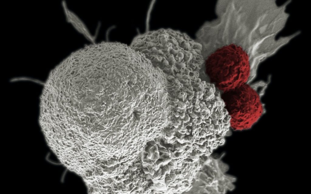
Shown here is a pseudo-colored scanning electron micrograph of an oral squamous cancer cell (white) being attacked by two cytotoxic T cells (red), part of a natural immune response. Photo by National Cancer Institute on Unsplash

Scientists have shown how tumour cells evade immunotherapy by forming temporary structures, where cells on the inside remain intact and can return to an individual state. These findings, published today in eLife, provide a novel theory as to how tumour cells avoid destruction by the immune system. They could also inform new treatments that combine immunotherapy with the timed inhibition of relevant signalling pathways in tumour cells.

“Cancer immunotherapy harnesses the body’s immune system to fight cancer. Despite its remarkable success, the majority of patients who receive immunotherapy will only see their tumours shrink in size temporarily before returning, and these relapsed tumours will likely be resistant to immunotherapy treatment,” said first author Amit Gutwillig, who was a PhD student at Tel Aviv University when the study was done.

To identify how tumours relapse after immunotherapy, Carmi and colleagues began by comparing the genetic sequences of whole genomes in primary and relapsed tumours in the same patient. Their analysis suggested that relapsed tumours do not change dramatically following immunotherapy.

Next, the team studied this process in breast cancer and melanoma, using mouse models in which immunotherapy-resistant tumours had relapsed. They administered the mice with cells from treated tumours and allowed these cells to reach a palpable size. The team found that the cells were equally susceptible to the same immunotherapy approach as the parent tumour, although they relapsed sooner.

To better characterise the tumour cells that survived in mice following immunotherapy, the researchers isolated and studied the live tumour cells. They found that most of the cells responded to the presence of T cells by organising into temporary formations. These were made up of clusters of several tumour cell nuclei, which are surrounded by a single, multilayered membrane and a meshwork of cortical actin filaments. The inner cell of the formation was dense and appeared to be compacted within another cell.

To show that this result was not due to the isolation of the melanoma cells, the team also analysed tumours with fluorescently labelled cell nuclei and membranes. They found that the cell-in-cell formation was more prevalent in immunotherapy-treated tumours, particularly in sites associated with tumour cell death. Further analysis indicated that roughly half of the tumour cells that survived immunotherapy were arranged in the cell-in-cell formation. Over time, these cells returned to a single-cell state, with similar structural features to those of the parental cell line.

The team next tested whether this phenomenon occurs in human cancers. To do this, they incubated tumour cell lines with pre-activated T cells from healthy donors. They discovered that the vast majority of breast, colon and melanoma tumour cells that survived T cell killing organised into the cell-in-cell structure. A three-day observation of T cells interacting with tumour cells showed that these structures were dynamic, with individual tumour cells constantly forming and disseminating from the structure.

Finally, they tested the clinical relevance of this discovery by analysing cancerous tissues from multiple organs of four stage 4 melanoma patients. These patients were undergoing surgical removal of primary and metastatic lymph nodes. The researchers found that in all four patients, the cell-in-cell formation was highly abundant in the T cell zone of the draining lymph nodes, but not in the primary tumours. Furthermore, in a patient with untreated recurrent melanoma, most of the cells in the primary tumour were single cells, whereas the recurrent tumours had an abundance of the cell-in-cell formations.

“This previously unknown mechanism of tumour resistance highlights a current limitation of immunotherapy,” said senior author Yaron Carmi at Tel Aviv University. “Over the past decade many clinical studies have used immunotherapy followed by chemotherapy. But our findings suggest that timed inhibition of relevant signalling pathways needs to occur alongside immunotherapy to prevent the tumour becoming resistant to subsequent treatments.”

Source: eLife

Simple, Painless Microneedle Tattoos

Photo by Benjamin Lehman on Unsplash

Researchers from the Georgia Institute of Technology have developed a low-cost patch of microneedles that can be applied just by pressing it into the skin, with none of the pain and blood of traditional tattooing. The team development presented their research in the journal iScience.

These tattoos, which can be self-administered, have many potential applications, from medical alerts to tracking neutered animals to cosmetics. 

“We’ve miniaturised the needle so that it’s painless, but still effectively deposits tattoo ink in the skin,” said principal investigator Mark Prausnitz “This could be a way not only to make medical tattoos more accessible, but also to create new opportunities for cosmetic tattoos because of the ease of administration.” 

Medical applications of tattoos include covering up scars, guiding radiotherapy, or restoring nipples after breast surgery. Tattoos also serve instead of bracelets as medical alerts to communicate serious medical conditions such as diabetes, epilepsy, or allergies.  

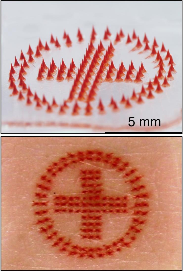
<p>Medical alert tattoo: microneedle patch (above) and tattoo on skin (below).</p><p>Credit: Song Li, Georgia Tech</p>
Medical alert tattoo: microneedle patch (above) and tattoo on skin (below). Credit: Song Li, Georgia Tech

Various cosmetic products using microneedles are already on the market – mostly for anti-ageing – but developing microneedle technology for tattoos is new. Prausnitz, a veteran in this area, has studied microneedle patches for years to painlessly administer drugs and vaccines to the skin without the need for hypodermic needles. 

“We saw this as an opportunity to leverage our work on microneedle technology to make tattoos more accessible,” Prausnitz said. “While some people are willing to accept the pain and time required for a tattoo, we thought others might prefer a tattoo that is simply pressed onto the skin and does not hurt.”   

Transforming tattooing 

Tattoos typically use large needles to puncture repeatedly into the skin to get a good image, a time-consuming and painful process. The Georgia Tech team has developed microneedles that are smaller than a grain of sand and are made of tattoo ink encased in a dissolvable matrix.  

“Because the microneedles are made of tattoo ink, they deposit the ink in the skin very efficiently,” said former Georgia Tech postdoctoral fellow Song Li, the lead author of the study. 

In this way, the microneedles can be pressed into the skin just once and then dissolve, leaving the ink in the skin after a few minutes without bleeding.   

Creating the tattoo 

Although most microneedle patches for pharmaceuticals or cosmetics have dozens or hundreds of microneedles arranged in a square or circle, microneedle patch tattoos imprint a design that can include letters, numbers, symbols, and images. By arranging the microneedles in a specific pattern, each microneedle acts like a pixel to create a tattoo image in any shape or pattern.  

The researchers start with a mold containing microneedles in a pattern that forms an image. They fill the microneedles in the mold with tattoo ink and add a patch backing for convenient handling. The resulting patch is then applied to the skin for a few minutes, during which time the microneedles dissolve and release the tattoo ink. Tattoo inks of various colors can be incorporated into the microneedles, including black-light ink that can only be seen when illuminated with ultraviolet light.  

Prausnitz’s lab has been researching microneedles for vaccine delivery for years and realised they could be equally applicable to tattoos. Prausnitz’s team started working on tattoos to identify spayed and neutered pets, but then realised the technology could be effective for people, too. 

The tattoos were also designed with privacy in mind. The researchers even created patches sensitive to environmental factors such as light or temperature changes, where the tattoo will only appear with ultraviolet light or higher temperatures. This provides patients with privacy, revealing the tattoo only when desired. 

The study showed that the tattoos could last for at least a year and are likely to be permanent, which also makes them viable cosmetic options for people who want an aesthetic tattoo without risk of infection or the pain associated with traditional tattoos. Microneedle tattoos could alternatively be loaded with temporary tattoo ink to address short-term needs in medicine and cosmetics.  

Microneedle patch tattoos can also be used to encode information in the skin of animals. Rather than clipping the ear or applying an ear tag to animals to indicate sterilisation status, a painless and discreet tattoo can be applied instead.  

However, the technology does not aim to put tattoo artists out of business.

“The goal isn’t to replace all tattoos, which are often works of beauty created by tattoo artists,” Prausnitz said. “Our goal is to create new opportunities for patients, pets, and people who want a painless tattoo that can be easily administered.”  

Source: Georgia Institute of Technology

A New Seated Exercise Using the Calf Muscle Boosts Metabolism

Photo by TheStandingDesk on Unsplash

A simple, groundbreaking exercise developed by researchers at the University at Houston can help boost metabolism in the sedentary office-based lifestyle that causes so many health problems. By using the soleus muscle in the calf, though accounting for only 1% of the body’s weight, the metabolic health of the rest of the body can be boosted – if this muscle activated in a very specific way.

Marc Hamilton, professor of Health and Human Performance at the University of Houston, has discovered such an approach for optimal activation: the “soleus pushup” (SPU) which effectively elevates muscle metabolism for hours, even while sitting. The soleus, one of the human body’s 600 muscles, is a posterior leg muscle that runs from just below the knee to the heel.

Prof Hamilton’s research, published in the journal iScience, suggests the soleus pushup’s ability to sustain an elevated oxidative metabolism to improve the regulation of blood glucose is more effective than any popular methods currently touted as a solution including exercise, weight loss and intermittent fasting. Oxidative metabolism burns metabolites like blood glucose or fats, but it partly depends on the immediate energy needs of the muscle when it’s working.

“We never dreamed that this muscle has this type of capacity. It’s been inside our bodies all along, but no one ever investigated how to use it to optimise our health, until now,” said Prof Hamilton. “When activated correctly, the soleus muscle can raise local oxidative metabolism to high levels for hours, not just minutes, and does so by using a different fuel mixture.”

Muscle biopsies had revealed that the soleus used minimal glycogen – the predominant carbohydrate for fuelling muscular exercise. Instead of breaking down glycogen, the soleus can use blood glucose and fats.

“The soleus’s lower-than-normal reliance on glycogen helps it work for hours effortlessly without fatiguing during this type of muscle activity, because there is a definite limit to muscular endurance caused by glycogen depletion,” he added. “As far as we know, this is the first concerted effort to develop a specialised type of contractile activity centred around optimising human metabolic processes.”

When the SPU was tested, the whole-body effects on blood chemistry included a 52% improvement in the excursion of blood glucose and 60% less insulin requirement over three hours after ingesting a glucose drink.

This new approach of keeping the soleus muscle metabolism going also doubles the normal rate of fat metabolism in the fasting period, reducing levels of VLDL triglyceride.

The soleus pushup

Building on years of research, Hamilton and his colleagues developed the soleus pushup, which activates the soleus muscle in a different way than standing or walking does. The SPU targets the soleus to increase oxygen consumption more than what’s possible with these other types of soleus activities, while also being resistant to fatigue.

While seated with feet flat on the floor and muscles relaxed, a soleus pushup is performed by the heel rising while the front of the foot stays put. When the heel gets to the top of its range of motion, the foot is passively released to come back down. The aim is to simultaneously shorten the calf muscle while the soleus is naturally activated by its motor neurons.

While the SPU movement might look like walking (though performed while seated) it is the exact opposite, the researchers say. The body is designed to minimise the amount of energy used in walking, because of how the soleus moves. Prof Hamilton’s method reverses that and makes the soleus use as much energy as possible for a long duration.

However, the method is very specific, and if you are trying this while seated at your desk right now, you may not be doing it in the right way.

“The soleus pushup looks simple from the outside, but sometimes what we see with our naked eye isn’t the whole story. It’s a very specific movement that right now requires wearable technology and experience to optimise the health benefits,” said Prof Hamilton.

Additional publications are in the works focused on how to instruct people to properly learn this singular movement, but without the sophisticated laboratory equipment used in this latest study.

The researchers are quick to point out that this is not some new fitness tip or diet of the month. It’s a potent physiological movement that capitalises on the unique features of the soleus.

Potential first step toward a health care breakthrough

Prof Hamilton said it is the “most important study” ever completed at his lab, and could be a solution to a variety of health problems caused by spending hours each day living with insufficient muscle metabolism caused by inactivity. The average American sits about 10 hours a day.

Inactivity is a major health risk, and a low low metabolic rate while seated is especially troublesome for people who are at high risk for age-associated metabolic diseases such as metabolic syndrome and type 2 diabetes.

Prof Hamilton said inactive muscles require less energy than most people seem to understand, saying it’s “one of the most fundamental, yet overlooked issues” guiding the way toward discovering metabolic solutions to assist in preventing some age associated chronic diseases.

“All of the 600 muscles combined normally contribute only about 15% of the whole-body oxidative metabolism in the three hours after ingesting carbohydrate. Despite the fact that the soleus is only 1% the body weight, it is capable of raising its metabolic rate during SPU contractions to easily double, even sometimes triple, the whole-body carbohydrate oxidation.

“We are unaware of any existing or promising pharmaceuticals that come close to raising and sustaining whole-body oxidative metabolism at this magnitude.”

Source: University of Houston

Elevated Cardiovascular Disease Risk in Adults with ADHD

Photo by Freestocksorg on Pexels

Adults with ADHD have a greater risk of developing a range of cardiovascular diseases than those without the condition, according to a large observational study. The study researchers say that these findings, published in the journal World Psychiatry, underscore the need to monitor cardiovascular health in people with ADHD.

Attention deficit/hyperactivity disorder (ADHD) is one of the most common neurodevelopmental disorders, with a global prevalence of around 2.5% in adults. It often exists in parallel with other psychiatric and physical conditions, some of which have been linked to an increased risk of cardiovascular disease (CVD). But whether ADHD is independently associated with overall and specific cardiovascular diseases has not received as much attention.

In the current study, led by Karolinska Institutet and Örebro University, the researchers investigated associations between ADHD and some 20 different cardiovascular diseases when separated from other known risk factors such as smoking and diabetes.

A doubled risk

“We found that adults with ADHD were more than twice as likely to develop at least one cardiovascular disease, compared with those without ADHD,” says the study’s first author Lin Li, postdoctoral researcher at Karolinska Institutet. “When we accounted for other well-established risk factors for CVDs, the association weakened but still remained significant, which indicates that ADHD is an independent risk factor for a wide range of cardiovascular diseases.”

The study accessed data of more than five million Swedish adults, of which some 37 000 had ADHD. After an average 11.8 years of follow-up, 38% of individuals with ADHD had at least one diagnosis of cardiovascular disease, compared with 24% of those without ADHD.

Risks were elevated for all types of cardiovascular diseases and especially high for cardiac arrest, haemorrhagic stroke and peripheral vascular diseases, with somewhat stronger associations in men than in women. Some psychiatric comorbidities, especially eating and substance use disorders, significantly increased the risk of cardiovascular disease in people with ADHD. Pharmacological treatments for ADHD, such as anti-anxiety drugs, did not significantly affect the association between ADHD and cardiovascular disease. A causal link could not be established due to the observational nature of the study, and limitations included a lack of information about confounding factors such as lifestyle.

Important information for clinicians

“Clinicians needs to carefully consider psychiatric comorbidity and lifestyle factors to help reduce the CVD risk in individuals with ADHD, but we also need more research to explore plausible biological mechanisms, such as shared genetic components for ADHD and cardiovascular disease,” said Henrik Larsson, the study’s last author, a professor at the School of Medical Sciences, Örebro University, and affiliated researcher at Karolinska Institutet.

Source: Karolinska Institutet