Month: July 2022

How Macrophages Control an Uncooperative Meal
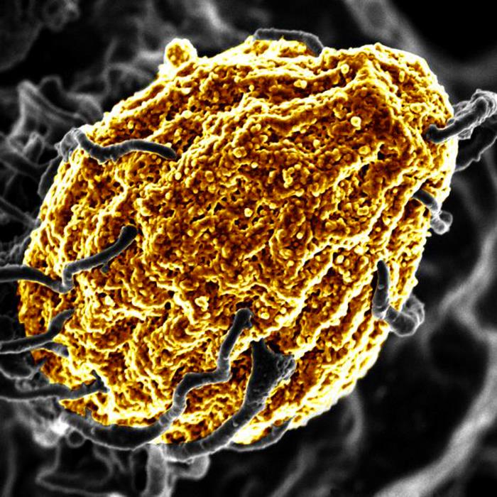
A macrophage digesting a yeast cell (yellow). Credit: NIH

Certain pathogens such as Salmonella have developed strategies to protect themselves from the macrophages’ digesting attempts, causing severe Typhoid infections and inflammations. Scientists report in Nature Metabolism how the inter-organellar crosstalk between phago-lysosomes and mitochondria restricts the growth of such bacteria inside macrophages.

Signals from the digestion cell organelle

As scavenger cells, macrophages have a very prominent digestion organelle, the phago-lysosome, where engulfed microorganisms are commonly degraded into pieces and become inactivated. “It has long been known that the molecule TFEB (Transcription factor EB) is important for the regulation of the phago-lysosomal system. More recent evidence also suggested that TFEB supports the defense against bacteria,” said Max Planck Institute group leader Angelika Rambold.

She and her team wanted to understand how exactly TFEB mediates its anti-bacterial role in macrophages. They confirmed earlier findings showing that a broad range of microbes, bacterial and inflammatory stimuli activate TFEB and thus the phago-lysosomal system.

“It made sense that pathogen signals trigger TFEB as macrophages need a more active digestion system quickly after they devour a meal of bacteria. But, interestingly, the experiments also revealed an additional strong effect of TFEB activation on another intracellular organelle system — mitochondria. This was completely unexpected and novel to us,” said Angelika Rambold.

Instructing mitochondria to increase anti-microbial activity

Composed of inner and outer membranes, mitochondria are the primary sites of cellular respiration and release energy from nutrients. Moreover, the mitochondria in immune cells were recently identified as sources of anti-microbial metabolites.

By using a broad experimental tool set, the investigators identified the pathway controlling an unexpected crosstalk between lysosomes and mitochondria. “Macrophages make use of extensive inter-organellar communication: the lysosome activates TFEB, which shuttles into the nucleus where it controls the transcription of a protein called IRG1. This protein is imported into mitochondria, where it acts as a major enzyme to produce the anti-microbial metabolite itaconate,” explained Angelika Rambold.

Exploiting organelle communication to control bacterial infections

The researchers investigated whether they could make use of this newly identified pathway to control bacterial growth. “We speculated that activating this pathway could be used to target certain bacterial species, such as Salmonella,” said Angelika Rambold. “Salmonella can escape the degradation by the phago-lysosomal system. They manage to grow inside macrophages, which can lead to the spreading of these bacteria to several organs in an infected body,” explained Alexander Westermann, collaborating scientist from the University of Würzburg.

When the researchers activated TFEB in infected macrophages in mice, the TFEB-Irg1-itaconate pathway inhibited the growth of Salmonella inside the cells. These data show that the lysosome-to-mitochondria interplay represents an antibacterial defense mechanism to protect the macrophage from being exploited as a bacterial growth niche.

In light of the increasing emergence of multi-drug resistant bacteria, with more than 10 million expected deaths per year by 2050 according to the various expert groups, it becomes important to identify new strategies to control bacterial infections that escape immune mechanisms. A promising path could be to use the TFEB-Irg1-itaconate pathway or itaconate itself to treat infections caused by itaconate-sensitive bacteria. According to the researchers from more work is needed to assess whether these new intervention points can be successfully applied to humans.

Source: Max Planck Institute of Immunobiology and Epigenetics

Increase in Cardiovascular Disease Diagnoses after COVID

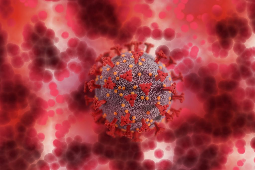
Image from Pixabay

A new study published in PLOS One found that COVID infection is associated with a nearly six-fold increase in cardiovascular disease (CVD) diagnoses over 12 months after the infection. 

The study analysed of UK electronic health records, comparing the risks of new diabetes mellitus (DM) and CVD diagnoses in the 12 months after infection. Researchers matched a cohort of 428 650 COVID patients matched to controls.

There was an 80% increased risk of DM diagnosis in the first month after COVID infection, a trend that has been echoed in previous studies, although those studies’ results seem to indicate a temporary form of the disease resulting from the acute stress of viral infection.

The findings showed that the largest increases were in pulmonary embolism (Relative Risk [RR] 11.51) and in atrial arrhythmias (RR 6.44). New CVD diagnoses rose five weeks after infection and incidence declined within 12 weeks to a year and returned to baseline or showed a net decrease. Increased risk for new DM diagnoses remained elevated by 27% for up to 12 weeks. 

“It’s definitely reassuring that over the longer timeframe, cardiovascular disease and diabetes risk does seem to return to baseline levels,” study author Emma Rezel-Potts, PhD, told The Guardian. “But we do have to be cautious in the acute period with cardiovascular disease and take note that the risk of diabetes seems to be elevated for several months, so that could be a good opportunity for risk prevention.” 

She also stressed that the findings could be explained by many factors. For example, the COVID patients in the study were more likely to be overweight and had more underlying health problems compared to uninfected controls, predisposing them to DM and CVD. Additionally, some may have had underlying conditions which were discovered when they were treated for COVID.

Source: The Guardian

Neural Plastic Changes can Help in Cervical Spinal Cord Injuries

MRI images of the brain
Photo by Anna Shvets on Pexels

By studying damage involving the connection between the brain’s hemispheres, researchers are finding new ways to leverage neural plasticity to promote functional recovery after a spinal cord injury.

In a study published in JCI Insight, the team of researchers used models in the lab to investigate a unilateral spinal cord injury similar to Brown-Sequard Syndrome, a rare neurological condition where damage to the spinal cord in a person results in weakness or paralysis on one side of the body and a loss of sensation on the opposite side.

Assistant Professor Wei Wu at Indiana University School of Medicine, said that the spinal cord injury model damaged the connection between the left hemisphere of the brain and the right side of the body, leading to significant loss of function in the right forelimb.

“The skilled function of upper limbs is very important for the quality of life in the patients with cervical spinal cord injury, but such functional recovery is very difficult to achieve in the severe injury,” said Asst Prof Wu, first author of the paper. “We found that the intact corticospinal system in the opposite side of the brain and spinal cord can be modulated to at least partially take over the control of the forelimb that is damaged by the spinal cord injury, resulting in a forelimb functional improvement.”

Since each hemisphere controls the opposite side of the body, researchers discovered a spontaneous shift of the neural circuits after injury from the left hemisphere to the right. Although there are connections between the right hemisphere of the brain and the right side of the body through some relayed pathways after injury, Asst Prof Wu said that’s not sufficient to support the motor recovery.

Using optogenetics to stimulate the right hemisphere of the brain, the researchers modulated the motor cortex. Additional neural circuits were shifted from the left side to the right side of the brain to dramatically increase and improve forelimb function.

“New circuits in the whisker, jaw forelimb and neck areas in the right hemisphere of the brain are recruited to control the right forelimb,” Asst Prof Wu said. “Interestingly, the beneficial neural plastic changes emerge both in the brain and the distal spinal cord after the optogenetic neuromodulation was applied on the motor cortex.”

Asst Prof Wu said results of the study showed significant improvement to the forelimb; however, there are still many challenges ahead, since complete digital recovery was not achieved.

The research team will continue explore this transhemispheric neural reorganisation to further improve the functional recovery after the spinal cord injury, Asst Prof Wu said. He hopes that these findings will be applied to treatments for spinal cord injuries.

Source: Indiana University School of Medicine

No Consensus on a Definition for ‘Growing Pains’

Photo by Monstera from Pexels

The phrase ‘growing pains’ is often used by people to describe muscle or joint pain in young people and health professionals also use the term. However a broad review of medical literature has found there is no consistent medical definition of the condition behind a diagnosis.

Researchers from the University of Sydney found there is no agreement in the literature on what growing pains really are, what they mean, how they are defined, and how they should be diagnosed.

Growing pains may be a medical misnomer, the researchers said – more than 93% of studies did not refer to growth when defining the condition. Similarly, age was not mentioned in more than 80% of studies’ definitions.

The findings have prompted the researchers to recommend the term growing pains not be used by clinicians and other researchers as a stand-alone diagnosis, until a clear definition backed by evidence has been established.

Growing pains are considered to be one of the most common causes of recurring musculoskeletal pain in children and adolescents. Some studies suggest up to a third of children experience the condition at some point in their life.

The term first arose in 1823 in a book called ‘Maladies de la Croissance’ (‘diseases of growth’).

“Thousands of kids are diagnosed with growing pains by their healthcare professional, but we were curious – what does that diagnosis really mean?” said lead author Dr Mary O’Keeffe from Institute for Musculoskeletal Health at the University of Sydney. 

In order to see how researchers defined the term, and if there were any detailed criteria that led to a diagnosis, the reviewers examined 147 studies that mentioned growing pains. The medical literature spanned many types of research, including systematic reviews, editorials, observational studies, case-control studies, and theses.

“What we found was a little concerning: that there is no consistency in the literature on what ‘growing pains’ means,” said Professor Steven Kamper, at the University of Sydney.  

“The definitions were really variable, vague and often contradictory. Some studies suggested growing pains happened in the arms, or in the lower body. Some said it was about muscles while other studies said joints.”

Only seven studies, less than 10% of the studies examined, mentioned growth related to the pain. More than 80% of the studies did not mention a young person’s age at the time ‘growing pains’ occurred.

There was no widespread agreement or detail on where the pain was located or when the pain happened.

Half of the studies referenced ‘growing pains’ as being located in the lower limb, while 28% reported specifically in the knees.

As for time of occurrence, 48% of studies reported the ‘growing pains’ happens during the evening or night and 42% reported it was recurring.

“What this study uncovered was while ‘growing pains’ is a very popular label used to diagnose musculoskeletal pain, it means very different things to different people,” said senior author Professor Steve Kamper.

“This level of uncertainty means clinicians don’t have a clear guide or criteria to know when the label ‘growing pains might be appropriate for a patient’.”

The study questioned whether growing pains are connected to growth itself in bone or muscle.

“There is a lack of evidence or inconsistent information on growing pains as a condition – and how it is associated with growth, or even the cause of the pain,” said Dr O’Keeffe.

“There is a real opportunity to understand this condition – given how widespread the use of the term is, or whether there is even a need to use this term.”

Source: EurekAlert!

A Novel Anticoagulant That can be ‘Deactivated’

Source: NCI

A new biomolecular anticoagulant platform reported in Nano Letters holds promise as a revolutionary advancement over the anticoagulants currently used during surgeries and other procedures. The technology is based around injectable fibre structures which can be quickly dissolved and excreted by the kidneys.

“We envision the uses of our new anticoagulant platform would be during coronary artery bypass surgeries, kidney dialysis, and a variety of vascular, surgical and coronary interventions,” said Kirill Afonin, leader of the team which invented the technology. “We are now investigating if there are potential future applications with cancer treatments to prevent metastasis and also in addressing the needs of malaria, which can cause coagulation issues.”

The team’s technology turns to programmable RNA-DNA anticoagulant fibres that, when injected into the bloodstream, form into modular structures that communicate with thrombin. The technology allows the structures to prevent blood clotting as it is needed and then be quickly eliminated via the renal system once their job is done.

The fibre structures use aptamers, short sequences of DNA or RNA designed to specifically bind and inactivate thrombin.

“Instead of having a single small molecule that deactivates thrombin,” Afonin said, “we now have a relatively large structure that has hundreds of the aptamers on its surface that can bind to thrombin and deactivate them. And because the structure becomes larger, it will circulate in the bloodstream for a significantly longer time than traditional options.”

The extended circulation in the bloodstream allows for a single injection, instead of multiple doses. The design also decreases the concentration of anticoagulants in the blood, resulting in less stress on the body’s renal and other systems, Afonin said.

This technology also introduces a novel “kill-switch” mechanism, which reverses the fibre structure’s anticoagulant function with a second injection. This lets makes the fibres able to be metabolised into materials that are tiny, harmless, inactive and easily excreted by the renal system.

The entire process takes place outside the cell, through extracellular communication with the thrombin. The researchers note that this is important as immunological reactions do not appear to occur, based on their extensive studies.

The team has tested and validated the platform in computer models, human blood and various animal models“We conducted proof-of-concept studies using freshly collected human blood from donors in the US and in Brazil to address a potential inter donor variability,” Afonin said.

The technology may provide a foundation for other biomedical applications that require communication via the extracellular environment in patients, he said. “Thrombin is just one potential application,” he said. “Whatever you want to deactivate extracellularly, without entering the cells, we believe you can. That potentially means that any blood protein, any cell surface receptors, maybe antibodies and toxins, are possible.”

The technique permits the design of structures of any shape desired, with the kill switch mechanism intact. “By changing the shape, we can have them go into different parts of the body, so we can change the distribution,” Afonin said. “It gets an extra layer of sophistication of what it can do.”

While the application is sophisticated, production of the structures is relatively easy. “The shelf life is amazingly good for these formulations,” Afonin said. “They’re very stable, so you can dry them, and we anticipate they will stay for years at ambient temperatures, which makes them very accessible to economically challenged areas of the world.”

Source: University of North Carolina

Blood Vascular Network Retains the ‘Memory’ of a Stroke

Credit: American Heart Association

A study into the structure of blood vascular network structure found that it is dynamic and can adapt to external factors, resulting in a kind of memory of certain events such as an ischaemic stroke. In particular, the study researchers found that rarely used connections incrementally weaken until they disappear eventually.

Researchers from the Max Planck Institute for Dynamics and Self-Organization in Göttingen and the Technical University of Munich used computer simulations to model vascular networks and identified adaptation rules for their connections.

“We found that the strength of a connection within a network depends on the local flow,” explained Karen Alim, corresponding author of the study. “This means that links with a low flow below a certain threshold will decay more and more until they eventually vanish,” she continued. Since the limited amount of material available to build the vascular system needs to be efficiently used, this mechanism offers an elegant way to streamline the vascular system.

Persistent changes in the network

Once a connection has become very weak due to a low flow rate, recovering that connection is very difficult. For example, a blood vessel blockage of the type that could lead to an ischaemic stroke. During an ischaemic stroke, some blood vessels in the affected region are weakened by the blockage.

“We found that in such a case, adaptations in the network are permanent and are maintained after the obstacle is removed. One can say that the network prefers to reroute the flow through existing stronger connections instead of re-growing weaker connections – even if the flow would require the opposite,” explained Komal Bhattacharyya, principal author of the study.

The researchers have thus shown that blood flow permanently changes even after successful removal of the clot. This memory capability of networks can also be found in other living systems: for example, the slime mould Physarum polycephalum uses its adaptive network to navigate its environment based on imprints by food stimuli, as demonstrated previously.

The study was published in Physical Review Letters.

Source: Max Planck Institute for Dynamics and Self-Organization

A Potent New Non-opioid Analgesic

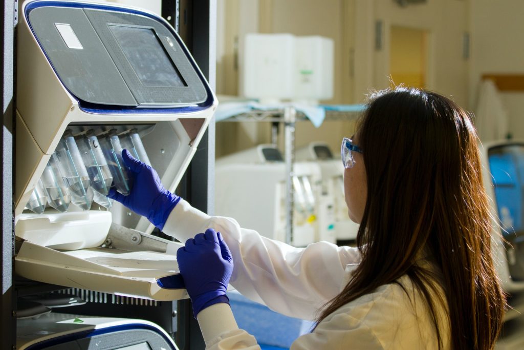
Woman using lab equipment
Source: NCI on Unsplash

Researchers co-led by University of Warwick have discovered a potent new non-opioid analgesic with potentially fewer side effects compared to other potent painkillers.

Their study found that a compound called BnOCPA (benzyloxy-cyclopentyladenosine), is a potent and selective analgesic which is non-addictive. BnOCPA also has a unique mode of action and potentially opens a new pipeline for the development of new analgesic drugs.

The research is published in Nature Communications.

Chronic pain has a negative impact on quality of life and many commonly prescribed analgesics come with side effects. Opioid drugs, such as morphine and oxycodone, can lead to addiction and are dangerous in overdose.

Drugs that act on G protein-coupled receptors (GPCRs) are one possibility, but their development is hampered by the propensity of GPCRs to couple to multiple intracellular signalling pathways. A unique feature of BnOCPA is that it only activates one type of GPCR, leading to very selective effects and thus reducing potential side effects.

University of Warwick’s Dr Mark Wall, who led the research said: “The selectivity and potency of BnOCPA make it truly unique and we hope that with further research it will be possible to generate potent painkillers to help patients cope with chronic pain.”

Source: University of Warwick

Study Shows that Not Everyone can Adjust to Shift Work

Photo by SJ Objio on Unsplash

A new study of French hospital workers have challenged the widely held belief that shift workers adjust to the night shift over time, using data drawn from wearable sensors.

By monitoring groups of the hospital workers working day or night shifts during their working and free time, the researchers have shown that not only does night work significantly disrupt both sleep quality and circadian rhythms, also that workers can experience such disruption even after years of night shift work.

Their findings, reported in eBioMedicine, are the most detailed analysis of the sleep and circadian rhythm profiles of shift workers yet attempted, and the first to also monitor body temperature. This key circadian rhythm is driven by the brain pacemaker clock, and coordinates the peripheral clocks in all organs.

In addition, the research demonstrates the value of telemonitoring technology for identifying early warning signs of disease risks associated with night-shift work opening up intervention opportunities to improve the health of workers.

The study compared 63 night-shift workers, working three or more nights of 10 hours each per week, and 77 day-shifters alternating morning and afternoon shifts at a single university hospital near Paris. Both groups wore accelerometers with chest surface temperature sensors throughout the day and night for a full week.

The accelerometer measured movement intensity and served as an estimate for participants’ sleep duration, how regular were their circadian rhythms, and whether that sleep was disrupted by movement. Patterns in the chest surface temperature gave a further indication of the participants’ circadian rhythm, which coordinates rest-activity phases, varying core body temperature, and an array of other bodily rhythms.

Analysis of interruptions to sleep and rhythmic variations in core body temperature showed that night-shift workers had less than half the median regularity and quality of sleep of their day-shift colleagues. 48% of the night-shift workers had a disrupted circadian temperature rhythm.

Using information from questionnaires on the participants’ chronotypes, they also found that the centre of sleep for those working the night shift didn’t correlate with their respective chronotype, ie their morningness or eveningness orientation. This meant they were not sleeping in synch with their internal clocks.

Even workers with years of being on night shifts still showed these negative effects on circadian and sleep health. The more years of night work they had, the more severe the circadian disruption – contradicting widely held assumptions about night work adaptation.

This helps explain why previous studies have shown an association between disrupted circadian rhythms with long term health risks, such as cancer and cardiovascular disease.

Professor Bärbel Finkenstädt from the University of Warwick Department of Statistics said: “There’s still an assumption that if you do night work, you adjust at some stage. But you don’t. We saw that most workers compensate in terms of quantity of sleep, but not in terms of quality during the work time.”

Dr Julia Brettschneider of the University of Warwick Department of Statistics said: “I think there’s a misunderstanding that night shift work is just an inconvenience, whereas it can be linked to serious health risks. We can’t avoid shift work for many professions, like healthcare workers, so we should be thinking about what can be done in terms of real-world adjustments to improve working conditions and schedules of shift workers. A better understanding of the biological mechanisms helps to find answers to this question.

“Together with our PhD student Yiyuan Zhang, we have developed a statistical analysis framework that enables the discovery of patterns and predictive factors in the complex data sets created by wearable tech.”

Professor Francis Lévi from Université Paris-Saclay further added: “Nearly 20% of the night workers could not even adjust their circadian rhythms during their free time, with the severity of impairment tending to increase with the number of years of night work. The telemonitoring technology, and analysis methods we have set up make it now possible to objectively evaluate circadian and sleep health in night workers in near real time, and design prevention measures for individual workers whenever necessary.”

In future research, the team may look at more long-term outcomes, such as diseases such as cancer that have been linked to circadian disruption.

Source: University of Warwick

Once-off Gene Therapy Reduces Haemophilia B Bleeding in Patients

https://www.pexels.com/photo/a-close-up-shot-of-bags-of-blood-4531306/
Photo by Charlie-Helen Robinson on Pexels

A single gene therapy injection could dramatically reduce the bleeding risk faced by people with haemophilia B, according to the results of a Phase I/II clinical trial published in the New England Journal of Medicine.

Low levels of the factor IX (FIX) protein, needed for clot formation, are behind haemophilia B. The FIX protein gene is on the X chromosome, so the severe form of haemophilia B is much more common in men, though it can occur in women due to X chromosome inactivation.

To prevent excessive bleeding, patients with haemophilia B typically need regular replacement therapy consisting of weekly injections of recombinant FIX. Despite advances in treatment, patients may continue to see debilitating joint damage.

The new treatment, from University College London, Royal Free Hospital and biotechnology company Freeline Therapeutics, is a type of adeno-associated virus (AAV) gene therapy candidate, called FLT180a, is being developed to treat severe and moderately severe cases of haemophilia B.

The Phase I/II multi-centre clinical trial, called B-AMAZE, and the related long-term follow up study found that a single treatment with FLT180a led to sustained production of FIX protein from the liver in 9 of 10 patients, across four different dose levels, removing the need for regular replacement therapy.

Out of 17 male patients aged 18 or over who underwent screening, 10 with severe or moderately severe haemophilia B took part in the 26-week trial of FLT180a, will be followed-up to assess safety and durability of FIX expression for 15 years.

Lead author Professor Pratima Chowdary of the Royal Free Hospital said: “Removing the need for haemophilia patients to regularly inject themselves with the missing protein is an important step in improving their quality of life. The long term follow up study will monitor the patients for durability of expression and surveillance for late effects.”

One patient, Elliott Mason, told the BBC: “I’ve not had any treatment since I had my therapy, it’s all a miracle really, well it’s science, but it feels quite miraculous to me.

“My life is completely normal, there’s nothing that I have to stop and think ‘how might my haemophilia affect this?’.”

AAV gene therapy works uses a packaging from the proteins found in the outer coat of the virus to deliver a functional copy of a gene directly to patient tissues. Newly synthesised proteins are released into the blood and a one-time infusion can have long-term effects.

Over several weeks to several months, patients took immunosuppressive drugs to prevent their immune systems from rejecting the therapy, and all reported known side effects.

Though the therapy was well tolerated, patients all experienced adverse events, with an abnormal blood clot in one who received the highest FLT180a dose and had the highest levels of FIX protein.

Professor Amit Nathwani, who co-authored the study, said: “Gene therapy is still a young field that pushes the boundaries of science for people with severe genetic diseases.

“The B-AMAZE long-term data add to the growing body of evidence that gene therapy has the potential to free patients from the challenges of having to adhere to lifelong therapy or could provide treatment where none exists today.”

In nine out of the ten patients, the treatment led to a sustained increase in FIX protein production, which led to a decrease in excessive bleeding. They also no longer required weekly injections of FIX protein.

After 26 weeks, five patients had normal levels of FIX protein, three had low but increased levels, and one patient treated at the highest dose had an abnormally high level.

Pamela Foulds, MD, Chief Medical Officer of Freeline, said: “The B-AMAZE long-term data continue to support our confidence that a single dose of FLT180a could protect people with haemophilia B from bleeding and the need for lifelong FIX replacement through durable expression of FIX at protective levels.”

Source: EurekAlert!

Antibody Cocktail Could Treat HIV while Minimising Escape Risk

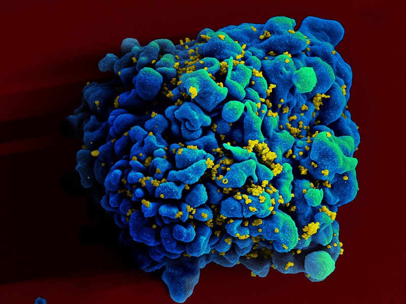
HIV Infecting a T9 Cell. Credit: NIH

Specifically designed cocktails of broadly neutralising antibodies (bNAbs) could help treat HIV while minimising the risk of the virus escaping treatment, researchers reported in eLife.

The study shows that computational approaches to selecting combinations of bNAbs based on viral genetics could help prevent viral escape, making HIV treatment more effective. It may also offer a strategy for designing effective combinations of bNAbs for treating other rapidly evolving pathogens.

bNAbs offer a promising new tool to treat or potentially cure infections with rapidly evolving viruses such as HIV. Clinical trials using a single bNAb to treat HIV have shown that some viral strains may survive the treatment and lead to a rebound of viruses in the blood. Combinations of bNAbs may therefore be a more effective approach, but finding the best combinations is a challenge. 

“For our study, we proposed using a computational approach to predict the effectiveness of bNAb combinations based on the HIV genetics,” said researcher Colin LaMont.

LaMont and colleagues analysed the genetics of HIV viruses collected over 10 years from 11 untreated patients with HIV, and used this data to predict which viral strains might be able to escape treatment with different bNAbs and whether dodging bNAbs had a survival cost. Next, using computational methods, they applied the knowledge gained to predict viral rebounds in three real-life trials of bNAbs.

Finally, the team used their computational approach to find a combination of bNAbs that is least likely to allow any virus to escape. They also found that some bNAbs, such as 10-1074, are better against diverse populations of viruses because mutations that allow viruses to escape also make the virus less likely to survive. Others, including PGT121, are more effective against less diverse viral populations because mutations that enable escape are rare. Overall, the results suggested that the optimal combination includes three bNAbs: PG9, PGT151 and VRC01. 

“We’ve shown the combination of PG9, PGT151 and VRC01 reduces the chance of viral rebound to less than 1%,” LaMont said. “It does this by targeting three different regions of the virus’ protective outer wrapping, or envelope.” 

“Combining bNAbs, administered via intravenous infusion every few months, with current antiretroviral therapies (ART) that require daily doses could further improve long-term HIV treatment success,” suggested senior author Armita Nourmohammad, Assistant Professor at the University of Washington.

ART hinders HIV multiplication and ability to create new variants, limiting the genetic diversity of the viral population and reducing the odds of bNAb escape variants emerging. The authors say that more studies are needed to confirm the potential benefits of combining ART and bNAbs. 

“Our study shows that leveraging genetic data can help us design more effective HIV therapies,” Asst Prof Nourmohammad concluded. “Our approach may also be useful for designing therapies against other rapidly evolving agents that cause disease, such as the Hepatitis C virus, drug-resistant bacteria, or cancer tumour cells.”  

Source: eLife Sciences