Using short-lived killifish to study how the immune system weakens with ageing, scientists have found that fewer types of antibodies are produced as organisms age. Published in eLife, the findings could lead to ways to rejuvenate the immune systems of older people.
The immune system has to constantly respond to new attacks from pathogens and remember them in order to be protected during the next infection. For this purpose, B cells build a library of information that can produce a variety of antibodies to recognise the pathogens.
“We wanted to know about the antibody repertoire in old age,” explained lead researcher Dario Riccardo Valenzano. “It is difficult to study a human being’s immune system over his or her entire life, because humans live a very long time. Moreover, in humans you can only study the antibodies in peripheral blood, as it is problematic to get samples from other tissues. For this reason, we used the killifish. It is very short-lived and we can get probes from different tissues.”
The shortest-lived vertebrates that can be kept in the laboratory, killifishes quickly age over their three to four month lifespan and have become the focus of ageing research in recent years due to these characteristics.
The researchers were able to accurately characterise all the antibodies that killifish produce. They found that older killifish have different types of antibodies in their blood than younger fish. They also had a lower diversity of antibodies throughout their bodies.
The discovery could lead to ways to rejuvenate the immune system. “If we have fewer different antibodies as we age, this could lead to a reduced ability to respond to infections. We now want to further investigate why the B cells lose their ability to produce diverse antibodies and whether they can possibly be rejuvenated in the killifish and thus regain this ability,” Valenzano said.
Being conceived via assisted reproductive technology (ART), such as IVF, could boost quality of life in adulthood, according to the results of a new study published in Human Fertility. The findings offer reassuring news for people who have been conceived with ART, and those who need to use the technology to conceive.
“Our findings suggest that being ART-conceived can provide some advantages on quality of life in adulthood, independent of other psychosocial factors,” said lead author Karin Hammarberg of Monash University. “Together with previous evidence that adults conceived by ART have similar physical health to those who were naturally conceived, this is reassuring for people who were conceived with ART—and those who need ART to conceive.”
In the more than four decades since the first birth following in vitro fertilization (IVF) in 1978, more than 8 million children have been born as a result of ART. In that time, many studies have evaluated the physical health, development and psychosocial well-being of ART-conceived children compared with those naturally conceived (NC). But currently, there is less known about the health and quality of life of adults who were conceived by ART.
The study recruited 193 young adults conceived through ART and 86 through NC. These participants completed questionnaires, which included a standardised quality of life measure (World Health Organization Quality of Life – Brief Assessment [WHOQoL-BREF]), when aged 18–28 years (T1) and again when aged 22–35 years (T2). The WHOQoL-BREF assesses four domains of quality of life: 1) physical 2) psychosocial 3) social relationships and 4) environment.
The researchers looked at the associations between factors present at T1 (mode of conception, the mother’s age when the participant was born, sexual orientation, family financial situation in secondary school, perceptions of own weight, number of close friends, frequency of vigorous exercise and quality of relationships with parents) and the scores on the four domains of WHOQoL-BREF at T2.
After making statistical adjustments to account for other psychosocial factors present in young adulthood, the results showed that being ART-conceived was strongly linked with higher scores (better quality of life) on both the social relationships and environment WHOQoL-BREF domains at T2. In addition, having less psychological distress, a more positive relationship with parents, a better financial situation, and perceptions of being about the right weight at T1 were associated with higher scores on one or more WHOQoL-BREF domains at T2.
“Children conceived via ART are nowadays a substantial part of the population—and it’s important to continue to evaluate the long-term effects of ART on their physical health and well-being as they progress through adolescence into adulthood,” said Hammarberg. “When accounting for other factors present in young adulthood, being ART-conceived appears to confer some advantages in quality of life. Perhaps unsurprisingly, we also found that, independently of how the person was conceived, having a better relationship with parents, less psychological distress, and a better family financial situation in young adulthood contributed to a better adult quality of life.”
This is the first study to explore the contributions of being conceived with ART and psychosocial factors present in young adulthood to the quality of life of adults. While the findings are reassuring, they should be interpreted with caution because many of those who took part in the first study did not take part in the follow-up study.
Before and after images for participants who received 36 weeks of treatment for alopecia areata with baricitinib. Credit: Yale University
Results from a new study in three patients with alopecia areata treated with a Janus kinase (JAK) inhibitor, baricitinib, were able to regrow hair. The study is based on Phase III clinical trials using baricitinib – a drug commonly used for arthritis – to treat alopecia areata, a skin disease characterised by loss of hair from the scalp and sometimes eyebrows and eyelashes.
“This is so exciting, because the data clearly show how effective baricitinib is,” said Dr. Brett King, an associate professor of dermatology at the Yale School of Medicine and lead author of the new study, published in the New England Journal of Medicine. “These large, controlled trials tell us that we can alleviate some of the suffering from this awful disease.”
Alopecia areata is an autoimmune disorder in which the body’s immune system attacks hair follicles, and which typically occurs in people under the age of 40. At present there is no FDA-approved treatment for the disease.
To test the drug, the researchers conducted two large, randomised trials involving a total of 1 200 people. The participants were adults with severe alopecia areata, who had lost at least half of their scalp hair.
For 36 weeks, participants were randomised to either a daily dose of either 4mg of baricitinib, 2mg of baricitinib, or a placebo. One-third of the patients who received the larger dose grew hair back.
According to the researchers, baricitinib works by disrupting the communication of immune cells involved in harming hair follicles. Baricitinib and other JAK inhibitors are routinely used to treat autoimmune forms of joint disease.
“Alopecia areata is a crazy journey, marked by chaos, confusion, and profound sadness for many who suffer from it,” King said. “It will be incredible to have a medicine to help people emerge on the other side, normalcy restored, recognizable again to themselves and those around them.”
Over the past decade, A/Prof King has developed methods for using JAK inhibitors to treat a variety of skin diseases — including eczema, vitiligo, granuloma annulare, sarcoidosis, and erosive lichen planus.
A/Prof King noted that the clinical trials involving baricitinib are ongoing, allowing researchers to assess the long-term effectiveness and safety of the treatment.
In a discovery that could greatly benefit the treatment of traumatic injuries, scientists have identified a cluster of cells in the brainstem that control the body’s response to severe blood loss
The collection of neurons that the researchers discovered drive a response that maintains blood pressure during blood loss. However, severe blood loss eventually results in cardiovascular collapse, a condition called ‘decompensated haemorrhage’, marked by an abrupt and dangerous loss of blood pressure that presages haemorrhagic shock, where the body’s organs begin to shut down.
“During blood loss, the brain coordinates a cardiovascular response that supports blood flow to critical organs, like the heart and brain,” said researcher George Souza, PhD. “Our study shows that the cardiovascular response to blood loss depends on changes in the activity of a few hundred neurons in the brainstem.”
The new results, published in Cell Reports, shed light on an important process the body uses to maintain its blood pressure. Neurons, termed adrenergic C1 neurons, monitor blood pressure and activate during blood loss, increasing vasodilatory nerve activity that maintains proper blood pressure.
The scientists utilised advanced imaging and a technique called optogenetics controls neurons using light. Their research revealed that the C1 neurons are hyperactive during blood loss, and this keeps blood pressure study. But these neurons become inactive with severe blood loss, resulting in cardiovascular collapse.
The scientists found that re-activating the C1 neurons in lab rats restored both blood pressure and heart rate before cardiovascular collapse could lead to haemorrhagic shock.
“Our study indicates that reactivating the brain pathways controlling blood pressure during decompensated haemorrhage effectively reverses cardiovascular collapse. We think this indicates that neuromodulation of the pathways described by our study could be a beneficial adjunct therapy for low blood pressure following blood loss,” explained lead researcher Stephen Abbott, PhD.
The researchers noted that more research is needed as several factors could also cause the C1 neurons’ drop in activity during the onset of decompensated haemorrhage.
“These findings illuminate the importance of the brain-body interactions during blood loss and provide a new perspective for the underlying cause of cardiovascular collapse,” Dr Abbott said.
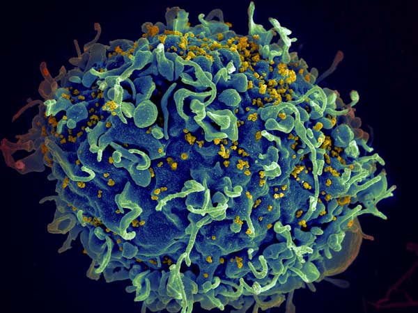
Even with antiretroviral therapy, HIV still lingers in the body, preventing complete cure. Now, new research published in PLOS Pathogens, revealed a possible answer to why HIV persists in the body: a lack of a certain protein in HIV patients’ killer T cells. The discovery also explained why people with HIV have less risk of developing multiple sclerosis (MS).
Because this protein, CD73 is responsible for migration and cell movement into the tissue, the lack of the protein compromises the ability of killer T cells to find and eliminate HIV-infected cells, explained immunologist Shokrollah Elahi, lead researcher of the study.
“This mechanism explains one potential reason for why HIV stays in human tissues forever,” he said, adding that the research also shows the complexity of HIV infection.
“This provides us the opportunity to come up with potential new treatments that would help killer T cells migrate better to gain access to the infected cells in different tissues.”
After spending three years identifying the role of CD73, Elahi turned his focus to understanding potential causes for the drastic reduction. He found it is partly due to the chronic inflammation that is common among people living with HIV.
“Following extensive studies, we discovered that chronic inflammation results in increased levels of a type of RNA found in cells and in blood, called microRNAs,” he explained. “These are very small types of RNA that can bind to messenger RNAs to block them from making CD73 protein. We found this was causing the CD73 gene to be suppressed.”
This discovery also helps explain why people with HIV have a lower risk of developing MS, Elahi noted.
“Our findings suggest that reduced or eliminated CD73 can be beneficial in HIV-infected individuals to protect them against MS. Therefore, targeting CD73 could be a novel potential therapeutic marker for MS patients.”
Elahi said the research could next look into seeing how to turn on the CD73 gene in patients with HIV and off in those with MS.
Olfactory dysfunction in COVID patients is common. Researchers recently searched the medical literature for studies reporting changes in olfactory structures detected through imaging tests of patients with COVID, and found that swelling in nasal passages is responsible for some temporary olfactory dysfunction. Their results in were published in The Laryngoscope.
A recent meta-analysis based on 83 studies provided high-quality evidence of a combined prevalence of olfactory dysfunction in 47.9% of COVID patients. Olfactory dysfunction is diagnosed more commonly among female patients and outpatients.
The researchers observed abnormality in olfactory clefts, which provide a crucial channel for airborne molecules to reach sensory olfactory neurons. The rate of abnormalities was nearly 16-fold higher in patients with COVID and olfactory dysfunction (63%) compared with controls (4%).
“Before this study, most scientists thought that the loss of smell in COVID was mainly due to inflammation and damage to the olfactory nerves. Now, we have compiled evidence from medical imaging that COVID loss of smell is also due to swelling and blockage of the passages in the nose that conduct smells,” said senior author Neville Wei Yang Teo, MRCS, MMed, of Singapore General Hospital.
“We think this is good news for patients who want to recover their sense of smell, since these blockages are expected to resolve with time, while nerve damage in comparison would likely be more difficult to recover from,” added lead author Claire Jing-Wen Tan, of the National University of Singapore. “These findings may not fully account for those who suffer from prolonged olfactory dysfunction, however, and further studies that evaluate patients in this group may provide more information.”
The data underpinning a controversial study of the use as vitamin C as a sepsis treatment may in fact be fraudulent, according to an analysis by an Australian physician and statistician, reports MedPage Today.
PhD student Kyle Sheldrick, MBBS, alleges that the pre- and post- comparison groups involved in the 94-patient study were too similar to be realistic.
In an interview with MedPage Today, Sheldrick said the case is “extreme”, stating that “This is probably the most obviously fake data I have seen. … These groups are more similar than would be probable.”
The paper, led by Paul Marik, MD – who led another COVID protocol study that has since been retracted – has been the subject of much debate in the intensive care community since it was published in 2017. The so-called HAT protocol was a simple regimen of hydrocortisone, ascorbic acid (vitamin C), and thiamine which could have saved many lives easily if it indeed worked. Obviously, there was much excitement worldwide about the significance of the findings – but not all were convinced.
“The effect size seemed just impossible,” said Nick Mark, MD, an ICU physician at Swedish Medical Center. “It seemed too good to be true.”
The trial was followed by larger studies, and so far none have shown shown a similar reduction in mortality, raising suspicions even further, Dr Mark said. With Sheldrick’s analysis, the penny dropped: “This was under our noses for 5 years,” Mark said. “This isn’t just a mistake. We know things can be done unethically, but to actually fake it? That it’s not just flawed, but perhaps actually fraudulent?”
Sheldrick told MedPage Today the key problem with the Marik paper is “probably the most common sign of fraud that we see, which is overly similar groups at baseline.” That is, people tend to fake data which do not vary enough from the average.
Sheldrick said he first looked at the study methods, which noted a pre- and post- comparison design, rather than a randomised or matched case-control design. With such a design, one would expect a more random distribution of baseline characteristics, but that wasn’t the case for the Marik paper, he said.
A further analysis with Fisher’s test showed that most P-values were 1, meaning they were distributed perfectly evenly across two time periods – and only one fell below 0.5. Instead, an even spread should be expected with an overall value of 0.5.
Sheldrick sent his findings to the journal CHEST and to Marik’s former employer Sentara Norfolk General Hospital, but had not heard back from either.
While Sentara Norfolk General Hospital did not respond to comment, and the journal CHEST could not confirm whether an investigation was underway but that it did take ethical concerns very seriously.
A spokesperson for Dr Marik emailed a statement to MedPage Today, claiming that the conclusions had been validated in several meta-analyses, and recommended the source examine “this and other research on the data before making false allegations on social media. Such claims are harmful and do not add to the public discourse.”
This wouldn’t be the first time concerns have been raised about data in a paper that Dr Marik co-authored. In November 2021, the Journal of Intensive Care Medicine (JICM) retracted an article by Marik and others on their MATH+ protocol for COVID. The retraction followed a communication that raised concerns about the accuracy of COVID mortality data from the hospital used in the article.
“It seems a bit improbable for someone to discover two miracle cures in three years,” Dr Mark commented to MedPage Today.
Dr Mark noted that the 2017 paper is widely cited, and even if the intervention was not directly harmful, the resources invested in subsequent large, high-quality trials of vitamin C and sepsis could have been better spent.
“While I’m really glad we did high-quality studies and had brilliant people working on this, it’s kind of a shame,” he said. “Instead of studying vitamin C based on a faulty premise, we could have spent our efforts elsewhere.”
An observational study with over 100 000 participants suggests that some artificial sweeteners are associated with increased cancer risk. The findings, published in PLOS Medicine, reflect other results from experimental studies.
The safety of artificial sweeteners has long been a subject of debate. To evaluate the potential carcinogenicity of artificial sweeteners, researchers analysed data from 102 865 French adults participating in the NutriNet-Santé study. The NutriNet-Santé study is an ongoing web-based cohort initiated in 2009 by the Nutritional Epidemiology Research Team (EREN). Participants enrolled voluntarily, and self-reported their medical history, sociodemographic, diet, lifestyle, and health data.
Researchers gathered data concerning artificial sweetener intake from 24-hour dietary records. After collecting cancer diagnosis information during follow-up, the researchers conducted statistical analyses to investigate the associations between artificial sweetener intakes and cancer risk. They also adjusted for a range of variables including age, sex, education, physical activity, medical history and dietary intake.
The researchers found that participants who consumed larger quantities of artificial sweeteners, particularly aspartame and acesulfame-K, had higher risk of overall cancer compared to non-consumers (hazard ratio 1.13). Higher risks were observed for breast cancer and obesity-related cancers.
In addition to being an observational study, there were a number of limitations; dietary intakes are self-reported. Selection bias may also have been a factor, as participants were more likely to be women, to have higher educational levels, and be more health-conscious. Additional research will be required to confirm the findings and clarify the underlying mechanisms.
According to the authors, “Our findings do not support the use of artificial sweeteners as safe alternatives for sugar in foods or beverages and provide important and novel information to address the controversies about their potential adverse health effects. While these results need to be replicated in other large-scale cohorts and underlying mechanisms clarified by experimental studies, they provide important and novel insights for the ongoing re-evaluation of food additive sweeteners by the European Food Safety Authority and other health agencies globally”.
First author Charlotte Debras added: “Results from the NutriNet-Santé cohort (n = 102 865) suggest that artificial sweeteners found in many food and beverage brands worldwide may be associated with increased cancer risk, in line with several experimental in vivo / in vitro studies. These findings provide novel information for the re-evaluation of these food additives by health agencies.”
Though light alcohol consumption may provide heart-related health benefits has been suggested observational research, a large study published in JAMA Network Open showed a link between all levels of alcohol intake and higher risks of cardiovascular disease. The researchers found that the supposed benefits of alcohol consumption may in fact be attributable to other healthy lifestyle factors common among light to moderate drinkers.
The study included 371 463 adult participants from the UK Biobank, average age 57 and consuming an average of 9.2 drinks per week. In line with previous findings, researchers found that the lowest heart disease risk was in light to moderate drinkers, followed by people who abstained from drinking. People who drank heavily had the highest risk. However, light to moderate drinkers also tended to have healthier lifestyles than abstainers, such as more physical activity and vegetable intake, and less smoking. One a few lifestyle factors were taken into account, any benefit associated with alcohol consumption was significantly reduced.
The study also used new techniques in Mendelian randomisation, which uses genetic variants to determine whether an observed link between an exposure and an outcome is consistent with a causal effect. “Newer and more advanced techniques in ‘non-linear Mendelian randomisation’ now permit the use of human genetic data to evaluate the direction and magnitude of disease risk associated with different levels of an exposure,” said senior author Krishna G. Aragam, MD, MS, a cardiologist at MGH and an associate scientist at the Broad Institute. “We therefore leveraged these new techniques and expansive genetic and phenotypic data from biobank populations to better understand the association between habitual alcohol intake and cardiovascular disease.”
When such genetic analyses were performed on samples taken from participants, they found that individuals with genetic variants that predicted higher alcohol consumption were indeed more likely to consume greater amounts of alcohol, and more likely to have hypertension and coronary artery disease. The analyses also revealed significant differences in cardiovascular risk across the spectrum of alcohol consumption for both males and females, with minimal risk increase when going from zero to seven drinks per week, much higher risk increases when progressing from seven to 14 drinks per week, and greatly increased risk for 21 or more drinks per week. Notably, the findings suggest a rise in cardiovascular risk even at “low risk” levels (ie below two drinks per day for men and one per day for women).
This discovery of an exponential rather than liner relationship between alcohol intake and cardiovascular risk is was supported by an additional analysis of data on 30 716 participants in the Mass General Brigham Biobank. Therefore, cutting back on large consumption of alcohol may have even more clinical benefits than cutting back on moderate amounts.
“The findings affirm that alcohol intake should not be recommended to improve cardiovascular health; rather, that reducing alcohol intake will likely reduce cardiovascular risk in all individuals, albeit to different extents based on one’s current level of consumption,” said Dr Aragam.
The amygdala, which is enlarged in two-year-old children with autism spectrum disorder (ASD), begins its accelerated growth between 6 and 12 months of age, suggests a study appearing in the American Journal of Psychiatry. The findings indicate that therapies to reduce the symptoms of ASD might have the greatest chance of success if they begin in the first year of life, before the amygdala begins its accelerated growth.
The amygdala, which is involved in processing emotions, such as interpreting facial expression in typically developing children increases substantially in volume from 7.5 to 18.5 years of age. The amygdala in children with autism is initially larger, but does not undergo the age-related increase observed in typically developing children.
The study included 408 infants, 270 of whom with an increased ASD risk from having an older sibling with ASD, 109 typically developing infants, and 29 infants with Fragile X syndrome. The researchers conducted MRI scans of the children at 6, 12 and 24 months of age. They found that the 58 infants who went on to develop ASD had a normal-sized amygdala at 6 months, but an enlarged amygdala at 12 months and 24 months. Moreover, the faster the rate of amygdala overgrowth, the greater the severity of ASD symptoms at 24 months. The infants with Fragile X syndrome had no differences in amygdala growth but enlargement of the caudate, which was linked to increased repetitive behaviours.
The researchers suggested that difficulty in processing sensory information during infancy may stress the amygdala, leading to its overgrowth.