Luallen Lab members pose in their lab at SDSU. Inset: Microscope image of Bordetella atropi (pink lines) infecting roundworm intestine (green). Credit: San Diego State University
Like something out of a horror movie, a new way that one type of bacteria invades tissue within a living organism has been identified by biologists from San Diego State University.
The study, published in Nature Communications, describes how a new species of bacteria, Bordetella atropi, invades its roundworm host. The name which comes from the Greek fate Atropos responsible for cutting the threads of life, is apt because the bacteria transforms into a long thread, growing up to 100 times the usual size of one bacterium in the span of 30 hours without dividing.
By altering the genes of B. atropi, the research team discovered that this invasive threading relies on the same genes and molecules that other bacteria use when they are in a nutrient-rich environment. However, these other bacteria only use this pathway to make subtly larger cells, whereas the B. atropi bacteria grows continuously.
Other bacteria often transform into threads, called filamentation, in response to dangerous environments or DNA damage. This lets them continue to grow in size, but delay cell division until they repair the damage inflicted by the stress.
Here, however, the researchers were the first to observe filamentation as a way of spreading from cell to cell in a living organism for a purpose other than the stress response. They believe that instead the new species is invading the host cells, detecting this rich environment and triggering filamentation in order to quickly infect more cells and access additional nutrients for their growth.
“We went from finding the worm in the ground, finding the bacteria, and carrying it all the way to the molecular mechanism of how the bacteria infects the worm,” explained Robert Luallen, biology professor and principal investigator of the study. “We’re seeing things that no one’s ever seen before.”
Though B. atropi does not infect humans, it is possible that human pathogens may also make use of its spreading mechanism. Separately, the nutrient-induced filamentation process might be used by other bacteria to form biofilms, which can coat the tubing of catheters and lead to complications for patients.
In a study published in Influenza and Other Respiratory Viruses, researchers were able to detect SARS-CoV-2 viral RNA in droplets from the exhaled breaths and coughs of COVID patients.
COVID is assumed to be transmitted mainly by respiratory droplets. However, probable aerosol transmission has been reported to occur under certain conditions. The researchers sought to address the lack of information on viral load in exhaled breath samples,as well as the size and concentration of exhaled endogenously generated droplets in relation to viral load. Additionally, the relationship between the viral load in upper airway diagnostic samples and aerosol samples needed to diagnose. For the study, researchers analysed exhalations by two different methods during 20 normal breaths, 10 airway opening breaths (which involves deep inhalation followed by relaxed exhalation), and 3 coughs.
PCR detection of SARS-CoV-2 RNA in aerosols was possible in 10 out of 25 participants. Viral RNA presence in aerosol was mainly detected in cough samples (8 samples), but also in normal breaths (4 samples) and in airway opening breaths (3 samples).
“Our data confirm findings from other researchers that SARS-CoV-2 can be detected in aerosol particles < 5µm and highlight the small amount of exhaled aerosol needed for detection. Of specific interest were findings from the airway opening maneuver, which is thought to generate particles mainly from the small airways,” said lead author Emilia Viklund, PhD student at the University of Gothenburg, in Sweden. “COVID causes a lot of damage in this region, and it would be of great interest to further explore the amount of exhaled virus and the course of disease, as well as the infectious potential of exhaled virus.”
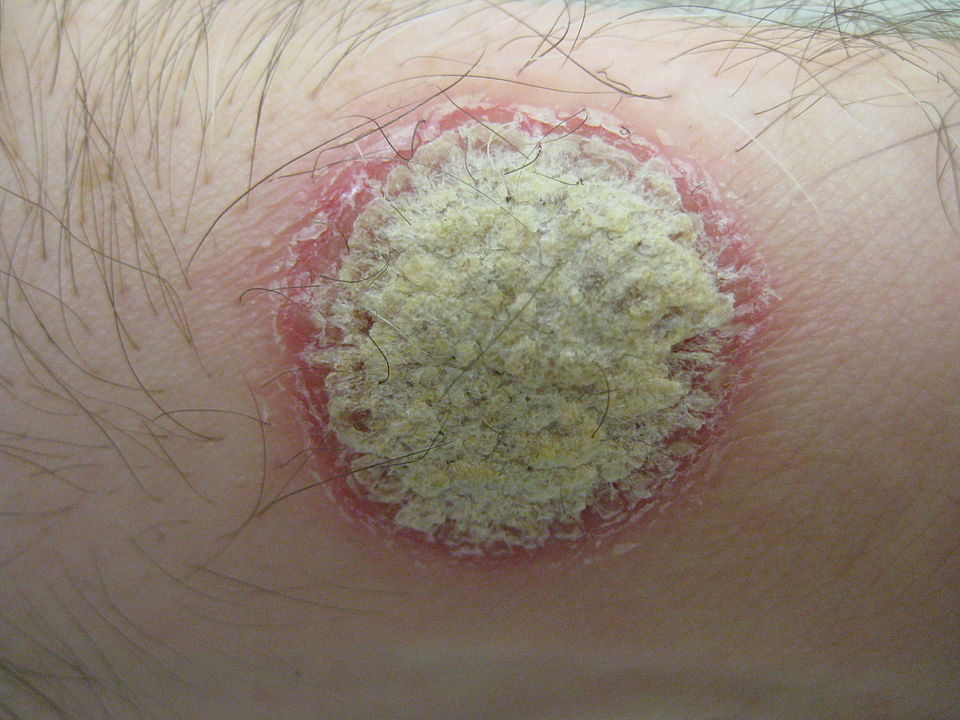
A study into the genetics of acne revealed 29 regions of the genome that underpin the condition, which could offer potential new treatment targets and may also help clinicians identify individuals at high risk of severe disease.
A common skin condition, acne is estimated to affect 80% of adolescents, with common features including spots and cysts, pigment changes and scarring. The face is the most common site, with the chest and back also frequently involved. The negative psychological consequences of acne are seen in all ages, but are of particular concern for many adolescents.
The research, published in Nature Communications, analysed nine genome wide association study datasets from patients around the world. These studies involved scanning the whole genomes of 20 165 people with acne and 595 231 without. The study identified 29 new genetic variants that are more common in people with acne. It also confirmed 14 of the 17 variants already known to be associated with the condition, which brings the total number of known variants to 46.
Professor Catherine Smith at St John’s Institute of Dermatology at Guy’s and St Thomas’ said: “Despite major treatment advances in other skin conditions, progress in acne has been limited. As well as suffering from the symptoms of acne, individuals describe consequent profound, negative impacts on their psychological and social wellbeing. It’s exciting that this work opens up potential avenues to find treatments for them.”
A number of genes associated with acne were identified, and are also linked to other skin and hair conditions. The team believe this will help to understand the causes of acne, which could be a mix of factors.
“We know that the causes of acne are complicated, with a mix of biological factors such as genetics and hormones, and environmental factors,” said Professor Michael Simpson at King’s College London. “Understanding the genetics of the condition will help us to disentangle some of these causes, and find the best way to treat the condition. This is a really promising area for further study, and opens up a lot of avenues for research.”
The study also uncovered a link between the genetic risk of acne and disease severity. Individuals with the highest genetic risk are more likely to have severe disease. While further research is required, this finding raises the potential to identify individuals at risk of severe disease for early intervention.
In a world first, Michel Roccati, a man with a completely severed spinal cord was able to walk again outside the lab with the help of a portable electrical stimulation system that causes his legs to take a step in conjunction with his intention to move and a walker to steady him.
This was a further development of a technology that in 2018 helped David M’zee, who had been left paralysed by a partial spinal cord injury suffered in a sports accident, to walk again. A research team led by Professors Grégoire Courtine, at École Polytechnique Fédérale de Lausanne (EPFL) and Jocelyne Bloch, at Lausanne University Hospital (CHUV) had developed an electrical stimulation system to help people with spinal cord injuries walk again.
They had wanted to see if electrodes could stimulate movement in the parts of the spine damaged so badly that signals no longer reach the nervous system from the brain. That pioneering study was detailed in Nature and Nature Neuroscience. Thanks to the electrodes making up for the weakness of the signals in his damaged spinal cord, M’zee was able to voluntarily move his legs and could walk several hundred metres at a time, sometimes without the aid of the rails on the treadmill.
Now a new milestone has just been reached with the technology, and the research team enhanced their system with more sophisticated implants controlled by advanced software. These implants can stimulate the region of the spinal cord that activates the trunk and leg muscles. Thanks to this new technology, three patients with complete spinal cord injury were able to walk again outside the lab. “Our stimulation algorithms are still based on imitating nature,” said Prof Courtine. “And our new, soft implanted leads are designed to be placed underneath the vertebrae, directly on the spinal cord. They can modulate the neurons regulating specific muscle groups. By controlling these implants, we can activate the spinal cord like the brain would do naturally to have the patient stand, walk, swim or ride a bike, for example.”
On a cold, snowy day last December, Michel Roccati – an Italian man who became paralysed after a motorcycle accident four years earlier – braved the icy wind to try out the system outdoors, in central Lausanne. He had recently undergone the surgical procedure in which Prof Bloch placed the new, implanted lead on his spinal cord.
Scientists attached two small remote controls to Michel’s walker and connected them wirelessly to a tablet that forwards the signals to a pacemaker in Michel’s abdomen. The pacemaker in turn relays the signals to the implanted spinal lead that stimulates specific neurons, causing Michel to move. Grasping the walker, Michel pressed a button corresponding to either the left or right leg with the firm intention of taking a step forward, and his feet rose and fell in short steps.
“The first few steps were incredible – a dream come true!” he says. “I’ve been through some pretty intense training in the past few months, and I’ve set myself a series of goals. For instance, I can now go up and down stairs, and I hope to be able to walk one kilometre by this spring.”
Two other patients have also successfully tested the new system, which is described in Nature Medicine. “Our breakthrough here is the longer, wider implanted leads with electrodes arranged in a way that corresponds exactly to the spinal nerve roots,” said Bloch. “That gives us precise control over the neurons regulating specific muscles.” Ultimately, it allows for greater selectivity and accuracy in controlling the motor sequences for a given activity.
While extensive training is necessary for patients to get comfortable using the device, the speed and scope of rehabilitation is amazing. “All three patients were able to stand, walk, pedal, swim and control their torso movements in just one day, after their implants were activated!” said Prof Courtine. “That’s thanks to the specific stimulation programs we wrote for each type of activity. Patients can select the desired activity on the tablet, and the corresponding protocols are relayed to the pacemaker in the abdomen.”
While the progress achievable in a single day is astonishing, the gains after several months are even more impressive. The three patients followed a training regimen based on the stimulation programs and were able to regain muscle mass, move around more independently, and take part in social activities like having a drink standing at a bar. What’s more, because the technology is miniaturized, the patients can perform their training exercises outdoors and not only inside a lab.
Presently there is one key limitation, Prof Bloch said: “We need at least six centimetres of healthy spinal cord under the lesion. That’s where we implant our electrodes.”
As for Roccati, after nine months of Lausanne-based rehab, he now lives independently in Italy. “I continued rehab at home, working alone, with all the devices,” he said. “And I see improvements every day.”
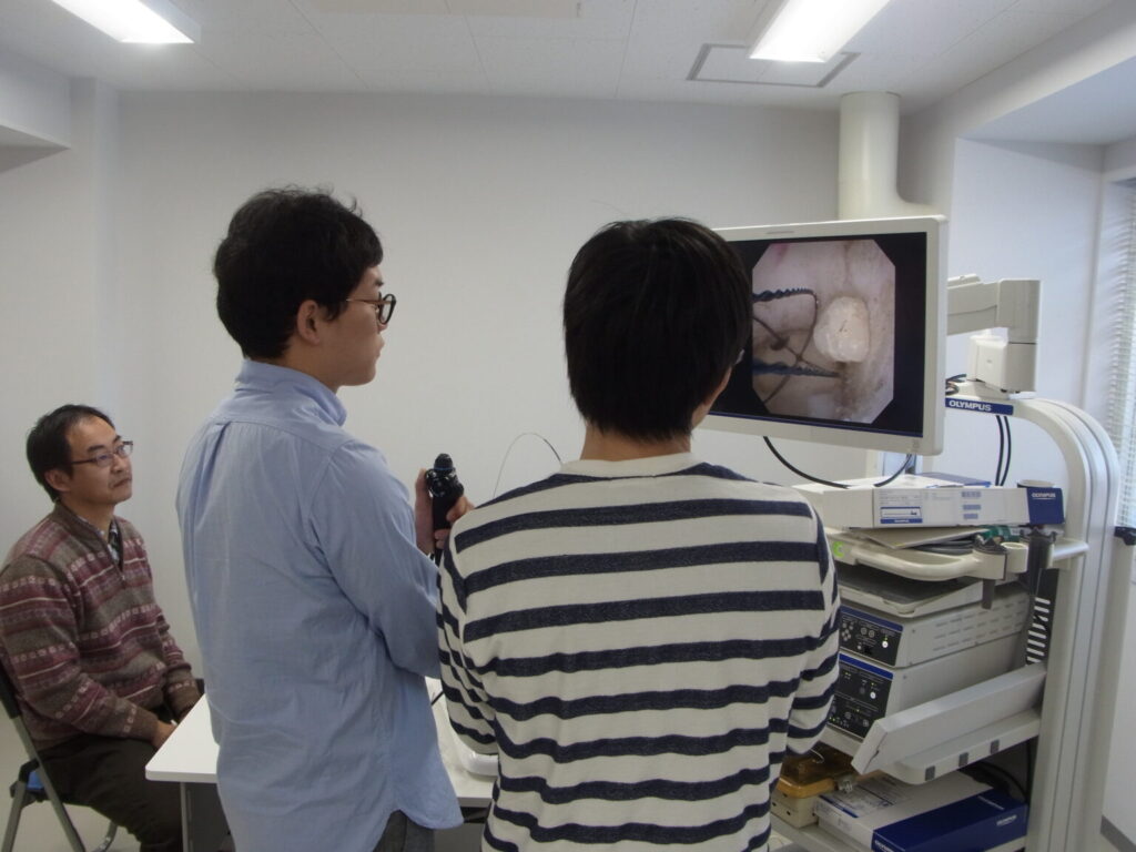
A surgical simulation unit used in the study. Credit: Takashige Abe.
An international trial found that while simulation-based training for ureteroscopy did not speed up surgeons general proficiency acquisition, it did increase skills in more complex surgeries, with fewer total complications and ureteric injuries. The results were published in the journal European Urology.
“To date, there have been limited data, mostly from small-scale studies conducted with medical students, assessing the transferability of surgical simulation,” said paper authors Takashige Abe, Associate Professor of Urology at Hokkaido University. The aim, he said, was to evaluate whether surgical residents undergoing additional simulation training can achieve proficiency sooner and with better patient outcomes, compared to usual operation room-based training.
The trial followed 65 participants in 10 countries for 18 months, or up to 25 procedures. A total of 32 participants received simulation-based training while 33 received conventional apprenticeship-style training. Both remained supervised by more experienced surgeons. Participants performed a total of 1140 surgeries, either semi-rigid or flexible ureteroscopy to remove ureteral or renal stones, respectively, demonstrating “mixed results” in proficiency.
“For our primary outcome measure, while we showed what might be deemed a clinically meaningful difference, it was not statistically significant,” Prof Abe said. “However, when stratified to each procedure type, there were higher rates of proficiency in the simulation-based training group when it came to the more technically challenging flexible ureteroscopy procedure.”
Prof Abe also noted that the simulation group scored higher on a standard assessment for each surgerythe other group.
“Simulation-based training led to higher overall proficiency scores than for conventional training, and fewer procedures were required to achieve proficiency in the complex form of the index procedure, with fewer serious complications overall,” Prof Abe said. “It is expected that the results of the trial will have a positive impact for advanced procedural training beyond the fields of surgery and urology in order to promote patients’ safety as well as better surgical outcomes.”
In a new study in the Canadian Medical Association Journal, researchers drawing on a provincial database report a small increased risk of congenital abnormalities in infants exposed to opioid medications in the first trimester of pregnancy.
Prescribed opioid pain medications are capable of crossing the placenta and have the potential to cause harm. In a study comparing placental crossing rates for various opioids, oxycodone, a commonly prescribed opioid for pain relief, was the fastest. About 2%–4% of foetuses are exposed to these drugs. To determine the association between opioid pain medications in early pregnancy and congenital abnormalities in infants, investigators analysed administrative health data from Ontario on almost 600 000 birth parent–infant pairs.
Among the infants included in the study, 2% (11 903) were exposed in utero to opioid analgesics, such as codeine, oxycodone, hydromorphone, tramadol, and morphine. Analysis showed a small increased risk of major anomalies with exposure to tramadol and morphine, and of minor anomalies with exposure to codeine, hydromorphone and oxycodone. Specific congenital anomalies observed included gastrointestinal and genital anomalies, neoplasms and tumours, and ankyloglossia.
This study adds to the evidence from previous studies in Sweden and Norway and also from a recent study of pregnant US Medicaid beneficiaries that suggested a small increased risk of congenital anomalies, an important finding for a pregnant person who may be prescribed opioids for pain relief.
“Both the potential for harm or distress to the pregnant person as a consequence of foregoing treatment and the subsequent risk to the infant must be considered for effective treatment,” the authors concluded. “These findings further quantify harms associated with prenatal exposure to opioid analgesics to inform treatment choices for pain in pregnancy.”
In women with generalised anxiety disorder (GAD), researchers using functional magnetic resonance imaging (fMRI) have identified an abnormal link between the heart and the prefrontal cortex.
The researchers were seeking to determine whether individuals suffering from GAD show dysfunction in the neural circuitry underlying cardiovascular arousal, and if it is associated with certain disorder-related symptoms such as anxiety and body sensation. To conduct the study, they completed a randomised clinical trial of 58 adult female participants (29 with GAD and 29 healthy controls).
During the study, they stimulated the cardiovascular system using isoproterenol, which mimics the effects of adrenaline but, unlike adrenaline, cannot cross the blood-brain-barrier to directly impact brain activity. Intravenous infusions of isoproterenol or saline were administered during fMRI, allowing them to assess whether the brains of patients with GAD differ in the processing of information received from the body, a function known as ‘interoception’. The main findings were that patients with GAD perceived their heartbeats to be more intense and had relatively higher heart rates and lower neural activity in the ventromedial prefrontal cortex. However, these were only observed during the lower of two dosages of isoproterenol: a key finding. Self-reported anxiety was higher only for those with GAD compared to healthy participants in response to either dose.
Lead author Adam Teed, a postdoctoral associate at Laureat Institute of Brain Research, said “administering isoproterenol allowed us to provide causal evidence that an abnormally sensitive cardiovascular system and an abnormally insensitive frontal cortex in GAD patients lowers their ability to regulate bodily arousal. This could help to explain why they experience anxiety so frequently and in a wide variety of contexts.” The authors hope that their study prompts further research into the ventromedial prefrontal cortex as a therapeutic target for novel treatments helping individuals with GAD to regulate physiological and emotional responses to stress.
In addition to this link, the observation of cardiovascular hypersensitivity in GAD patients was also noteworthy. This is because the DSM-5 describes autonomic symptoms such as sweating, rapid heart rate, or shortness of breath, as being less prominent in GAD than other anxiety disorders, such as panic disorder. As senior author Sahib Khalsa, MD, PhD, a psychiatrist and principal investigator at LIBR explains, “this study shows us that anxiety is not only something that happens within our brains but within our bodies as well.”
Thus abnormal autonomic nervous system functioning is not only a factor in GAD, but it occurs in combination with abnormal functioning of certain areas of the brain. Dr Khalsa believes that this finding is the most important research outcome: “it is the interaction between our brain and body that may be essential for determining whether an innocuous situation creates a state of fear in individuals with GAD. We need to better understand how this abnormal physiological response relates to the functional impairments that commonly interfere with the daily lives of such individuals.”
In a new publication in the journal Science, researchers propose that NFTs, or nonfungible tokens, could help patients assert better control over their personal health information.
NFTs, or nonfungible tokens, created using blockchain technology, have been a big sensation in the art world as they serve as a platform to buy and sell digital art backed by a digital contract. Now, an international multidisciplinary team of scholars in ethics, law and informatics led by bioethicists have written one of the first commentaries on how this new emerging technology could be repurposed for the healthcare industry. NFT digital contracts could provide an opportunity for patients to specify who can access their personal health information and to track how it is shared.
“Our personal health information is completely outside of our control in terms of what happens to it once it is digitised into an electronic health record and how it gets commercialised and exchanged from there,” said Dr Kristin Kostick-Quenet, first author of the paper. “NFTs could be used to democratise health data and help individuals regain control and participate more in decisions about who can see and use their health information.”
“In the era of big data, health information is its own currency; it has become commodified and profitable,” said Dr Amy McGuire, senior author of the paper and Leon Jaworski Professor of Biomedical Ethics and director of the Center for Medical Ethics and Health Policy at Baylor. “Using NFTs for health data is the perfect storm between a huge market place that’s evolving and the popularity of cryptocurrency, but there are also many ethical, legal and social implications to consider.”
Presently, NFTs are still vulnerable to data security flaws, privacy issues, and disputes over intellectual property rights, the researchers noted. The complexity of NFTs may also prevent the average person from properly making use of them. The researchers believe it is important to consider potential benefits and challenges as NFTs emerge as a potential avenue to transform the world of health data.
A comprehensive review has uncovered “clear evidence” of associations between atopic dermatitis (AD) and a range of comorbid conditions, which has informed updated clinical guidelines for AD.
An expert panel reported on the results of a wide-ranging review in the Journal of the American Academy of Dermatology. They found evidence linking AD to certain allergic, atopic, and immune-mediated conditions, as well as mental health problems, bone diseases, and skin infections. There is some evidence which supports associations between AD and substance use, attention deficit-hyperactivity disorder (ADHD), and some elements of metabolic syndrome. Less compelling evidence suggests AD has links to some cardiovascular conditions. There is inconclusive evidence for associations between adult AD and autism spectrum disorders, myocardial infarction, stroke, and metabolic syndrome.
“Atopic dermatitis is one of several atopic diseases, meaning that there are internal sensitivities that can help drive the disease in the organ of choice, including asthma, allergic rhinoconjunctivitis, and food allergies, among others,” Dawn M.R. Davis, MD, co-chair of the guideline panel, told MedPage Today. “We always knew there was an association between atopic dermatitis and the other atopic diseases, but we lacked the evidence. Fortunately, because we’re getting more attention and more research is being performed in these areas, we now have data to back up our suspicions regarding the associations between atopic dermatitis and other atopic diseases.”
“Thanks to research by our colleagues, we discovered several other comorbidities that we did not expect, including skin diseases like alopecia areata and urticaria, as well as mental health conditions, including depression, anxiety, and substance use,” she continued. “We also have some evidence of associations with metabolic conditions, such as disorders of bone metabolism, and cardiovascular diseases.”
This review was conducted in concert with updating the American Academy of Dermatology clinical guideline on AD. The quantity and depth of data also warranted a separate guideline component for recognition of comorbidities associated with AD. The main goal was to increase awareness of the associations.
“The goal of this guideline is to plant a seed in the mind of providers and to empower and validate patients, so they can have a customised, individualised, robust discussion about how their particular circumstances relate to any of the risk factors,” said Dr Davis.
Key findings and statements in the guideline include:
The association between AD and asthma is well established, but the “atopic march” explanation remains unproven
“Clear evidence” of an association between AD and food allergy, but estimated prevalence of food allergy in adults with AD remains low
Epidemiologic studies “consistently show” an association between AD and alopecia areata, but there are limited data on severity of alopecia or treatment response
Analysis of four studies showed that AD doubles the odds ratio of depression though reasons for this are unclear
A “potential association” between AD and substance use/abuse (limited evidence)
Accumulating evidence suggests small associations between AD and hypertension, peripheral and coronary artery disease, congestive heart failure, and acute clinical events
A “small association” suggested between adult AD with obesity and dyslipidemia; however, limited data have pointed to a possible inverse association with diabetes
Several studies have shown associations with increased risk of osteoporosis and fracture in adults with AD, possibly linked by systemic inflammation.
“To date, research on AD-associated comorbidities has focused on identifying potential associations in epidemiologic studies,” the guideline authors wrote. “There is currently no conclusive evidence demonstrating that screening for comorbid conditions associated with AD improves patient outcomes. For the evidence of AD associations to be put into action, research is required on whether screening or management of these comorbidities among adults with AD beyond what is recommended for the general population is beneficial.”
Besides the comorbidities document, other documents will be published over two years which will address topical therapy, systemic treatment and phototherapy, and paediatric AD.
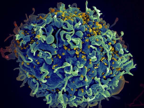
A new, more virulent and more damaging HIV variant has been discovered in the Netherlands.
Viral mutations are a source of concern because they can affect transmissibility and other factors. There have been fears of this happening in HIV-1, and now a new, highly virulent HIV strain in the Netherlands has been identified in a study. The results are published today in Science.
Prior to antiretroviral treatment, individuals infected with the new “VB variant” (for virulent subtype B) showed significant differences compared with individuals infected with other HIV variants:
A viral load between 3.5 and 5.5 times higher.
A doubled rate of CD4 cell decline (the hallmark of immune system damage by HIV), placing them at risk of developing AIDS much more rapidly.
Increased risk of transmitting the virus to others.
Fortunately, individuals with the VB variant had similar immune system recovery and survival to individuals with other HIV variants. However, because the VB variant causes a faster drop in immune system strength, early diagnosis and treatment is critical.
Researching the mechanism that causes the VB variant to be more transmissible and damaging to the immune system could lead to new targets for next-generation antiretroviral drugs. The VB variant is characterised by many mutations spread throughout the genome, meaning that a single genetic cause cannot currently be identified
Lead author Dr Chris Wymant said: ‘Before this study, the genetics of the HIV virus were known to be relevant for virulence, implying that the evolution of a new variant could change its impact on health. Discovery of the VB variant demonstrated this, providing a rare example of the risk posed by viral virulence evolution.’
Senior author Professor Christophe Fraser added: ‘Our findings emphasise the importance of World Health Organization guidance that individuals at risk of acquiring HIV have access to regular testing to allow early diagnosis, followed by immediate treatment. This limits the amount of time HIV can damage an individual’s immune system and jeopardise their health. It also ensures that HIV is suppressed as quickly as possible, which prevents transmission to other individuals.’
The VB variant was first identified in 17 HIV positive individuals from the BEEHIVE project, an ongoing study which collects samples from across Europe and Uganda. Since 15 of these people came from the Netherlands, the researchers then analysed data from a cohort of over 6700 HIV positive individuals in the Netherlands. This identified an additional 92 individuals with the variant, from all regions of the Netherlands, bringing the total to 109.
The researchers estimate that the VB variant first arose during the late 1980s and 1990s in the Netherlands, spreading more quickly than other HIV variants during the 2000s. However its spread has been declining since around 2010. The research team believe that the VB variant arose in spite of widespread treatment in the Netherlands, not because of it, since effective treatment can suppress transmission.
Since individuals with the VB variant are demographically similar, the spread is likely due to the properties of the virus itself.