Month: February 2022

CVD Risk Warning for Paracetamol That Contains Sodium

Graphical abstract from European Heart Journal editorial: sodium hidden in medication warrants warning labels by drug companies

Clinicians have recommended avoiding effervescent, soluble paracetamol that contains sodium, following findings from a large study that shows a link with a significantly increased risk of cardiovascular disease (CVD) and mortality in people who have hypertension and even in people with normal blood pressure.

The study of nearly 300 000 patients registered with UK GPs was published in the European Heart Journal.

Sodium is often used to help drugs such as paracetamol dissolve and disintegrate in water. However, effervescent and soluble formulations of 0.5g tablets of paracetamol can contain 0.44 and 0.39g of sodium respectively. If a person took the maximum daily dose of two 0.5g tablets every six hours, they would consume 3.5 and 3.1g of sodium respectively – a dose that exceeds the WHO-recommended total daily intake of 2g a day. In 2018, 170 people per 10 000 of the population in the UK were using sodium-containing medications, with a higher proportion among women. There are alternative formulations that contain little or no sodium.

Excessive salt in the diet remains a major public health problem and is associated with an increased risk of cardiovascular disease (CVD) and death among patients with hypertension. However, there is inconsistent evidence showing an increased risk in normotensive individuals.

Professor Chao Zeng led a team which analysed data from a medical database of UK GPs’ records. They looked at 4532 hypertensive patients who had been prescribed sodium-containing paracetamol and compared them with 146 866 hypertensive patients who had been prescribed sodium-free paracetamol. They also compared 5351 normotensive patients who were prescribed sodium-containing paracetamol with 141 948 normotensive patients prescribed sodium-free paracetamol. The patients were aged 60-90 years and followed up for one year.

The researchers found the risk of heart attack, stroke or heart failure after one year for patients with high blood pressure taking sodium-containing paracetamol was 5.6% (122 cases of CVD), while it was 4.6% (3051 CVD cases) among those taking sodium-free paracetamol. Mortality risk was also higher; the one-year risk was 7.6% (404 deaths) and 6.1% (5510 deaths), respectively.

A similar increased risk was seen among normotensive patients. Among those taking sodium-containing paracetamol, the one-year CVD risk was 4.4% (105 cases of CVD) and 3.7% (2079 cases of CVD) among those taking sodium-free containing paracetamol. The risk of dying was 7.3% (517 deaths) and 5.9% (5190 deaths), respectively.

Prof Zeng said: “We also found that the risk of cardiovascular disease and death increased as the duration of sodium-containing paracetamol intake increased. The risk of cardiovascular disease increased by a quarter for patients with high blood pressure who had one prescription of sodium-containing paracetamol, and it increased by nearly a half for patients who had five or more prescriptions of sodium-containing paracetamol. We saw similar increases in people without high blood pressure. The risk of death also increased with increasing doses of sodium-containing paracetamol in both patients with and without high blood pressure.”

Prof. Zeng said that clinicians and patients should be aware of the risks associated with sodium-containing paracetamol and avoid unnecessary consumption, especially when the medication is taken for a long period of time.

“Given that the pain relief effect of non-sodium-containing paracetamol is similar to that of sodium-containing paracetamol, clinicians may prescribe non-sodium-containing paracetamol to their patients to minimise the risk of cardiovascular disease and death. People should pay attention not only to salt intake in their food but also not overlook hidden salt intake from the medication in their cabinet,” he said.

“Although the US Food and Drug Administration requires that all over-the-counter medications should label the sodium content, no warning has been issued about the potentially detrimental effect of sodium-containing paracetamol on the risks of hypertension, cardiovascular disease and death. Our results suggest re-visiting the safety profile of effervescent and soluble paracetamol.”

Being an observational study it can only show only that there is an association between salt in paracetamol and CVD and deaths, rather than that salt causes these events. Other limitations include a lack of data on dietary intake of salt and excretion of salt from urinary samples. The use of over-the-counter paracetamol was not also recorded, however by restricting the study to those over 60 who qualify for free prescriptions in the UK, the risk of this is minimised.

Source: EurekAlert!

Indoor Air Pollution Linked to Pneumonia in Children

Streptococcus pneumoniae. Credit: CDC

A new study published in The Lancet Global Health, highlights the impact indoor air pollution can have on the development of child pneumonia, showing that increases in airborne particulate matter results in greater carriage of Streptococcus pneumoniae.

Streptococcus pneumoniae is a major human pathogen causing more than two million deaths per year; more than HIV/AIDS, measles and malaria combined, but it is also part of the normal microbial community of the nasopharynx. It is the leading cause of death due to infectious disease in children under five years of age; in sub-Saharan Africa, the burden of pneumococcal carriage and pneumonia is especially high.

Household air pollution from solid fuels increases the risk of childhood pneumonia. Nasopharyngeal carriage of S. pneumoniae is a necessary step in the development of pneumococcal pneumonia. More than 2.6 billion people are exposed to household air pollution worldwide. Inefficient indoor biomass burning is estimated to cause 3.8 million premature deaths annually and approximately 45% of all pneumonia deaths in children aged younger than five years. However, a causal pathway between household air pollution and pneumonia had not yet been identified.

In order to understand the connection between exposure to household air pollution and the risk of childhood pneumonia researchers from the UK, Malawi and the United States conducted the MSCAPE (Malawi Streptococcus pneumoniae Carriage and Air Pollution Exposure) study embedded in the ongoing CAPS (Child And Pneumonia Study) trial. The MSCAPE study assessed the impact of PM2.5, the single most important health-damaging pollutant in household air pollution, on the prevalence of pneumococcal carriage in a large sample of 485 Malawian children.

Through exposure-response analysis, a statistically significant 10% increase in risk of S. pneumoniae carriage in children was observed for a unit increase (deciles) of exposure to PM2.5 (ranging from 3.9 μg/m³ to 617.0 μg/m³).

Dr. Mukesh Dherani, the study principal investigator, indicated: “This study provides us with greater insight into the impact household air pollution can have on the development of child pneumonia. These findings provide important new evidence of intermediary steps in the causal pathway of household air pollution exposure to pneumonia and provide a platform for future mechanistic studies.”

Study author Professor Dan Pope said: “Moving forward further studies, particularly new randomized controlled trials comparing clean fuels (e.g. liquefied petroleum gas) with biomass fuels, with detailed measurements of PM2.5 exposure, and studies of mechanisms underlying increased pneumococcal carriage, are required to strengthen causal evidence for this component of the pathway from household air pollution exposure to ALRI in children.”

Professor Nigel Bruce, co-principal investigator, stated: “This study provides further important evidence that emphasises the need to accelerate to cleaner fuels, such as LPG, which are now being promoted by many governments across the continent in order to meet SDG7 by 2030.”

Source: University of Liverpool

Investigating Vaping’s Impact on Gum Disease

Photo by Chiara summer on Unsplash

A series of new studies investigates how e-cigarettes alter oral health and may be contributing to gum disease. The latest, published in mBio, finds that e-cigarette users have a distinctive oral microbiome that is less healthy than nonsmokers but potentially healthier than cigarette smokers, and measures worsening gum disease over time.

“To our knowledge, this is the first longitudinal study of oral health and e-cigarette use. We are now beginning to understand how e-cigarettes and the chemicals they contain are changing the oral microbiome and disrupting the balance of bacteria,” said co-lead researcher Prof Deepak Saxena.

While cigarette smoking is known to increase gum disease risk, much less is known about the impact of e-cigarette use on oral health, especially in the long term.

The researchers studied the oral health of 84 adults from three groups: cigarette smokers, e-cigarette users, and people who have never smoked. Gum disease was assessed through two dental exams six months apart, during which plaque samples were taken to analyse the bacteria present.

Gum health changes
All participants had some gum disease at the start of the study, with cigarette smokers having the most severe disease, followed by e-cigarette users. After six months, the researchers observed that gum disease had worsened in some participants in each group, including several e-cigarette users.

Clinical attachment loss is a key indicator of gum disease, measured by gum ligament and tissue separating from a tooth’s surface, leading the gum to recede and form pockets. These pockets are bacterial breeding grounds and can lead to worsening gum disease. In a study of the same participants published in Frontiers in Oral Health, the research team found that clinical attachment loss was significantly worse only in the e-cigarette smokers after six months.

A unique microbiome
Analysis of the bacteria in plaque samples showed that e-cigarette users had a different oral microbiome than smokers and nonsmokers, in line with their earlier results.

While all groups shared roughly a fifth of the types of bacteria, the bacterial makeup for e-cigarette users had strikingly more in common with cigarette smokers than nonsmokers. Several types of bacteria, including Selenomonas, Leptotrichia, and Saccharibacteria, were abundant in both smokers and vapers compared to nonsmokers. Several other bacteria – including Fusobacterium and Bacteroidales, linked to gum disease – were particularly dominant in the mouths of e-cigarette users.

When plaque samples were gathered and analysed in the six-month follow-up, the researchers found greater diversity in bacteria for all groups studied, yet each group maintained its own distinct microbiome.

“Vaping appears to be driving unique patterns in bacteria and influencing the growth of some bacteria in a manner akin to cigarette smoking, but with its own profile and risks to oral health,” said Fangxi Xu, study co-first author.

An altered immune response
The researchers found that the distinct microbiome in e-cigarette users was correlated with clinical measures of gum disease and changes to the host immune environment. In particular, vaping was associated with different levels of cytokines. Certain cytokines are linked to an imbalance in oral bacteria and can worsen gum disease by making people prone to inflammation and infection.

TNFα, a cytokine that causes inflammation, was significantly elevated among e-cigarette users. In contrast, cytokines IL-4 and IL-1β were lower among e-cigarette users; because IL-4 tends to be reduced in people with gum disease and increases after treatment, it suggests that certain bacteria in the mouths of e-cigarette users are worsening inflammation.

The researchers concluded that the distinct oral microbiome of e-cigarette users elicits altered immune responses, which along with clinical markers for gum disease illustrate how vaping presents its own challenge to oral health.

“E-cigarette use is a relatively new human habit,” said Scott Thomas, study co-first author. “Unlike smoking, which has been studied extensively for decades, we know little about the health consequences of e-cigarette use and are just starting to understand how the unique microbiome promoted by vaping impacts oral health and disease.”

Source: New York University

Blood Test for Alzheimer’s Proves Highly Accurate

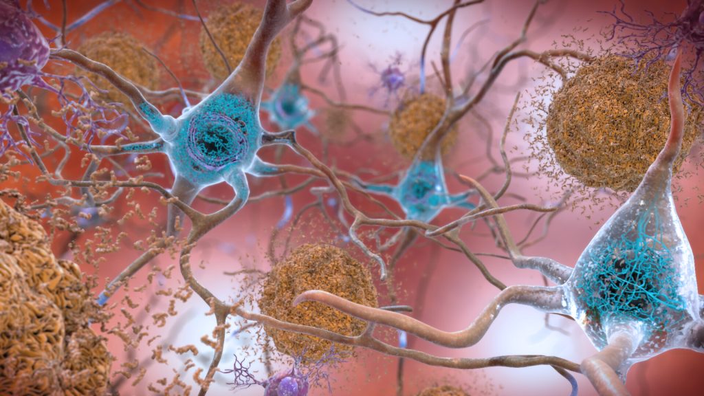
Plaques and neurons. Source: NIAH

A study in the journal Neurology has shown that a less expensive blood test to detect Alzheimer’s is highly accurate at early detection, providing further evidence that the test should be considered for routine screening and diagnosis. 

“Our study shows that the blood test provides a robust measure for detecting amyloid plaques associated with Alzheimer’s disease, even among patients not yet experiencing cognitive declines,” said senior author Professor Randall J. Bateman, MD.

“A blood test for Alzheimer’s provides a huge boost for Alzheimer’s research and diagnosis, drastically cutting the time and cost of identifying patients for clinical trials and spurring the development of new treatment options,” Prof Bateman said. “As new drugs become available, a blood test could determine who might benefit from treatment, including those at very early stages of the disease.”

Developed by Prof Bateman and colleagues, the blood test assesses whether amyloid plaques have begun accumulating in the brain based on the ratio of the levels of the amyloid beta proteins Aβ42 and Aβ40 in the blood.

The gold standard PET scan evaluation requires a radioactive brain scan, at an average cost of $5000–$8000 (R75 000–R120 000) per scan. Another common test, which analyses levels of amyloid-beta and tau protein in cerebrospinal fluid, costs about $1000 (R15 000) but requires a spinal tap process.

This study estimates that prescreening with a $500 (R7500) blood test could halve both the cost and the time it takes to enrol patients in clinical trials that use PET scans. Using only blood testing for screening could be done in under six months, a tenth or less of the cost. The test is currently only available in the US and Europe.

The current study shows that the blood test remains highly accurate, even when performed in different labs following different protocols, and in different cohorts across three continents.

Scientists didn’t know if small differences in sampling methods (such as anticoagulant use) could have a big impact on test accuracy because results are based on subtle shifts in amyloid beta protein levels in the blood. Subtle interfernece in these amyloid protein ratios could have triggered a false negative or positive result.

To confirm the test’s accuracy, researchers tested blood samples from current Alzheimer’s studies in the United States, Australia and Sweden, each of which uses different protocols for the processing of blood samples and related brain imaging.

Findings from this study confirmed that the Aβ42/Aβ40 blood test using a high-precision immunoprecipitation mass spectrometry technique developed at Washington University provides highly accurate and consistent results for both cognitively impaired and unimpaired individuals across all three studies.

When blood amyloid levels were combined with another major Alzheimer’s risk factor – the presence of the genetic variant APOE4 – the blood test accuracy was 88% compared to brain imaging and 93% when compared to spinal tap.

“These results suggest the test can be useful in identifying nonimpaired patients who may be at risk for future dementia, offering them the opportunity to get enrolled in clinical trials when early intervention has the potential to do the most good,” Prof Bateman said. “A negative test result also could help doctors rule out Alzheimer’s in patients whose impairments may be related to some other health issue, disease or medication.”

Source: Washington University School of Medicine

Scientists Unravel Neurological Origins of the Placebo Effect

Researchers at Massachusetts General Hospital (MGH) have discovered a network of brain regions activated by the placebo effect overlaps with several regions targeted by brain-stimulation therapy for depression.

The findings of this study, published in Molecular Psychiatry, will help in understanding the neurobiology of placebo effects and could inform how brain stimulation trial results are interpreted. In addition, this could provide insights on how to harness placebo effects for the treatment of a variety of conditions.

The placebo effect occurs when a patient’s symptoms improve because they expect a therapy to help (due to a variety of factors), but not from the specific effects of the treatment itself. Recent research indicates that there is a neurological basis for the placebo effect, with imaging studies identifying a pattern of changes that happen in certain brain regions when a person experiences this phenomenon.

The use of brain-stimulation techniques for patients with depression that doesn’t respond adequately to medication or psychotherapy has gained wider use in recent years. Transcranial magnetic stimulation (TMS) delivers electromagnetic pulses to the brain, and its effect on brain activity has been established over the last three decades in animal and human research studies, with several TMS devices approved by the Food and Drug Administration for treating depression. In addition, for treatment depression, deep brain stimulation (DBS, which requires an implanted device) has shown some promise.

Senior author Emiliano Santarnecchi, PhD, saw studies of brain stimulation as a unique opportunity to learn more about the neurobiology of the placebo effect. Santarnecchi and his co-investigators conducted a meta-analysis and review of neuroimaging studies involving healthy subjects and patients to create a “map” of brain regions activated by the placebo effect. They also analysed studies of people treated with TMS and DBS for depression to identify brain regions targeted by the therapies. The team found that several sites in the brain that are activated by the placebo effect overlap with brain regions targeted by TMS and DBS.

Dr Santarnecchi and his colleagues believe that this overlap has critical importance in interpreting the results of research on brain stimulation for conditions such as depression. In clinical trials, a significant portion of depression patients receiving brain stimulation improve — but so do many patients receiving placebo (sham) treatment, in which no stimulation is administered, which has led to confusion over the therapy’s benefits.

A possible explanation is “that there is a significant placebo effect when you do any form of brain stimulation intervention,” said Dr Santarnecchi. TMS involves a clinical setting, with loud clicks as the pulse is delivered. “So the patient thinks, ‘Wow, they are really activating my brain’, so you get a lot of expectation,” said Dr Santarnecchi.

Elevated placebo effects associated with brain stimulation may create problems when studying the intervention, said first author Matthew Burke, MD, a cognitive neurologist. If brain stimulation and the placebo effect overlap in activating the same brain regions, then those circuits could be maximally activated by placebo effects, which could make it difficult to show any additional benefit from TMS or DBS, said Dr Burke. If so, this could explain the disparity of results in neurostimulation treatment of depression. Screening out placebo from brain stimulation’s direct impact on brain activity will help in designing studies where the real potential of techniques such as TMS will be more easily quantified, thus improving the effect of treatment protocols.

The findings from this study also suggest broad applications for the placebo effect, said Dr Santarnecchi. “We think this is an important starting point for understanding the placebo effect in general, and learning how to modulate and harness it, including using it as a potential therapeutic tool by intentionally activating brain regions of the placebo network to elicit positive effects on symptoms,” he said.

Dr Santarnecchi and his colleagues are currently designing trials that they hope will “disentangle” the effects of brain stimulation from placebo effects and offer insights about how they can be leveraged in clinical settings.

Source: Massachusetts General Hospital

Sound Waves Used to Regrow Bone

Image by Pawel Czerwinski on Unsplash

In a significant advance for the field of tissue engineering, researchers have used sound waves to turn stem cells into bone cells, a technology which may help regrow bone lost by cancer or disease.

Described in the journal Small, the innovative stem cell treatment from researchers at RMIT University offers a smart way forward for overcoming some of the field’s biggest challenges, through the precision power of high-frequency sound waves.

Tissue engineering is an emerging field that aims to rebuild bone and muscle by harnessing the human body’s natural ability to heal itself. A key challenge in regrowing bone is having sufficient amounts of bone cells that can thrive once implanted in the target area.

So far, turning stem cells into bone cells has needed complicated and expensive equipment, making widespread clinical use unrealistic.

The few clinical trials trying to regrow bone mostly used stem cells painfully extracted from a patient’s bone marrow.

In a new study published in the journal Small, the RMIT research team showed stem cells treated with high-frequency sound waves turned into bone cells quickly and efficiently.

Importantly, the treatment was effective on multiple types of cells including fat-derived stem cells, which are far less painful to extract from a patient.

Co-lead researcher Dr Amy Gelmi said the new approach was faster and simpler than other methods.

“The sound waves cut the treatment time usually required to get stem cells to begin to turn into bone cells by several days,” said Dr Gelmi. “This method also doesn’t require any special ‘bone-inducing’ drugs and it’s very easy to apply to the stem cells.

“Our study found this new approach has strong potential to be used for treating the stem cells, before we either coat them onto an implant or inject them directly into the body for tissue engineering.”

The high-frequency sound waves used in the stem cell treatment were generated on a low-cost microchip device developed by RMIT.

Co-lead researcher Distinguished Professor Leslie Yeo and his team have spent over a decade researching the interaction of sound waves at frequencies above 10MHz with different materials.

The sound wave-generating device they developed can be used to precisely manipulate cells, fluids or materials.

“We can use the sound waves to apply just the right amount of pressure in the right places to the stem cells, to trigger the change process,” Prof Yeo said.

“Our device is cheap and simple to use, so could easily be upscaled for treating large numbers of cells simultaneously – vital for effective tissue engineering.”

The next stage in the research is investigating methods to upscale the platform, working towards the development of practical bioreactors to drive efficient stem cell differentiation.

Source: RMIT

Hypertension Risk for Women After Sexual Assault or Harassment

Photo by Sydney Sims on Unsplash

A new study has found that women who have experienced sexual assault or harassment are at higher long-term risk of developing hypertension than women who have not.

The study appears in the Journal of the American Heart Association.

In the US, nearly 43% of women aged 20 and older have hypertension. Defined as a blood pressure of 130/80mmHg or higher, hypertension is a major risk factor for cardiovascular disease – the number one killer of women, causing one in three deaths each year.

“We know that experiences of sexual violence in the form of sexual assault and workplace sexual harassment are common, and that women are disproportionately victims of such violence, with 13–44% of women reporting sexual assault and up to 80% of women reporting workplace sexual harassment,” said study author Rebecca B. Lawn, PhD. “However, exposure to sexual violence is not widely recognized as a contributor to women’s cardiovascular health. We felt it was important to investigate the relationship among common forms of sexual violence with the risk of developing hypertension. These links could help in the early identification of factors that influence women’s long-term cardiovascular health.”

In this study, researchers analysed data over the course of seven years beginning with a 2008 follow-up of the Nurses’ Health Study II, an ongoing cohort study of US women. The 2008 follow-up measured the incidence of sexual violence and other trauma exposure, as well as post-traumatic stress disorder (PTSD) and symptoms of depression, among a subset of 54 703 of the study’s original participants.

From that subset, Lawn and colleagues analysed data for 33 127 women (95% non-Hispanic white women; average age of 53 years at the beginning of the 2008 follow-up) who had no history of hypertension or had not taken medication for high blood pressure as of the start of the 2008 follow-up.

The analyses found:

  • At the seven-year follow-up in 2015, about 1 in 5 (nearly 7100) of the women self-reported they had developed hypertension, validated with medical records.
  • Sexual assault and workplace sexual harassment were common, with lifetime prevalence of 23% for sexual assault and 12% for workplace sexual harassment; 6% of women reported experiencing both.

Compared to women with no history of sexual assault or harassment, women who reported having experienced both had the greatest increased hypertension risk (21%), followed women who reported experiencing workplace sexual harassment (15%) and an women who reported experiencing sexual assault (11%).

“We did not find any association of increased risk for hypertension among women who had a history of other types of trauma and who did not experience sexual violence, suggesting that increased hypertension risk does not appear to be associated with all trauma exposure,” Dr Lawn said. “Our finding that experiencing both sexual assault and workplace sexual harassment had the highest risk of hypertension underscores the potential compounding effects of multiple sexual violence exposures on women’s long-term cardiovascular health.”

Dr Lawn observed screening for partner violence by primary care clinicians is becoming more common, sexual violence overall is not recognised as a risk factor among women for developing cardiovascular disease.

“These results suggest that screening for a broader range of experiences of sexual violence in routine health care, including sexual harassment in the workplace, as well as verbal harassment or assault, and being aware of and treating potential cardiovascular health consequences may be beneficial for women’s long-term health,” she said. “Reducing sexual violence against women, which is important in its own right, may also provide a strategy for improving women’s lifetime cardiovascular health.”

There are several limitations to the study, including memory biases in recall of sexual violence. The sexual assault and harassment had no measures of severity or timing. Most of the women in the study were white women in the nursing field, limiting generalisability.

“We hope future studies will examine these questions with more detailed information on sexual and other forms of violence. These questions need to be investigated in more diverse groups of people of various ages, races and ethnic backgrounds and gender,” Dr Lawn said. “Although women are disproportionately victims of sexual violence, men are also victims and the physical health implications of experiences of sexual violence against men warrants further investigation.”

Source: American Heart Association

Greenspaces in Hospitals Calm Patients and Visitors

It can be stressful and time-consuming for patients and visitors to become accustomed to navigating large, unfamiliar hospitals, and so an architecture researcher tested a simple remedy: to let nature in with the use of indoor greenspaces and large windows.

Research conducted by West Virginia University’s associate professor Shan Jiang showed that introducing nature into large hospitals can humanise the institutional environment and reduce the stress of patients, visitors and healthcare providers.

Prof Jiang made use of immersive virtual environments for a controlled experiment that asked participants to complete various wayfinding tasks in a simulated hospital.

Though participants saw the same layout, one group encountered large windows and nature views among the corridor walls. The control group meanwhile saw solid walls without any daylight or views of nature, more like a typical modern hospital. Participants in the greenspace group used shorter time and walked less distance to complete wayfinding tasks.

“In terms of spatial orientation and wayfinding, window views of nature and small gardens can effectively break down the tedious interiors of large hospital blocks,” Prof Jiang said, “and serve as landmarks to aid people’s wayfinding and improve their spatial experience.”

In the greenspace group, participants’ mood states, particularly anger and confusion, were also found to be “significantly relieved”.

Prof Jiang’s findings are published in the Health Environments Research and Design Journal.

Based on prior research, it’s estimated that a patient or hospital visitor must go through at least seven steps in the wayfinding process to arrive at the final destination. WVU’s Center for Health Design cites wayfinding issues as an environmental stressor and a concerning topic in healthcare design.

Prof Jiang said that she was prompted to do the study by those factors, coupled with her own personal experiences (her family members have worked in healthcare) and others’ accounts of feeling lost in hospitals.

“Large hospitals can be visually welcoming but the functionality and internal circulation are indeed complex and confusing,” she said.

Greenspaces positioned at key decision points, such as main corridors or junctions, can help improve navigation.

With a background in landscape architecture, Jiang has been interested in the immediate surroundings of people in a smaller scope, particularly the indoor-outdoor relationship and the boundaries between architecture and landscapes.

Gardens and plants also tend to have strong therapeutic effects on people, she found.

“You may explain such therapeutic effects from multiple perspectives: people’s colour/hue preferences tend to range from blue to green, nature and plants are positive distractions that could restore people’s attentional fatigue, and human beings could have developed genetic preference of greenery from evolutionary perspectives,” Prof Jiang said. “All mechanisms together contribute to the positive experience when looking at gardens and nature views.”

Prof Jiang noted that many European hospitals have successfully integrated “hospital in a park” concepts. In the United States, the Lucile Packard Children’s Hospital Stanford in California has patios and window nooks in every patient room, and most rooms have direct views of a large healing garden, she said. The Alder Hey Children’s Hospital in the UK was literally built in a park.

Source: West Virginia University

Do People’s Lives ‘Flash Before Their Eyes’ When They Die?

Source: Pixabay CC0

By chance, neuroscientists were able to record the activity of a dying human brain and discovered brain wave patterns similar to dreaming, memory recall, and meditation. An analysis of this case, reported in Frontiers in Aging Neuroscience suggests a possible explanation for near-death experiences.

Imagine reliving your entire life in the space of seconds. Like a flash of lightning, you are outside of your body, watching memorable moments you lived through. This process, known as ‘life recall’, can be similar to what it’s like to have a near-death experience. What happens inside your brain during these experiences and after death are questions that have puzzled neuroscientists for centuries. However, the present study suggests that your brain may remain active and coordinated during and even after the transition to death, and may in fact be programmed to orchestrate the whole ordeal.

When an 87-year-old patient developed epilepsy, Dr Raul Vicente of the University of Tartu, Estonia and colleagues used continuous electroencephalography (EEG) to detect the seizures and treat the patient. During these recordings, the patient had a heart attack and passed away. This unexpected event allowed the scientists to record the activity of a dying human brain for the first time ever.

“We measured 900 seconds of brain activity around the time of death and set a specific focus to investigate what happened in the 30 seconds before and after the heart stopped beating,” said Dr Ajmal Zemmar, a neurosurgeon at the University of Louisville, US, who organised the study.

“Just before and after the heart stopped working, we saw changes in a specific band of neural oscillations, so-called gamma oscillations, but also in others such as delta, theta, alpha, and beta oscillations.”

Brain oscillations (aka ‘brain waves’) are patterns of rhythmic brain activity normally present in living human brains. These different types of oscillations, including gamma, are involved in high-cognitive functions, such as concentrating, dreaming, meditation, memory retrieval, information processing, and conscious perception, just like those associated with memory flashbacks.

“Through generating oscillations involved in memory retrieval, the brain may be playing a last recall of important life events just before we die, similar to the ones reported in near-death experiences,” Dr Zemmar speculated. “These findings challenge our understanding of when exactly life ends and generate important subsequent questions, such as those related to the timing of organ donation.”

Though this is the first study to ever measure live brain activity during the process of dying in humans, similar changes in gamma oscillations have been previously recorded in rats kept in controlled environments. This raises the possibility that, during death, the brain organises and executes a biological response that could be conserved across species.

The interepretation of this however is complicated by the fact that these measurements are based on a single case and stem from the brain of a patient who had suffered injury, seizures and swelling. Nonetheless, Dr Zemmar plans to investigate more cases and sees these results as a source of hope.

“As a neurosurgeon, I deal with loss at times. It is indescribably difficult to deliver the news of death to distraught family members,” he said.

“Something we may learn from this research is: although our loved ones have their eyes closed and are ready to leave us to rest, their brains may be replaying some of the nicest moments they experienced in their lives.”

Source: Frontiers

Heart Attack Survivors at Lower Risk of Parkinson’s

Credit: American Heart Association

Heart attack survivors may be slightly less likely to develop Parkinson’s disease later in life, according to new research published in the Journal of the American Heart Association.

Parkinson’s disease (PD) is a common neurodegenerative disorder. While a number of non-motor manifestations arise, the typical clinical features involve a movement disorder consisting of bradykinesia, resting tremor, and rigidity, with postural instability occurring at a later stage. The cause of PD is not known, but a number of genetic risk factors have now been characterised, as well as several genes which cause rare familial forms of PD. Secondary parkinsonism, which has symptoms similar to Parkinson’s disease, may be caused by stroke, psychiatric or cardiovascular medications, or other illness.

“We have previously found that following a heart attack, the risk of neurovascular complications such as ischaemic stroke or vascular dementia is markedly increased, so the finding of a lower risk of Parkinson’s disease was somewhat surprising,” said lead study author Jens Sundbøll, MD, PhD. “These findings indicate that the risk of Parkinson’s disease is at least not increased following a heart attack and should not be a worry for patients or a preventive focus for clinicians at follow-up.

“It is not known whether this inverse relationship with risk of Parkinson’s disease extends to people who have had a heart attack. Therefore, we examined the long-term risk of Parkinson’s disease and secondary parkinsonism among heart attack survivors,” Dr Sundbøll said.

Drawing on Danish National Health Service data, the researchers compared the risk of PD and secondary parkinsonism among roughly 182 000 patients who had a first-time heart attack between 1995 and 2016 (average age 71 years old; 62% male) and more than 909 000 matched controls. 

Over a maximum continual follow-up of 21 years, after adjusting for a wide range of potential confounding factors, the analysis found that, when compared to the control group:

  • there was a 20% lower risk of PD among people who had a heart attack; and
  • a 28% lower risk of secondary parkinsonism among those who had a heart attack.

“For physicians treating patients following a heart attack, these results indicate that cardiac rehabilitation should be focused on preventing ischaemic stroke, vascular dementia and other cardiovascular diseases such as a new heart attack and heart failure, since the risk of Parkinson’s appears to be decreased in these patients, in comparison to the general population,” Dr Sundbøll said.

Certain risk factors are common to both heart attack and PD, with higher risk found among elderly men and lower risk among people who drink more coffee and are more physically active. Interestingly, however, some classic heart attack risk factors – such as smoking, high cholesterol, hypertension and Type 2 diabetes – are associated with a reduced risk of PD.

In general, more heart attack patients smoke and have high cholesterol, either of which may explain the slightly reduced risk of PD among heart attack survivors.

“There are very few diseases in this world in which smoking decreases risk: Parkinson’s disease is one, and ulcerative colitis is another. Smoking increases the risk of the most common diseases including cancer, cardiovascular disease and pulmonary disease and is definitely not good for your health,” Dr Sundbøll noted.

One limitation of the study is that there was not enough information about smoking and high cholesterol levels among the participants, which may have influenced the findings. The study participants were almost entirely white, limiting the generalisability to other ethnic groups.

Source: American Heart Association